27 Normal abdominal CT: a retrospective study of renal hila angles
Radiol Bras. 2009 Jan/Fev;42(1):27–29 Original Article • Artigo Original
Normal abdominal computed tomography: a retrospective
study of renal hila angles*
Tomografia computadorizada de abdome normal: estudo retrospectivo das angulações dos hilos renais
Makoto Sakate1, Alzira Teruio Yida Sakate1, Seizo Yamashita2, Altamir dos Santos Teixeira2, Luciano Barbosa3, Luis Antonio Correia4
OBJECTIVE: To obtain mean angulation values for renal hila in relation to the horizontal plane traced over the right and left spinal erector muscles, considering the center of the lumbar vertebral spine as a parameter for measuring the renal hila angles. MATERIALS AND METHODS: The authors have analyzed 250 abdominal computed tomography studies of both male and female healthy individuals (128 men with mean age 52.45 ± 17.42 years, and 122 women with mean age 54.39 ± 18.27 years), corresponding to 500 renal hila evaluated. The mean angulation of each hilum in relation to the horizontal plane was obtained taking acute angles into consideration. RESULTS: The comparative study have not found any statistically significant difference in acute angles of renal hila between male and female individuals. The statistical analysis demonstrated limits of 40.40° and 44.54° for mean right hilum angulation and 39.91° and 43.23° for mean left hilum angulation, with a confidence interval of 95% CONCLUSION: Renal hila present similar angulation independently of sex. Higher angulation values correspond to hyper-rotation or excessive rotation, and lower angulation values, to incomplete or reverse rotation.
Keywords: Congenital anomalies; Renal pelvis; Urinary tract; Helical computed tomography.
OBJETIVO: Obter valores da angulação média dos hilos renais em relação ao plano horizontal, traçado sobre músculos eretores da espinha direito e esquerdo, considerando como parâmetro de referência para as medi-das medi-das angulações o centro da coluna vertebral lombar. MATERIAIS E MÉTODOS: Foram analisados 250 exames de tomografia computadorizada de abdomes considerados normais de indivíduos de ambos os se-xos, sendo 128 masculinos (idade média de 52,45 ± 17,42 anos) e 122 femininos (idade média de 54,39 ± 18,27 anos), totalizando 500 hilos renais estudados. A angulação média de cada hilo renal em relação ao plano horizontal foi obtida, sendo considerados sempre os ângulos agudos. RESULTADOS: O estudo com-parativo entre os sexos mostrou que não houve diferença estatisticamente significante em relação aos ân-gulos agudos dos hilos renais. A análise estatística mostrou, com intervalo de confiança de 95%, para média do ângulo direito os limites de 40,40° e 44,54° e para o hilo renal esquerdo os limites de 39,91° e 43,23°. CONCLUSÃO: Os hilos renais, independentemente do sexo, apresentam angulações semelhantes. Valores angulares maiores terão anomalia de hiper-rotação ou hiper-rotação exagerada e valores menores terão ano-malia de rotação incompleta ou rotação invertida.
Unitermos: Anomalias congênitas; Pelve renal; Trato urinário; Tomografia computadorizada helicoidal.
Abstract
Resumo
* Study developed at Faculdade de Medicina de Botucatu – Universidade Estadual Paulista “Júlio de Mesquita Filho” (Unesp), Botucatu, SP, Brazil.
1. PhDs, Assistant Professors at Faculdade de Medicina de Botucatu – Universidade Estadual Paulista “Júlio de Mesquita Filho” (Unesp), Botucatu, SP, Brazil.
2. Assistant Professors at Faculdade de Medicina de Botucatu – Universidade Estadual Paulista “Júlio de Mesquita Filho” (Unesp), Botucatu, SP, Brazil.
3. Assistant Professor at Instituto de Biociências – Universidade Estadual Paulista “Júlio de Mesquita Filho” (Unesp), Botucatu, SP, Brazil.
4. Full Professor at Faculdade de Medicina de Botucatu – Universidade Estadual Paulista “Júlio de Mesquita Filho” (Unesp), Botucatu, SP, Brazil.
Mailing address: Dr. Makoto Sakate. Rua Aleixo Varoli, 651, Jardim Paraíso. Botucatu, SP, 18610-295, Brazil. E-mail: [email protected]
Received May 8, 2008. Accepted after revision November 18, 2008.
Rotational anomalies are classified into non-rotation, incomplete, reverse and ex-cessive rotation. Non-rotation (renal hilum turned anteriorly) and incomplete rotation (renal hilum between the anterior and me-dial regions) are most frequently found. Reverse and excessive rotations are rarely found(1).
Late in the embryonic phase, the renal hila are anteriorly positioned and, in the early fetal phase, during the ascent of the kidneys to its normal bed, they rotate me-dially with their respective hila, lying in an intermediate position between medial and Sakate M, Yida-Sakate AT, Yamashita S, Teixeira AS, Barbosa L, Correia LA. Normal abdominal computed tomography: a retrospective study of renal hila angles. Radiol Bras. 2009;42(1):27–29.
INTRODUCTION
Diagnostic imaging specialists have a qualitative view on the normal positioning of renal hila at abdominal computed to-mography (CT); however, the authors have not found in the literature quantitative pa-rameters for defining such positioning as related to vascularization (inferior vena cava and abdominal aorta) in a healthy patient. Quantitative (angular) parameters of nor-mal renal hila are necessary for a better definition of congenital abnormalities, es-pecially regarding rotation of renal hila.
28
Sakate M et al.
Radiol Bras. 2009 Jan/Fev;42(1):27–29
anterior(2). However, angular values or
ana-tomical parameters are not available for defining the normal positioning of renal hila.
In the search for a better definition of anomalies of renal hila rotation, the present study is aimed at evaluating individuals with normal abdomens for obtaining quan-titative values of mean angulation values for renal hila in relation to the horizontal plane traced over the right and left spinal erector muscles, considering the center of the lumbar vertebral spine as a parameter for measurement of renal hila angles.
MATERIALS AND METHODS
The present study evaluated retrospec-tively 250 abdominal CT studies with oral and intravenous contrast enhancement of both male (128) and female (122) healthy individuals in the age range between 17 and 93 years (mean 52.45 ± 17.42 years) for men, and 21 and 94 years (mean 54.34 ± 18.27 years) for women.
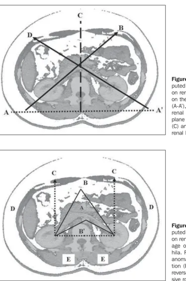
All of the patients included in this study presented with normal renal function, with no renal or adjacent disorders, correspond-ing to a total of 500 renal hila (250 right and 250 left renal hila) evaluated. On magnified abdominal CT cross-sectional views dem-onstrating renal hila, two lines were drawn: one horizontal line over the right and left spinal erector muscles, and another, per-pendicular to the horizontal plane, passing on the middle of the lumbar vertebral body. This horizontal line was considered as the zero degree (0°) angle, and the perpendicu-lar line as an anguperpendicu-lar reference parameter. The renal hila angulation was calculated from the center of the lumbar vertebra, so the angle calculation for each renal hilum in relation to the horizontal plane has ever taken acute angles into consideration (Fig-ure 1).
The research project was approved by the Committee for Ethics in Research of the Institution, and a term of free and informed consent was signed by the individuals in-cluded in the present study.
The t-Student test was utilized for com-parison of average ages. Averages were ob-tained, and a 95% confidence interval was constructed for the average of right and left renal hila angles(3).
RESULTS
Comparative study between sexes showed that there was no statistically sig-nificant difference (p = 0.40376) related to renal hila acute angles.
Statistical analysis of 500 renal hila angles calculated from the center of the lumbar vertebra on the horizontal plane determined on the right and left spinal erec-tor muscles demonstrated the following results: mean 42.47° and ± 16.64 standard-deviation for right renal hila angle, and mean 41.57° and ± 13.29° standard devia-tion for left renal hila angle. The confi-dence interval of 95% demonstrated limits of 40.40° and 44.54° for mean angle of the right renal hila, and 39.91° and 43.23° for mean angle of left renal hila, that is to say that every interval has a 95% probability of including the average for the study popu-lation (Figure 2).
DISCUSSION
According to the literature, the kidneys positioning and anatomic orientation do not present any rotational variation with aging for both male and female individu-als(4).
The upper urinary tract anatomy is dem-onstrated by CT slices of total or upper abdomen. On the unenhanced phase, the renal contour and the calyceal system ap-pear well-defined at CT images because of the peri- and pararenal fat and the renal sinuses(5,6). After intravenous iodinated
contrast agent bolus injection, enhance-ment of the kidneys is observed during the nephrographic phase, and subsequently the renal hila can be bilaterally identified be-tween the anterior and medial regions(7).
Computed tomography allows a noticeable topographic definition of the renal vessels (artery and vein), renal parenchyma and
Figure 1. Abdominal com-puted tomography, axial slice on renal hila. Imaginary plane on the spinal erector muscles (A-A’), inclination of the right renal hilum (B), reference plane of renal hila angulation (C) and inclination of the left renal hilum (D).
29 Normal abdominal CT: a retrospective study of renal hila angles
Radiol Bras. 2009 Jan/Fev;42(1):27–29
renal hila(8). Three or four minutes
follow-ing intravenous contrast injection, the re-nal parenchyma becomes homogeneous and the pyelocalyceal system is filled with contrast, corresponding to the pyolographic phase(9,10).
The pyelographic phase allows a spatial visualization and topographic determina-tion of normal renal hila, as well as renal abnormalities in relation to anatomic struc-tures. Congenital renal malformations may present as abnormalities of surface, vol-ume, number, fusion, migration with or without fusion, and rotation. Rotational abnormalities have been identified at CT by the direction of the opening of the diver-gent renal hilum in the medial region, with-out utilizing any quantitative parameter for inferring a degree or anatomic structure for reference(11).
Most frequently, rotational anomalies are associated with renal ectopia although this phenomenon may occur on its normal bed. The four types of anomalies described are the following: non-rotation, incomplete rotation, reverse rotation and excessive rotation; among them, non-rotation (renal pelvis turned towards the ventral region) and incomplete rotation (renal hilum be-tween the anterior and medial regions of the abdomen) are most frequently found(1,12).
Reverse and excessive rotations are rarely found. In reverse rotation, the renal vessels are observed anteriorly to the kid-ney, and in excessive rotation, posteriorly. These anomalies are responsible for mis-takes in the interpretation of excretory urography studies where an organ may be confused with an abnormal mass and inad-vertently being surgically removed(2,13,14).
However, with the introduction of com-puted tomography, these anomalies have become more perceptible, allowing the vi-sualization and characterization of rota-tional details.
The most significant aspect to be taken
into consideration in cases of malpositioned kidneys, frequently associated with rota-tional anomalies, is the higher predisposi-tion to excretory pathway obstrucpredisposi-tion. Obstruction generally occurs in the uretero-pyelic junction with dilatation of the renal pelvis (hydronephrosis) as a result from the compression by aberrant vessels or by the abnormal position of the hilum itself, pre-disposing to complications caused by dila-tation and urinary stasis(14).
The present study has allowed the quan-tification of renal hila positioning in nor-mal abdomens in terms of mean angular values. This interval is within the classifi-cation of congenital abnormality corre-sponding to incomplete rotation, although not occupying the whole angular range for the mentioned anomaly (Figure 2). Higher angular values suggest non-rotation, in-complete rotation or reverse rotation, and lower angular values, hyper-rotation (or ex-cessive rotation).
Upon the above considerations, a new definition of rotation anomalies in terms of angular values is necessary for patients submitted to abdominal CT, with changes in the parameters reported in the literature.
CONCLUSION
The evaluation of angular rotation of right and left renal hila in relation to a hori-zontal plane over the spinal erector muscles, with the center of the vertebral body as an angular reference in patients with no evidence of renal or adjacent ab-normalities has demonstrated two signifi-cant findings: firstly, in the confidence in-terval of 95%, the average between the right and left hila angles is within the clas-sification of congenital abnormality corre-sponding to incomplete rotation; and sec-ondly, this abnormality should be divided into normal and abnormality of incomplete rotation.
REFERENCES
1. Emmet JL, Witten DM. Embryology of the geni-tourinary tract. In: Emmet JL, Witten DM, editors. Clinical urography. An atlas and textbook of roentgenologic diagnosis. 3rd ed. Philadelphia: WB Saunders; 1971. p. 1313–48.
2. Netter FH. Apparent “ascent and rotation” of the kidneys in embryologic development. In: Netter FH, Shapter RK, Yonkman FF, editors. The Netter collection of medical illustrations – Kidneys, ure-ters and urinary bladder. 2nd ed. Vol. 6. Roches-ter: Ciba; 1975. p. 35.
3. Zar JH. Biostatistical analysis. New Jersey: Pren-tice-Hall; 1996.
4. Meschan I, Parker MD. Urinary tract. In: Meschan I, editor. Roentgen signs in diagnostic imaging. Abdomen. 2nd ed. Philadelphia: WB Saunders; 1986. p. 145–316.
5. Parienty RA, Pradel J. Radiological evaluation of the peri- and pararenal spaces by computed tomog-raphy. Crit Rev Diagn Imaging. 1983;20:1–26.
6. Prassopoulos P, Cavouras D. Renal parenchymal thickness in children measured by computed to-mography. Eur Urol. 1994;25:51–4.
7. Emmet JL, Witten DM. The normal urogram. In: Emmet JL, Witten DM, editors. Clinical urogra-phy. An atlas and textbook of roentgenologic di-agnosis. 3rd ed. Philadelphia: WB Saunders; 1971. p. 267–339.
8. Dalla Palma L, Rossi M. Advances in radiologi-cal anatomy of the kidney. Br J Radiol. 1982;55: 404–12.
9. Dalla Palma L, Bazzocchi M, Cressa C, et al. Ra-diological anatomy of the kidney revisited. Br J Radiol. 1990;63:680–90.
10. Yokoyama M, Watanabe K, Inatsuki S, et al. Mea-surement of renal parenchymal volume using computed tomography. J Comput Assist Tomogr. 1982;6:975–7.
11. Yokoyama M, Watanabe K, Inatsuki S, et al. Com-puterized tomography of the kidney: tissue-plasma ratio of contrast enhancement with bolus injection and renal function. J Urol. 1982;127: 721–3.
12. Netter FH. Anomalies in rotations. In: Netter FH, Shapter RK, Yonkman FF, editors. The Netter col-lection of medical illustrations – Kidneys, ureters and urinary bladder. 2nd ed. Vol. 6. Rochester: Ciba; 1975. p. 230.
13. Emmet JL, Witten DM. Anomalies of the geni-tourinary tract. In: Emmet JL, Witten DM, editors. Clinical urography. An atlas and textbook of roentgenologic diagnosis. 3rd ed. Philadelphia: WB Saunders; 1971. p. 1349–603.