25
Swallowing quantitative analysis software
Radiol Bras. 2008;41(1):25–28 Original Article
Swallowing quantitative analysis software*
Software para análise quantitativa da deglutiçãoAndré Augusto Spadotto1
, Ana Rita Gatto2
, Paula Cristina Cola3
, Arlindo Neto Montagnoli4 , Arthur Oscar Schelp5
, Roberta Gonçalves da Silva6
, Seizo Yamashita7
, José Carlos Pereira8 , Maria Aparecida Coelho de Arruda Henry9
OBJECTIVE: The present paper is aimed at introducing a software to allow a detailed analysis of the swal-lowing dynamics. MATERIALS AND METHODS: The sample included ten (six male and four female) stroke patients, with mean age of 57.6 years. Swallowing videofluoroscopy was performed and images were digi-tized for posterior analysis of the pharyngeal transit time with the aid of a chronometer and the software. RESULTS: Differences were observed in the average pharyngeal swallowing transit time as a result of mea-surements with chronometer and software. CONCLUSION: This software is a useful tool for the analysis of parameters such as swallowing time and speed, allowing a better understanding of the swallowing dynamics, both in the clinical approach of patients with oropharyngeal dysphagia and for scientific research purposes. Keywords: Swallowing videofluoroscopy; Software; Quantitative analysis.
OBJETIVO: Apresentar um software que permita uma análise detalhada da dinâmica da deglutição.
MATE-RIAIS E MÉTODOS: Participaram deste estudo dez indivíduos após acidente vascular encefálico, sendo seis do gênero masculino, com idade média de 57,6 anos. Foi realizada videofluoroscopia da deglutição e as imagens foram digitalizadas em microcomputador, com posterior análise do tempo do trânsito faríngeo da deglutição, por meio de um cronômetro e do software. RESULTADOS: O tempo médio do trânsito faríngeo
da deglutição apresentou-se diferente quando comparados os métodos utilizados (cronômetro e software).
CONCLUSÃO: Este software é um instrumento de análise dos parâmetros tempo e velocidade da deglutição,
propiciando melhor compreensão da dinâmica da deglutição, com reflexos tanto na abordagem clínica dos pacientes com disfagia como para fins de pesquisa científica.
Unitermos: Videofluoroscopia da deglutição; Software; Análise quantitativa.
Abstract
Resumo
* Study developed in the Department of Neurology and Psy-chiatry at Faculdade de Medicina de Botucatu da Universidade Estadual Paulista Júlio de Mesquita Filho (Unesp), Botucatu, SP, Brazil.
1. Master, Fellow PhD Degree in Electrical Engineering at Universidade de São Paulo (USP), São Carlos, SP, Brazil.
2. Speech-language Therapist, Supplementary Education fo-cused on Phonoaudiology Applied to Neurology, Universidade Estadual Paulista Júlio de Mesquita Filho (Unesp), Botucatu, SP, Brazil.
3. Master, Speech-language Therapist, Universidade Estadual Paulista Júlio de Mesquita Filho (Unesp), Botucatu, SP, Brazil.
4. PhD, Teacher at Universidade Estadual Paulista Júlio de Mesquita Filho (Unesp), Rio Claro, SP, Brazil.
5. PhD, Teacher, Department of Neurology and Psychiatry, Universidade Estadual Paulista Júlio de Mesquita Filho (Unesp), Botucatu, SP, Brazil.
6. PhD, Teacher, Department of Phonoaudiology, Universidade Estadual Paulista Júlio de Mesquita Filho (Unesp), Marília, SP, Brazil.
7. Master, Professor of the Discipline of Radiodiagnosis at Faculdade de Medicina de Botucatu da Universidade Estadual Paulista Júlio de Mesquita Filho (Unesp), Botucatu, SP, Brazil.
8. Titular Professor, Teacher, Department of Electrical Engineer-ing, Universidade de São Paulo (USP), São Carlos, SP, Brazil.
9. PhD, Teacher, Department of Surgery at Faculdade de Me-dicina de Botucatu daUniversidade Estadual Paulista Júlio de Mesquita Filho (Unesp), Botucatu, SP, Brazil.
Mailing address: André Augusto Spadotto. Faculdade de Me-dicina de Botucatu, Departamento de Neurologia e Psiquiatria. Distrito de Rubião Júnior, s/nº. Caixa Postal 540. Botucatu, SP, Brazil, 18618-970. E-mail: [email protected]
Received May 9, 2007. Accepted after revision July 19, 2007.
The clinical evaluation of the degluti-tion depends on the investigator’s knowl-edge about the anatomical structures and neurophysiologic processes involved in this function which are essential for under-standing the phases interrelation and for aiding in the clinical and therapeutic ratio-nale(3). The clinical evaluation should
in-clude anamnesis data and specific proce-dures to assess the phases of the swallow-ing function; some cases, however, require objective examinations to aid in the defi-nition of the therapeutic conduct.
Fluoroscopy allows a real-time visual-ization of internal structures, but is a lim-ited method in cases where further evalua-tion is required. The necessity of recording the fluoroscopic images has lead to the development of videofluoroscopy where the fluoroscopic images are recorded on videotapes or other recording medias. This process has reduced the radiation exposure both for patients and radiologists(4).
Spadotto AA, Gatto AR, Cola PC, Montagnoli AN, Schelp AO, Silva RG, Yamashita S, Pereira JC, Henry MACA. Software para análise quantitativa da deglutição. Radiol Bras. 2008;41(1):25–28.
INTRODUCTION
The swallowing dynamics includes the coordination and interaction of several muscles and nerves involved in the four phases of this process: oral preparatory phase, oral phase, pharyngeal phase and esophageal phase(1). These interrelated
phases compose a complex dynamic pro-cess based on a sophisticated neuromotor control. The phases synchronism allows the food transit from the mouth to the stom-ach without trstom-acheal penetration or aspira-tion.
Any alteration in the swallowing dy-namics is called oropharyngeal dysphagia. Dysphagia may be related to neurological, mechanical, psychogenic, iatrogenic or id-iopathic disorders. Buchholz(2) has defined
neurogenic oropharyngeal dysphagia as a result from a motor and sensorial disorder, affecting the oral and pharyngeal swallow-ing phases.
Radiol Bras. Jan/Fev 2008;41(1)
26
Spadotto AA et al.
Radiol Bras. 2008;41(1):25–28 Presently, swallowing
videofluoros-copy is considered as the method of choice in the evaluation of the swallowing dynam-ics allowing visualization of the whole pro-cess (oral preparatory, oral, pharyngeal and esophageal phases). However, it is impor-tant to note that this method allows only a qualitative evaluation of the swallowing dynamics.
The two-dimensional videofluoros-copic images are defined by the interaction between X-rays and the different densities of the several structures of the region evalu-ated, allowing their recording in a VHS tape. The identification of the structures and the understanding of their real function depend on an appropriate knowledge of anatomy and capacity of identifying the dynamic structures repositioning through their densities displacement.
Digital radiological equipment allow the acquisition of images with a better qual-ity; however, the non-availability of such equipment does not hinder an objective evaluation of the deglutition mechanism. The literature has described softwares for images digitization allowing a more accu-rate analysis besides reducing the cost.
The present study was aimed at intro-ducing a software for acquisition of quan-titative parameters to allow a more objec-tive analysis of the swallowing dynamics, covering aspects such as the transit time over the whole process and residual area in the pharyngeal recess by means of video-fluoroscopic images.
MATERIALS AND METHODS
Patients
The present study included ten (six male and four female) right-handed patients in the age range between 44 and 82 years (mean age = 57.6 years), who had suffered cortical cerebral vascular accident (CVA). Exclusion criteria were the following: patients with hemorrhagic CVA with de-crease in the consciousness level, and pa-tients presenting with unstable clinical con-ditions confirmed by medical evaluation.
The present study protocol was ap-proved by the Committee for Ethics in Research of Hospital das Clínicas da Universidade Estadual Paulista Júlio de Mesquita Filho (Unesp), and all patients
included in the protocol signed a term of free an informed consent.
Method
The methodology included a clinical-neurological evaluation, involving anam-nesis and personal antecedents, and also neuroimaging studies (computed tomogra-phy and/or magnetic resonance imaging). The studies requested as part of the assis-tance routine were interpreted by clini-cians, neuroradiologist and neurologist, and also by a speech-language therapist. The patients were also submitted to video-fluoroscopy for evaluation of the swallow-ing dynamics.
The videofluoroscopic studies were cooperatively performed by a radiologist and a speech-language therapist. The radio-logical evaluation of the deglutition in-volves a fluoroscopic study with degluti-tion of food modified with barium sulfate (contrast agent).
The equipment was a remote control General Electric Prestilix model 1600X, 1000 mA, 130 kV. The coupled collimator allowed 35 cm × 43 cm maximum aper-ture, with possibility of total shutter. The examination table remained at 90º angle for these examinations. The images were trans-mitted to a Sony PVM-95E video monitor, and simultaneously to a Panasonic SVHS AG 7400 video cassette recorder coupled with a Leson Sm-58 microphone for audio recording, improving the documentation of the method utilized at each deglutition and, consequently, facilitating the later analysis of the images.
Videofluoroscopic images were ac-quired in lateral projection views, with the patients seated upright in a feeding posi-tion, imaging from the oral cavity to the esophagus. Each patient was observed dur-ing the deglutition of 5 ml food with a pasty consistency offered in a spoon.
Digitization was performed at an acqui-sition rate of 29.97 frames/second to allow the evaluation of the bolus positioning at each 33 ms. Later, these images were pro-cessed in a computer. At a first moment, the bolus transit time was evaluated with the aid of a manual chronometer, and after, with a software developed by post-gradu-ation students of the Department of Neu-rology and Psychiatry of Unesp, Botucatu,
SP, and Department of Electrical Engineer-ing of Universidade de São Paulo, São Carlos, SP. This software allows the record-ing of the transit time in milliseconds by means of the frames analysis on the screen and the swallowing events sequencing.
A comparative analysis was performed between the results from the measurements with the manual chronometer and the soft-ware. The analysis of the chronometer measurements considered the onset and the end of the bolus transit through the pharyn-geal phase, corresponding to the duration of each phase. On the other hand, with the software, this analysis was based on the frames counting and also on the onset and end of the pharyngeal phase. The pharyn-geal phase onset corresponded to the mo-ment where the bolus reached the posterior nasal spine, at the end of the hard palate and beginning of the soft palate; the end of the pharyngeal phase was marked during the bolus transit through the upper esophageal sphincter(5).
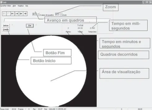
The software itself is very simple, and its confidence level is exclusively operator-dependent. In the case of the present study, the operators were the speech-language therapists who were able to evaluate the swallowing dynamics by means of video-fluoroscopy. A status bar including the present status and several information on the study as a whole is displayed at the bottom of the main window.
The main resource of this software is the tool for time measurement already exhaus-tively tested. Additionally, there is a second tool to evaluate the residual area, but this tool is yet to be improved and validated.
In the evaluation of the time elapsed between the swallowing events, where a quantitative data is required, the applica-tion utilizes funcapplica-tions based on the frames comprising the study (film format file) for time calculation. On the progression bar, the observer may choose an initial point in time, making more precise adjustments by means of the manual advancement on a step-by-step basis, changing the number of frames to be advanced at each step of the process.
ap-27
Swallowing quantitative analysis software
Radiol Bras. 2008;41(1):25–28
plication offers two additional options: one, to record the end of the swallowing event; and another to clear the saved data. The end of the swallowing event record-ing only should be performed when the exact frame corresponding to the end of the event is displayed. Then, the application will display the time elapsed in three ways: The first one, shows the number of frames of the event, the second, displays the time in seconds, and the third displays a more precise timing in milliseconds.
Another resource of the application is the zoom option, to enlarge the visualiza-tion area for each frame. The details of this process are shown on Figure 1.
RESULTS
Different values were found in the com-parison between the mean pharyngeal tran-sit times measured with the chronometer and the software. The patients in this group presented mean transit times of 4.6 s and 2.5 s respectively with the chronometer and the software (Figure 2). This difference may be understood as the latency for visual recognition and the manual chronometer activation. Obviously the level of attention of the investigator also affects the measure-ments accuracy.
It could be observed that the software allows a more accurate measurement, con-sidering the possibility of the precise detail-ing and identification of the onset and the end of the pharyngeal phase. The divergent values corroborate the necessity of more precise tools to evaluate the pharyngeal transit time by means of videofluoroscopic images. In the present case, the software has allowed this evaluation.
DISCUSSION
The initial idea for the development of this tool resulted from the need for quanti-fying and measuring the swallowing pro-cess, not as whole, but as segments, mea-suring the transit time at each swallowing phase. The software allows a dynamic analysis (real-time execution), and a static (frame to frame) analysis of the deglutition. Te divergent values between chronom-eter and software measurements demon-strated that the transit time evaluation based
only on videofluoroscopic images is not accurate. More precise tools utilizing spe-cific applications allow a more accurate, fast and reproducible measurement, and are essential both for utilization in the clinical assistance and new academic studies and literature reviews.
Accuracy becomes significant when the objective is to evaluate the efficacy of therapeutic procedures in the rehabilitation process involving dysphagic patients. The rehabilitation follow-up becomes more ef-fective with the utilization of the software. In the analysis of the swallowing dynam-ics, the differences in the transit time for each phase are minimal, and therefore
in-appropriate for visual and mechanical analysis.
Considering the poor quality of the video images and the absence of a digital equipment, there was the necessity of the software fore a more accurate quantitative analysis of the swallowing phases timing. Studies about videofluoroscopy, com-paring methods of swallowing dynamics quantitative analyses have not been found in the literature.
Studies like the one developed by Kendall et al.(5) utilize application
soft-wares to measure the bolus transit time over the different swallowing phases in healthy individuals. In the present study, post-CVA Figure 1. Visualization of some of the tools available in the application software.
28
Spadotto AA et al.
Radiol Bras. 2008;41(1):25–28 patients presented a higher mean
pharyn-geal transit time when compared with the healthy individuals of that study, where the mean pharyngeal transit time with a paste consistency bolus is 0.91 s. This difference is a result of the presence of oropharyngeal dysphagia in the patients evaluated in this study.
It may be observed that, for timing analyses in swallowing videofluoroscopy, it is necessary to utilize a software to allow a precise identification of internal struc-tures involved in the onset and end of each swallowing phase. According to Kendall et al.(5), the pharyngeal phase onset
corre-sponds to the moment where the bolus reaches the posterior nasal spine, at the end of the hard palate and beginning of the soft palate. An accurate determination of the different swallowing phases with a manual chronometer is unfeasible.
Martin-Harris et al.(6), in a study with
digitized videofluoroscopy, have analyzed the movements of anatomical structures such as the hyoid bone during the degluti-tion, correlating their timing with sex and racial groups. In this study the analysis is limited by the method (measurement with-out the utilization of a software), consid-ering the accuracy and swiftness of the swallowing movements whose individual-ization is unfeasible.
Other authors, such as Santoro et al.(7),
have reported a quantitative evaluation of the swallowing dynamics in a healthy
eld-erly population utilizing swallowing endo-scopy with the Adobe Premiere 6.0 appli-cation for images editing. In this study, the moments for starting the recording of the oral and pharyngeal phases are determined, with a mean transit time for the pharyngeal phase = 867.8 ± 157.8 ms (minimum 567 ms and maximum 1133 ms). However, consid-ering that the data are related to the swal-lowing endoscopy, the comparison with the method described in the present study was not feasible. The mentioned application includes tools for time elapsed evaluation, besides other complex video features con-sidered as non-relevant in our experiment whose main objective was to be focused on the video in execution for identification of relevant aspects. For this reason, the appli-cation software described in the present study was developed with a more clean and clear interface for timing evaluation.
The absence of similar application softwares does not allow a comparative analysis. The sole analysis method com-mercially available utilizes morphological and functional parameters similar to those of the application software described in the present study, but applies different soft-wares and records.
CONCLUSION
This application software offers a com-prehensive tool for parametric analysis of the swallowing timing and speed, resulting
in a better understanding of the swallow-ing dynamics, affectswallow-ing both the clinical approach to patients with oropharyngeal dysfunction and the scientific research. Additionally, the resources of this applica-tion allow an individualized interpretaapplica-tion of the swallowing phases which otherwise would be unfeasible with the conventional, subjective analysis methods subject to interobserver variations. Further studies will be necessary to minimize or eliminate the subjectivity in the determination of transition points between the different swallowing phases.
REFERENCES
1. Logemann JA. Evaluation and treatment of swal-lowing disorders. Austin: Pro-ed Inc.; 1983. 2. Buchholz DW. Dysphagia associated with
neuro-logical disorders. Acta Otorhinolaryngol Belg. 1994;48:143–55.
3. Silva RG. Disfagia neurogênica em adultos: uma proposta para avaliação clínica. In: Furkim AM, Santini CS. Disfagias orofaríngeas. São Paulo: Pró fono; 1999. p.35–48.
4. Costa MMB, Nova JLL, Carlos MT, et al. Video-fluoroscopia – um novo método. Radiol Bras. 1992;25:11–8.
5. Kendall KA, McKenzie S, Leonard RJ, et al. Tim-ing of events in normal swallowTim-ing: a videofluor-oscopic study. Dysphagia. 2000;15:74–83. 6. Martin-Harris B, Michel Y, Castell DO.
Physi-ologic model of oropharyngeal swallowing revis-ited. Otolaryngol Head Neck Surg. 2005;133: 234–40.