Evaluation of shear bond strength
of brackets bonded with orthodontic
fluoride-releasing composite resins
Marcia Cristina Rastelli*, Ulisses Coelho**, Emígdio Enrique Orellana Jimenez***
Objective: To evaluate the shear bond strength of stainless steel brackets bonded with fluo-ride releasing composite resins, comparing them with a conventional resin and to analyze
the amount of resin left on the enamel surface. Methods:Sixty premolars were randomly
divided into three groups: Group I – Concise (3M), Group II – Ultrabond (Aditek do Brasil) and Group III – Rely-a-Bond (Reliance). After bonding, the samples were thermocycled (500 cycles) at 5ºC and 55ºC temperatures. After 48 hours they were subjected to shear bond strength testing, in the occluso-gingival direction, using an MTS 810 Universal Testing
Machine with load speed of 0.5 mm/min. Results: The results demonstrated a mean shear
bond strength of 24.54 ± 6.98 MPa for Group I, 11.53 ± 6.20 MPa for Group II, and 16.46 ± 5.72 MPa for Group III. Analysis of Variance (ANOVA) determined a statistical difference in the mean shear bond strengths between groups (p < 0.001). The Tukey test evidenced that the averages of the three groups were significantly different (p < 0.05), with the highest values for Group I and the lowest for Group II. The Kruskal-Wallis test did not show sig-nificant differences in the amount of resin left on the enamel in any of the three groups (p = 0.361). Conclusion: All materials exhibited adequate adhesive bond strength for clinical use. Concise exhibited the highest degree of shear bond strength but no significant differ-ences were found in Adhesive Remnant Index (ARI) between the groups.
Abstract
Keywords: Shear bond strength. Brackets. Composite luoride resin.
* MSc inGeneral Practice, Universidade Estadual de Ponta Grossa – PR.
** MSc and PhD in Orthodontics, School of Dentistry, Araraquara – UNESP. Post-Doctor of Bioengeneering, Universidade Federal Tecnológica do Paraná. Associate Professor in Orthodontics, Universidade Estadual de Ponta Grossa. Professor of Orthodontics and Dentofacial Orthopedics, Escola de Aperfeiçoamento Proissional da Associação Brasileira de Odontologia de Ponta Grossa.
INTRODUCTION
Several advances have contributed to the improvement of the technique of bonding orth-odontic accessories, such as, the introduction of enamel acid etching by Buonocore,7 and its
asso-ciation with composite resins based on bisphe-nol A glycidyl methacrylate (Bis-GMA). As a re-sult, this technique has become the method of choice for bonding orthodontic accessories.11,12
However, during treatment with fixed orth-odontic appliances certain problems may occur, such as: (1) fractures or even loss of enamel, which may be related to the pretreatment of the enamel surface during prophylaxis27 and/or
dur-ing phosphoric acid etchdur-ing;6 (2) additional loss
of enamel during bracket debonding, removal of debris from the tooth, or rebonding procedures;2
and (3) decalcification of the enamel around the brackets, which is considered the most common problem in patients undergoing orthodontic treatment with fixed appliances.2,4,14,17,23,26
The presence of brackets and resin predispos-es to a greater accumulation of plaque around the brackets,2 which can cause white spot
le-sions likely to occur after the first four weeks of orthodontic treatment.17 These changes appear
mainly in the cervical region of upper incisors.1
The risk of demineralization can be coun-tered by performing plaque control and fluo-ride application.1,17 However, it has been found
that the fluoride toothpaste brushing pro-gram did not prevent enamel decalcification around the brackets because the effectiveness of plaque control depends on the daily routine followed by the patient.16
Given the fact that it is difficult to secure patient compliance in plaque control and the use of fluoride, and due to the inconvenient effects caused by the unsightly white spots, researchers started to develop adhesives with the addition of fluoride to prevent enamel de-mineralization around the brackets.25 These
materials were investigated for their fluoride
releasing efficacy, which has been confirmed by several studies.9,16,23,26,28,29
Fluoride inhibits bacterial activity and can remineralize enamel.25 However, such materials
are relatively new and the need therefore arises to ascertain that the bond strength is sufficient to meet clinical needs, and also whether or not the fluoride comprised in these materials de-creases its strength. For these reasons, the au-thors set out to evaluate the shear bond strength of stainless steel brackets bonded with fluoride-releasing resins, compare them with conven-tional resins and assess the amount of adhesive left on the enamel surface.
MATERIAL AND METHODS
This study used 60 freshly extracted perma-nent premolars—all extractions indicated for orthodontic purposes—of patients aged between 12 and 14 years. The design of this study was submitted to and approved by the Ethics Com-mittee of the Ponta Grossa State University.
After extraction, the teeth were cleaned with a scalpel blade n° 11, spatula LeCron and a spray of bicarbonate, washed and stored in chilled dis-tilled water changed weekly. Prior to the prepara-tion of the specimens, the teeth were immersed in a 0.5% chloramine solution for disinfection for 48 hours in a closed container, as directed by the ISO/TS 11405 (2003) standard. A 6.5X magnification stereomicroscope was used to se-lect teeth with the following characteristics: A healthy enamel surface or at least an intact facial surface, i.e., should not present decay, decalcifi-cation, restorations, cracks, fractures, and should not have undergone any treatment with chemi-cal agents, such as formaldehyde, hydrogen per-oxide, alcohol or thymol. The teeth selected for this study were healthy and free of any flaws that might impair adhesion.
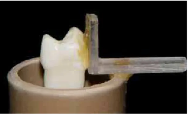
made from two 2 mm thick acrylic sheets. Each acrylic sheet was 5 mm wide, one measuring 10 mm in length and one 20 mm. These sheets were glued with universal instant adhesive. Each tooth was attached to the acrylic square with sticky wax while keeping the buccal sur-face parallel to the sursur-face of the square and the cemento-enamel junction was used as the lower limit. The tooth-square set was bonded with sticky wax to a PVC tube measuring 25 mm in diameter and 35 mm in height (Fig 1).
The crown was centered and the root com-pletely inserted inside the tube, which was filled with hard plaster type IV (SS White, Rio de Janeiro, Brazil). After the hard plaster had set the square was removed. The bonding area ran perpendicular to the base of the PVC tube to keep the buccal surface parallel to the force during the shear bond strength test. All traces of wax and plaster were removed from the sam-ples, which were stored in distilled water for 24 hours in a closed container.
Prior to bonding, buccal surface prophylaxis was performed using a rubber cup and pumice and water, ensuring that the rubber cup was re-placed following five prophylaxis procedures. The teeth were washed with water sprays for 15 seconds and dried with moisture-free air sprays for 15 seconds.12,24
Buccal surface enamel etching was performed
with 37% phosphoric acid gel (Dentalville, Jo-inville, Brazil) for 30 seconds in all groups. The buccal surfaces were then washed with air and water sprays for 20 seconds and dried with mois-ture-free air sprays for 10 seconds.
Premolar stainless steel brackets (Morelli, Lot No. 664362) were bonded with the following orthodontic resins: Concise (3M/ESPE, Dental Products Division, St. Paul, Minnesota, USA - Lot No. 17093), Ultrabond with fluoride (Aditek do Brazil, Cravinhos, São Paulo, Lot No. 9776) and Rely-a-Bond with fluoride (Reliance Orthodon-tic Products, Itasca, Illinois, Lot No. 046602). The brackets were pre-adjusted with -7º torque, 0° angulation and had a 13.02 mm2 base area,
which was automatically obtained using Solid-works software (SolidWorks Corp., USA), ac-cording to the manufacturer’s instructions. The samples were divided into three groups with twenty sampling units, according to the orth-odontic resin that was used.
After etching the enamel, a sealant—specific for each group—was applied, followed by the resin. Bonding was then performed according to manufacturer’s recommendations.
During bonding, an ABZ-0179 (Ormco Corp., USA) positioner was used at a distance of 4 mm from the occlusal surface to the bracket slot to standardize bracket positioning. A stan-dard seating pressure of 300 grams was used throughout bonding of all teeth, with the aid of a Correx dynamometer (Haag-Streit, Swit-zerland).3,4,5 Excess resin was removed with an
explorer probe prior to polymerization.
After bonding, the samples were stored for 24 hours in distilled water at room tempera-ture in sealed plastic containers and labeled according to each group. The samples were then subjected to thermocycling in an MSCT-3 machine (Marcelo Nucci ME, Brazil), applying 500 cycles at 5°C (± 3°C) and 55°C (± 3°C) temperatures. Each cycle was performed for 20 seconds with 7-second intervals.
After a 48-hour interval, counted from the end of thermocycling, the samples were subjected to shear bond strength tests in the occluso-cervical direction and with the chisel positioned at the tooth-bracket interface. The tests conformed to the ISO/TS 11405 (2003) standard and were per-formed with a universal electronic machine for mechanical tests (MTS 810, MTS Systems Corp., USA), with 1 kN load cell, and crosshead speed of 0.5 mm/min. The breaking loads were recorded in Newtons and converted to Megapascal. This con-version was carried out automatically by the test machine itself, or else it could have been calcu-lated using the following formula: R = F/A, where R = shear bond strength in Megapascal, F = break-ing load or debondbreak-ing force in Newtons, and A = bracket base area in mm2.
After debonding, the teeth with their respec-tive brackets were stored in individual plastic bags for later analysis of the amount of adhesive remnant. The teeth and brackets were examined with the help of a stereomicroscope using 40X magnification and classified according to the ad-hesive remnant index (ARI) proposed by Artun and Bergland1, with scores of 0 to 3, indicating:
• Score 0 = no adhesive remnant left on the tooth.
• Score 1 = less than 50% adhesive remnant left on the tooth.
• Score 2 = more than 50% adhesive rem-nant left on the tooth.
• Score 3 = 100% adhesive remnant left on the tooth.
Statistical analysis
Analysis of Variance (ANOVA) is a useful statistical procedure, provided that certain con-ditions are met, such as: (1) data should be ob-tained randomly and independently—which is true on this study; (2) there should be homoge-neity of variance between experimental groups and residuals should be within a normal range. Homogeneity of Variance, i.e., the requirement
that the variances or standard deviations of the bond strength measurements be equivalent across the three experimental groups, was tested using Levene statistics. Normality of Residuals, which can be defined as estimates of experimen-tal errors determined by the difference between each bond strength measurement and the aver-age of the group to which each measurement belongs, was tested using Shapiro-Wilk statis-tics. A 5% significance level was adopted.
Analysis of Variance was utilized to assess shear bond strength of brackets bonded with two resins, both containing fluoride (Ultra-bond and Rely-a-Bond) and a conventional resin (Concise). Analysis was complemented by the Tukey test for multiple comparison of means in pairs.
In addition, 95% confidence intervals were constructed for the population means of the experimental groups. These intervals allow re-searchers to quantify the differences between the means since the tests only indicate whether or not there is evidence that these differences are significant at 5%.
The Kruskal-Wallis nonparametric test was used—at 5% significance level—to evaluate the adhesive remnant index.
RESULTS
Table 1 shows the means and standard de-viations in MPa, according to the experimental groups analyzed: Group I - Concise (3M/ESPE), Group II - Ultrabond with fluoride (Aditek do Brasil) and Group III - Rely-a-Bond with fluo-ride (Reliance Orthodontic Products).
The result of the Levene Statistics (p = 0.366) and the result of the Shapiro-Wilk Statistics (p = 0.164) demonstrated that there was homogeneity of variance and normality of residuals since the p values are greater than 0.05 (Table 2), which ensured that analysis of variance could be applied.
be-groups were significantly different. Group I (Concise) had a significantly higher mean than the means of the fluoride-releasing resin Groups (p < 0.001), while group III (Rely-a-Bond) had a significantly higher mean (p = 0.044) than group II (Ultrabond).
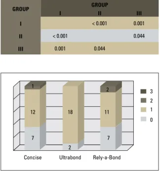
Figure 2 presents the observed frequencies of ARI scores for each resin used for bonding. There was no score 3 and only one or two scores 2. Although Ultrabond showed a tendency to have more scores 1 (and consequently fewer scores 0) compared with other resins, the Krus-kal-Wallis test showed no statistically signifi-cant difference between the three procedures in terms of debonding (p = 0.361).
DISCUSSION
Many researchers have investigated alterna-tive materials to the use of conventional resins with the purpose of preventing enamel decal-cification around the brackets—through the release of fluoride for a prolonged period of time—thus increasing enamel strength and pro-moting its remineralization. These authors have also investigated whether these materials have an adequate shear bond strength.3,4,8,10,11,13,14,24,25
Fluoride-releasing resins are a new generation of preventive orthodontic materials for bracket bonding, which combine the appropriate enam-el-bonding physical properties and fluoride re-leasing agents. They also provide clinically desir-able shear bond strength features, easy cleaning after bonding and easily removable residual ma-terials in debonding procedures.25
Practitioners should be aware of the prop-erties of resins used for bracket bonding, es-pecially with respect to their efficiency during accessory placement.3 This feature is essential
as an orthodontic resin must be capable of keeping accessories firmly adhered to the teeth throughout treatment, resisting masticatory forces as well as those generated by orthodon-tic mechanics.21,24 The minimum shear strength
Sample
Group
I II III
mean 24.54 11.53 16.46
standard deviation 6.98 6.20 5.72
TABLE 1 - Mean and standard deviation by experimental group.
effect DeGreeS of
freeDom rmS f p
Group 2 862.66 21.59 < 0.001
residuals 57 39.95
Homogeneity of variances: p = 0.366 (Levene). Normality of residuals: p = 0.164 (Shapiro-Wilk).
TABLE 2 - Summary of analysis of variance applied to compare the study groups in terms of shear bond strength.
Group Group
I II III
I < 0.001 0.001
II < 0.001 0.044
III 0.001 0.044
TABLE 3 - p values of the Tukey test for comparison of shear bond strength means between groups.
FIGURE 2 - Graphical representation of the frequencies of ARI scores. Concise Ultrabond
0 1 2 3
Rely-a-Bond
12 18 11
7 7
2
2 1
tween the means of shear bond strength be-tween the groups (p < 0.001).
of any adhesive should be between 60 Kgf/cm2
(5.88 MPa) and 80 Kgf/cm2 (7.84 MPa) if it
is to meet clinical needs.21,22 When the results
of this study were compared with the values of reference,21,22 all adhesives showed strength
values suitable for clinical use.
Several factors can affect the final outcome of shear bond strength tests. Therefore, in an attempt to achieve more reliable results the methods were standardized according to the ISO/TS11405 (2003) standard, which is specif-ic for shear tests and recommends that to obtain a pure shear stress it is necessary that the action of the force be parallel to the tooth surface.
This study compared two fluoride-releasing composite resins (Ultrabond and Rely-a-bond) and a conventional composite resin (Concise). All were employed as per manufacturer’s rec-ommendations. It is a known fact that improper manipulation and/or the use of inadequate quan-tities of resin may affect shear bond test results.
The results show that the three groups are significantly different from one another. Group I (Concise = 24.54 ± 6.98 MPa) had the highest shear bond strength mean compared with the other groups. These findings corroborate the work of Kawakami et al13 (48 hours = 20.10
± 1.44 MPa and 10 days = 20.62 ± 1.53 MPa), and Meister15 (29.99 ± 15.89 MPa), which also
found higher shear bond strength values when using Concise.
Kawakami et al13 evaluated Concise using
48-hour and 10-day periods after the polymer-ization of the material. They related their re-sults to the time used for acid etching, whether or not etching had been performed and the time consumed in debonding brackets, since full po-lymerization does not occur before a period of 24 hours has elapsed. Within 10 days there was an increase in shear bond strength but for Con-cise no statistically significant difference was found in both periods. Meister15 ascribed their
results to method standardization and the use of
premolar-specific brackets given their better fit to the tooth surface.
Concise exhibited the highest shear bond strength due to its high filler content since the content of inorganic particles directly influ-ences the resistance of composite resins.12 The
results found by Correr Sobrinho et al10 (after
10 min = 6.22 ± 0.28 MPa and after 24 hours = 7.73 ± 0.21 MPa) were lower than those found in this study. This is probably due to the short-er time taken to debond the brackets, which delayed polymerization. Nevertheless, Concise still showed higher shear bond strength com-pared with the other materials.
Group III (Rely-a-Bond = 16.46 ± 5.72 MPa) showed a significantly higher shear bond strength mean than Group II (Ultrabond = 11.53 ± 6.20 MPa). This difference becomes more pronounced when these two groups (II and III) were compared with Group I (Concise = 24.54 ± 6.98 MPa).
The results of Ultrabond (Group II) and Re-ly-a-Bond (Group III) were smaller and could be explained as follows. Since these are 1-paste resins the catalyst is applied to the tooth and to the base of the brackets while the paste is placed on the base of the brackets. Since these are chemical polymerization materials and are not manipulated prior to use the catalyst is mixed with the base paste only by the seating pressure exerted on the bracket during bracket placement, this procedure can lead to incom-plete polymerization of some portions of the material, which compromises its strength and makes it difficult to attain the homogeneity of results for this bonding system.
When the results for the fluoride-releasing resins used in this study were observed—Ul-trabond (Group II = 11.53 ± 6.20 MPa) and Rely-a-Bond (Group III = 16.46 ± 5.72 MPa)— they were found to be similar to those obtained by Sinha et al,25 who used a fluoride-releasing
Simplício24 also found similar results when
us-ing a self-curus-ing resin (Rely-a-Bond = 13.16 ± 4.87 MPa). Komori and Ishikawa,14 however,
found a different result for the same self-curing resin (Rely-a-Bond = 25.7 ± 3.6 MPa).
As regards the adhesive remnant index, bonding failures were found to occur more fre-quently at the adhesive-enamel interface in all three groups assessed since there was little or no adhesive left on the teeth after debonding. Moreover, there was no damage to the enamel surface after debonding, with the exception of two samples of Group 1 (Concise), which showed fractures on the enamel. Penido et al18
also noted a greater number of fractures at the adhesive-enamel interface in an in vitro study. However, in an in vivo study, Penido et al18
found that bonding failures occurred at the ad-hesive-bracket interface, and remarked that this type of fracture, often found in clinical prac-tice, is the most desirable since any fracture at the adhesive-enamel interface can damage the enamel. This is due to the entanglement of the resin in the bracket mesh, which makes this area more brittle. Pithon et al19,20 found that
the fracture occurred at the adhesive-bracket interface and underscored the importance that
bonding materials allow for a greater amount of adhesive to be left on the tooth surface af-ter bracket removal as this will provide greaaf-ter security and maintain tooth integrity while preventing enamel damage. Removal of resin remnants is not a difficult procedure. It is part and parcel of the orthodontic office routine. Nevertheless, it does require skill as it can also damage the enamel.
CONCLUSIONS
A careful review of the results yields the fol-lowing conclusions:
1. All materials tested in this investigation have adequate shear bond strength to meet clin-ical needs, i.e., sufficient strength to withstand the stresses generated by orthodontic mechanics and chewing. However, Concise showed greater resistance than the other two resins (Rely-a-Bond and Ultrabond).
2. Regarding the adhesive remnant index, no difference was found between the groups, and although the fractures occurred at the adhe-sive-enamel interface, no damage was found to have been caused to the enamel surface after debonding, except in two samples of Group 1 (Concise), which exhibited enamel fractures.
1. Årtun J, Bergland S. Clinical trials with crystal growth conditioning as an alternative to acid-etch enamel pretreatment. Am J Orthod. 1984 Apr;85(4):333-40.
2. Årtun J, Brobakken BO. Prevalence of carious white spots after orthodontic treatment with multiband appliances. Eur J Orthod. 1986 Nov; 8(4):229-34.
3. Bishara SE, Vonwald L, Laffoon JF, Jakobsen JR. Effect of altering the type of enamel conditioner on the shear bond strength of a resin-reinforced glass ionomer adhesive. Am J Orthod Dentofacial Orthop. 2000 Sep;118(3):288-94.
4. Bishara SE, Soliman M, Laffoon J, Warren JJ. Effect of antimicrobial monomer-containing adhesive on shear bond strength of orthodontic brackets. Angle Orthod. 2005 May;75(3):397-9.
REfERENCES
5. Bishara SE, Soliman M, Laffoon J, Warren JJ. Effect of changing a test parameter on the shear bond strength of orthodontic brackets. Angle Orthod. 2005 Sep;75(5):832-5.
6. Brown CR, Way DC. Enamel loss during orthodontic bonding and subsequent loss during removal of illed and unilled adhesives. Am J Orthod. 1978 Dec;74(6):663-71.
7. Buonocore MG. A simple method of increasing the adhesion of acrylic illing material to enamel surface. J Dent Res. 1955 Dec;34(6):849-53.
Contact address Marcia Cristina Rastelli Rua Santana, 276, Centro
CEP: 84.010-320 – Ponta Grossa / PR, Brazil E-mail: [email protected] Submitted: December 2006
Revised and accepted: September 2009 9. Cohen WJ, Wiltshire WA, Dawes C, Lavelle CLB. Long-term in vitro
luoride release and rerelease from orthodontic bonding materials containing luoride. Am J Orthod Dentofacial Orthop. 2003 Nov;124(5):571-6.
10. Correr Sobrinho L, Correr GM, Consani S, Sinhoreti MAC, Consani RLX. Inluência do tempo pós-ixação na resistência ao cisalhamento de braquetes colados com diferentes materiais. Pesqui Odontol Bras. 2002 jan-mar;16(1):43-9.
11. Graf I, Jacobi BE. Bond strength of various luoride-releasing orthodontic bonding systems – Experimental study. J Orofac Orthop. 2000 May;61(3):191-8.
12. Ianni Filho D, Silva TBC, Simplício AHM, Loffredo LCM, Ribeiro RP. Avaliação in vitro da força de adesão de materiais de colagem em Ortodontia: ensaios mecânicos de cisalhamento. Rev Dental Press Ortod Ortop Facial. 2004 jan-fev;9(1):39-48.
13. Kawakami RY, Pinto AS, Gonçalves JR, Sakima MT, Gandini LG. Avaliação “in vitro” do padrão de descolagem na interface de ixação de materiais adesivos ortodônticos ao esmalte de dentes inclusos: resistência ao cisalhamento após 48 horas e 10 dias. Rev Dental Press Ortod Ortop Facial. 2003 nov-dez;8(6):43-61. 14. Komori A, Ishikawa H. Evaluation of a resin-reinforced glass
ionomer cement for use as an orthodontic bonding agent. Angle Orthod. 1997 Jun;67(3):189-96.
15. Meister ER. Avaliação “in vitro” da resistência adesiva ao cisalhamento na colagem de braquetes usando dois tipos de resinas. [tese]. Ponta Grossa: Universidade Estadual de Ponta Grossa; 2004.
16. Øgaard B, Rezk-Lega F, Ruben J, Arends J. Cariostatic effect and luoride release from a visible light-curing adhesive for bonding of orthodontics brackets. Am J Orthod Dentofacial Orthop. 1992 Apr;101(4):303-7.
17. O’Reilly MM, Featherstone JDB. Demineralization and remineralization around orthodontic appliances: an in vivo study. Am J Orthod Dentofacial Orthop. 1987 Jul;92(1):33-40. 18. Penido SMMO, Penido CVSR, Pinto AS, Sakima T, Fontana
CR. Estudo in vivo e in vitro com e sem termociclagem, da resistência ao cisalhamento de braquetes colados com fonte de luz halógena. Rev Dental Press Ortod Ortop Facial. 2008 maio-jun;13(3):66-76.
19. Pithon MM, Santos RL, Oliveira MV, Ruellas ACO. Estudo comparativo in vitro da resistência ao cisalhamento da colagem e do índice de remanescente adesivo entre os compósitos Concise e Fill Magic. Rev Dental Press Ortod Ortop Facial. 2006 jul-ago;11(4):76-80.
20. Pithon MM, Bernardes LAA, Ruellas ACO, Romano FL. Avaliação da resistência ao cisalhamento do compósito Right-On em diferentes condições de esmalte. Rev Dental Press Ortod Ortop Facial. 2008 maio-jun;13(3):60-5.
21. Reynolds IR. A review of direct orthodontic bonding. Br J Orthod. 1975;2(3):171-8.
22. Reynolds IR, von Fraunhofer JA. Direct bonding in orthodontics: a comparison of attachments. Br J Orthod. 1977 Apr;4(2):65-9. 23. Rix D, Foley TF, Banting D, Mamandras A. A comparison of
luoride release by resin-modiied GIC and polyacid modiied composite resin. Am J Orthod Dentofacial Orthop. 2001 Oct;120(4):398-405.
24. Simplício AHM. Avaliação in vitro de materiais utilizados para colagem ortodôntica – potencial cariostático, resistência ao cisalhamento e padrão de descolagem. [tese]. Araraquara: Universidade Estadual Paulista Júlio de Mesquita Filho; 2000. 25. Sinha PK, Nanda RS, Duncanson MG Jr, Hosier MJ. In vitro
evaluation of matrix-bound luoride-releasing orthodontic bonding adhesives. Am J Orthod Dentofacial Orthop. 1997 Mar;111(3):276-82.
26. Staley RN, Mack SJ, Wefel JS, Vargas MA, Jakobsen JR. Effect of brushing on luoride from 3 bracket adhesives. Am J Orthod Dentofacial Orthop. 2004 Sep;126(3):331-6.
27. Thompson RE, Way DC. Enamel loss due to prophylaxis and multiple bonding/debonding of orthodontic attachments. Am J Orthod. 1981 Mar;79(3):282-95.
28. Wheeler AW, Foley TF, Mamandras A. Comparison of luoride release protocols for in-vitro testing of 3 orthodontic adhesives. Am J Orthod Dentofacial Orthop. 2002 Mar;121(3):301-9. 29. Wilson RM, Donly KJ. Demineralization around orthodontic