Cephalometric evaluation of vertical
and anteroposterior changes associated
with the use of bonded rapid maxillary
expansion appliance
Moara De Rossi*, Maria Bernadete Sasso Stuani**, Léa Assed Bezerra da Silva***
Abstract
Introduction: Bonded rapid maxillary expansion appliances have been suggested to control increases in the vertical dimension of the face after rapid maxillary expansion but there is still no consensus in the literature concerning its actual effectiveness. Objective: The purpose of this study was to evaluate the vertical and anteroposterior cephalometric changes associ-ated with maxillary expansion performed using bonded rapid maxillary expansion appliances.
Methods: The sample consisted of 25 children of both genders, aged between 6 and 10 years old, with skeletal posterior crossbite. After maxillary expansion, the expansion appliance itself was used for fixed retention. Were analyzed lateral teleradiographs taken prior to treatment onset and after removal of the expansion appliance. Conclusion: Based on the results, it can be concluded that the use of bonded rapid maxillary expansion appliance did not significantly alter the children’s vertical and anteroposterior cephalometric measurements.
Keywords: Bonded rapid maxillary expansion appliance. Rapid maxillary expansion. Cephalometry.
* PhD in Pediatric Dentistry, FOP / UNICAMP. MSc in Pediatric Dentistry, FORP / USP. ** Professor of Orthodontics, FORP / USP.
*** Professor and Chair of the Department of Child, Preventive and Social Dentistry, FORP / USP.
INtrOduCtION
Rapid maxillary expansion (RME) is a wide-ly accepted procedure recommended for the correction of maxillary atresia related to poste-rior crossbite.7,8 The opening of the midpalatal suture causes increases in maxillary width and dental arch perimeter, allowing the coordina-tion of the upper and lower basal bones and crossbite correction. As well as the correction of transverse discrepancy, however, RME also pro-motes changes such as inferior displacement of
the maxilla, extrusion and inclination of maxil-lary and mandibular molars, clockwise rotation of the mandible, with a resulting increase in fa-cial height and anterior open bite.4,14,15,20,21,26
Bonded rapid maxillary expansion appliance have been proposed to control the side effects of RME, which may be associated with adverse increases in anterior facial height, especially in individuals with a predominantly vertical growth pattern and a tendency towards open bite.2,10,17,18,20,22,24 No consensus has been found in the literature, however, concerning the RME-related vertical and anteroposterior effects pro-duced with this type of appliance.2,7,9,13,19,20,24,25
The purpose of this study was to evaluate lateral teleradiographs for possible vertical and anteroposterior changes resulting from the use of bonded rapid maxillary expansion appliance for the correction of skeletal posterior cross-bite in children.
MAtErIAL ANd MEtHOdS Sample
The sample comprised 25 children (13 girls and 12 boys), irrespective of gender, race or social class, with a mean age of 8 years and 5 months (ranging from 6 years and 11 months to 10 years and 11 months) presenting with maxil-lary atresia and either unilateral or bilateral pos-terior crossbite, indicated for maxillary expan-sion as the first stage of orthodontic treatment. Maxillary atresia was detected based on clini-cal parameters characterized by the presence of posterior crossbite associated with a deep palate, “V”-shaped maxillary arch and reduced transverse maxillary dimensions compared with the mandible. This study was approved by the Research Ethics Committee of the Ribeirão Pre-to School of Dentistry, University of São Paulo (FORP / USP - Case No 2003.1.1067.58.8), and the children’s parents and/or guardians signed a consent form, according to resolution 196/96 of the Brazilian Health Council.
The children included in the sample had received no previous orthodontic treatment and exhibited good general and oral health. Their upper and lower first permanent molars
had erupted and were in occlusion. The orth-odontic documentation comprised panoramic and occlusal X-rays, lateral and frontal cepha-lometric radiographs, intraoral photographs and study models.
rapid maxillary expansion
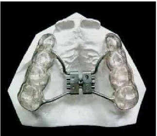
RME was performed using bonded rapid maxillary expansion appliance, made from col-orless acrylic resin covering the posterior teeth (Jet; Artigos Odontológicos Clássico Ltda, São Paulo, SP, Brazil) and a palatal expansion screw (split screw, 9 mm, code 65.05.011; Dental Morelli, Sorocaba, SP, Brazil) positioned on the midpalatine raphe at about 2 mm from the palate and between the primary second molars (Fig 1). The appliance was adjusted in the pa-tient’s mouth in order to ensure as many bilat-eral occlusal contacts as possible, and was then attached using dual-curing acrylic resin cement adhesive (Rely X: 3M do Brasil Ltda., Produtos Dentários, Sumaré, SP, Brazil).
Activation was carried out by the children’s parents and/or guardians and amounted to ¼ turn of the screw every 12 hours, starting one week after appliance installation. When cross-bite overcorrection was observed, i.e., when the palatal cusps of the upper posterior teeth were occluding on the buccal cusps of the lower pos-terior teeth, the expander screw was fixed with acrylic resin and a new occlusal adjustment was made. The average interval time between acti-vations was 20 days (ranging between 14 and 26 days) and the appliance remained in the pa-tients’ mouth as fixed retention for a minimum of 90 days (107 days average, ranging from 90 to 124 days). After this period, the appliance was removed and patients wore a removable re-tainer (acrylic plate with a Hawley labial clasp and retention clasps) for 6 months.
Cephalometric evaluation
Lateral teleradiographs were taken before treatment onset (T1) and after removal of the expansion appliance (T2). The cephalometric radiographs were performed in standardized fashion by a single technician in the Laboratory of Analysis and Control of Dental Radiographic Images (LACIRO), at FORP-USP.
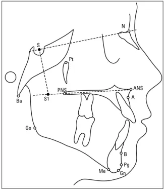
The cephalometric tracings were performed manually by the same experienced and calibrat-ed examiner. The following cephalometric land-marks were located and marked on the lateral cephalograms (Fig 2):
• Sella (S): Virtual point located at the geo-metric center of the sella turcica.
• Nasion (N): The anterior-most point of the frontonasal suture.
• Subspinal Point (A): The deepest point of the subspinal concavity.
• Supramental Point (B): The deepest point of the supramental concavity.
• Anterior Nasal Spine Point (ANS): Lo-cated at the anterosuperior end of the maxilla.
• Posterior Nasal Spine Point (PNS): Locat-ed at the posterior end of the maxilla. • Basion (Ba): Lowest point of the image of
the anterior margin of the foramen magnum. • Pterygoid Point (Pt): Posterior-most and
superior-most point in the upper contour of the pterygomaxillary fissure.
• Pogonion (Pg): Anterior-most point of the bony chin.
• Gnathion (Gn): The anterior-most and inferior-most point of the mandibular symphysis, as determined by bisecting the angle formed by the lower margin of the mandibular body and the facial line (NPg). • Menton (Me): Located at the intersection
of the outer contour of the mandibular symphysis and the inferior margin of the mandibular body.
• Gonion (Go): Located in the outer contour of the gonial angle, determined by bisecting the angle between the mandibular ramus and the lower margin of the mandibular body.
FIGURE 2 - Lateral cephalogram and location of cephalometric landmarks.
S
N
Pt
Go
A
B Pg Gn Me
PNS ANS
6 4
8
7
9 3 10
12 13
11 2
1
5
permanent molars and intersecting the upper and lower incisors.
To assess the anteroposterior behavior of the apical bases, the following cephalometric mea-surements were used (Fig 3):
• SNA Angle: Formed by intersecting the SN and NA lines. Measures the position of the maxilla relative to the anterior cra-nial base.
• SNB Angle: Formed by intersecting the SN and NB lines. It measures the position of the mandible relative to the anterior cranial base.
• ANB Angle: Determined by the differ-ence between SNA and SNB. It measures the anteroposterior relationship between maxilla and mandible.
To assess the vertical behavior of the api-cal bases, we used the following cephalometric measurements (Fig 3 and 4):
• S-S1: linear measurement determined by the junction of the S and S1 landmarks. • Point S1: Connection point between a line
drawn from Point S—perpendicularly to the SN line—and the palatal plane (junc-tion of ANS and PNS).
After locating and marking the landmarks the following lines and planes of orientation were traced:
• S-N Line: Connecting S to N. • N-A Line: Connecting N to A. • N-B Line: Connecting N to B. • S-Gn Line: Connecting S to Gn. • Ba-N Line: Connecting Ba to N. • Pt-Gn Line: Connecting Pt to Gn. • N-ANS Line: Connecting N to ANS. • ANS-Me Line: Connecting ANS to Me. • N-Me Line: Connecting N to Me.
• Steiner’s mandibular plane (GoGn): De-termined by Go and Gn.
• Palatal plane (PP): Determined by ANS and PNS.
• Occlusal Plane (Ploc): Determined by intersecting the landmarks of the first
FIGURE 3 - Lateral cephalogram and location of the vertical and antero-posterior angular cephalometric measurements: (1) SNA angle, (2) SNB angle, (3) ANB angle, (4) SN.PP angle, (5) PP.GoGn angle, (6) SN.GoGn angle, (7) SN.Ploc angle; (8) SN.Gn angle; (9) Facial Axis.
intersecting the BaN and PtGn lines. Shows the direction of mandibular growth. • N-ANS: Linear measurement determined
by the junction of the N and ANS land-marks. Reflects the anterior superior height of the face.
• ANS-Me: Linear measurement deter-mined by the junction of the Me and ANS landmarks. Reflects the anteroinferior height of the face.
• N-Me: Linear measurement determined by the junction of the N and Me land-marks. Reflects the anterior facial height.
data analysis and statistics
The cephalometric data were statistically analyzed using SPSS software version 10.0 for Windows (SPSS Inc., Chicago, IL, USA) and the paired t-test was used to compare pre and post-expansion.
• SN.PP Angle: Formed by intersecting the PP plane with the SN line. Reflects the degree of inclination of the maxilla rela-tive to the anterior skull base.
• PP.GoGn Angle: Formed by intersecting the PP plane with the GoGn line. Reflects the inclination of the mandible relative to the palatal plane.
• SN.GoGn Angle: Formed by intersecting the GoGn plane with the SN line. Reflects the degree of inclination of the mandible relative to the anterior cranial base. • SN.Ploc Angle: Formed by intersecting the
SN line with the occlusal plane. Reflects the degree of inclination of the maxilla relative to the anterior cranial base. • SN.Gn Angle: “Y”-growth axis, formed by
intersecting the SN and SGn lines, shows the direction of mandibular growth. • Facial Axis (BaN.PtGn Angle): Formed by
Pre-expansion (T1)
Post-expansion (T2)
Difference (T2-T1)
Paired t-test
MEASUREMENTS mean s.d. mean s.d. mean s.d. variation “p” values
Anteroposterior
SNA (degrees) 80.76 4.40 81.12 4.31 0.36 1.93 -0.43 to 1.15 0.361
SNB (degrees) 77.24 4.77 77.44 4.69 0.20 1.32 -0.34 to 0.74 0.457
ANB (degrees) 3.52 2.48 3.68 2.86 0.16 1.46 -0.44 to 0.76 0.590
Vertical
SN.PP (degrees) 7.88 3.44 7.40 3.31 -0.48 1.75 -1.20 to 0.24 0.158
PP.GoGn (degrees) 29.40 4.17 29.92 3.35 0.52 2.16 -0.37 to 1.41 0.241
SN.GoGn (degrees) 37.28 5.31 37.36 4.79 0.08 1.60 -0.58 to 0.74 0.805
SN.Ploc (degrees) 19.24 3.97 19.00 4.67 -0.24 2.87 -1.42 to 0.94 0.680
SN.Gn (degrees) 68.88 4.52 68.92 4.61 0.04 1.05 -0.39 to 0.47 0.852
Facial Axis (degrees) 85.16 3.28 85.04 4.01 -0.12 2.12 -0.99 to 0.75 0.780
N-ANS (mm) 45.96 2.92 46.52 3.76 0.56 1.41 -0.02 to 1.14 0.060
ANS-Me (mm) 63.08 4.06 63.72 3.92 0.64 1.97 -0.17 to 1.45 0.119
N-Me (mm) 106.72 5.07 107.76 5.24 1.04 1.83 0.28 to 1.79 0.009*
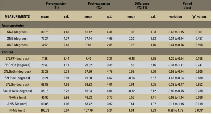
TABLE 1 - Mean, standard deviation and statistical significance of the cephalometric variables before and after expansion (n = 25).
To obtain method error, 10 radiographs were retraced of 10 different, randomly selected pa-tients after a minimum three month interval time. Dahlberg’s formula11 was applied to esti-mate error magnitude and the paired t-test to detect statistical significance.
rESuLtS
The values (mean and standard deviation) of each cephalometric variable measured before treatment (T1) and after expansion and remov-al of the expansion appliance (T2) are shown in Table 1. The mean, standard deviation, variation in the difference between the values of T1 and T2 and statistical significance (“p” values) can be found in Table 1.
In assessing the anteroposterior behavior of the apical bases after maxillary expansion an in-crease in the means of the SNA (0.36°), SNB (0.20°) and ANB (0.16°) angles was observed, although the changes were not statistically sig-nificant (p > 0.01).
In assessing the vertical behavior of the api-cal bases after maxillary expansion an increase in the means of variables PP.GoGn (0.52°), SN.GoGn (0.08°) and SN.Gn (0.04°) and a decrease in SN.PP (-0.48°), SN.Ploc (-0.24°) and Facial Axis (-0.12°) were observed. These changes, however, were not statistically signifi-cant (p > 0.01).
As for the behavior of the facial heights, after maxillary expansion an increase in the means of variables N-ANS (0.56 mm), ANS-Me (0.64 mm) and N-ANS-Me (1.04 mm) was not-ed, with a statistically significant increase (p < 0.01) only for N-Me.
Method error was greater than 0.5 mm and statistically significant (p < 0.05) only for the anterior facial height measurement (N-Me).
dISCuSSION
Since the RME early studies, several inves-tigations have evaluated transverse, vertical
and anteroposterior cephalometric changes as-sociated with the opening of the sutures us-ing different types of appliances. Currently, in view of RME’s positive and proven results, it has become a widely accepted procedure used to increase the transverse dimension of the maxilla. On the other hand, the literature is not unanimous about the actual vertical and anteroposterior orthopedic effects associated with the RME and its potential benefits or harm in orthodontic treatment.
This study showed that, with the exception of N-Me, no vertical change exceeded 1° or 1 mm. Thus, in addition to a lack of statistical sig-nificance, the vertical changes occurring after RME—when using the bonded rapid maxillary expansion appliance—are also devoid of clinical significance. Although the 1.04 mm increase in anterior face height (N-Me) was statistically sig-nificant (p < 0.01), this change does not cause any clinical losses. Moreover, such change may be related to the method error, which was 0.8 mm and proved significant (p < 0.05) for the anterior face height measurement (N-Me).
Thus, it was found that RME—when per-formed using the bonded rapid maxillary expan-sion appliances—did not cause posteroinferior mandibular displacement, nor did it increase the children’s anterior facial height. Contrary to these findings, studies conducted with Haas and Hyrax style appliances show that RME fosters inferior displacement of the maxilla, alveolar process inclination, extrusion and buccal incli-nation of posterior teeth, which result in pos-teroinferior mandibular rotation and increased lower anterior facial height.4,14,15,21,26
with conventional Haas and Hyrax type expand-ers2,10,12,17,18,20,22,23. In agreement with the present study, Asanza et al2 did not see a significant in-crease in anteroinferior facial height (ANS-Me) after RME had been performed using bonded rapid maxillary expansion appliances. Accord-ing to the authors, both inferior displacement of the maxilla and mandibular plane inclination are greater with Hyrax-type appliances. In Sarver and Johnston’s view,20 inferior displacement of the maxilla and mandible is decreased when bonded rapid maxillary expansion appliances are used due to the action of the levator muscles and stretching of soft tissues provided by the occlusal acrylic.
As regards anteroposterior skeletal changes after RME, anterior maxillary displacement was observed by several authors who used conven-tional expansion appliance (like Haas and Hy-rax) and bonded rapid maxillary expansion ap-pliances.2,6-9,13,14,15,21,26 Bramante and Almeida7 found no significant differences in anteroposte-rior changes with the use of Haas/Hyrax-type appliances or bonded rapid maxillary expansion appliances. Sarver and Johnston20 and Johnson et al,16 on the other hand, found that anterior max-illary displacement increased when the appliance was used with orthodontic bands, suggesting the use of bonded rapid maxillary expansion appli-ances to restrict maxillary movement, which is undesirable in patients presenting with skeletal Class II malocclusion.
In the present study it was observed that, following RME, a slight displacement of the maxilla and mandible occurred as could be at-tested by an increase of 0.36° in the SNA angle and 0.20° in SNB. Clockwise mandibular rota-tion was negligible and insufficient to displace point B posteriorly, which justifies the fact that the SNB did not decrease. The fact that SNA underwent a considerable increment relative to SNB caused a 0.16º increase in ANB. Skele-tal anteroposterior changes, however, were not statistically significant.
Contrary to these results, Sarver and John-ston20 and Asanza et al2 reported posterior maxillary displacement after the use of bonded rapid maxillary expansion appliances. In this study, although SNA increased in most pa-tients, there were cases where SNA decreased and cases where SNA remained stable (ranging from 1.15° to -0.43°), as must have been the case with Sarver and Johnston,20 who found an average 0.75º decrease in SNA, and Asanza et al,2 whose average SNA decrease was 0.66° (ranging from -3.6º to 1.7º). Thus, any diver-gence in the results can be explained by the variability of the samples used in each study.
Haas14,15 and Biederman5 reported anterior maxillary displacement after RME, which aids in the correction of skeletal Class III malocclu-sion and anterior crossbite. After the retention period, however, values tend to revert close to those found at the start.7,9,13,14 The relapse of an-teroposterior cephalometric changes after RME using Haas-type appliance was also found using Hyrax-type and bonded rapid maxillary expan-sion appliances.7,9,19 The maxilla is projected an-teriorly as an immediate response to therapy, but throughout the retention period it tends to re-turn to the starting position, which may explain the fact that anterior maxillary displacement was significant in some studies where analysis was carried out immediately after expander ac-tivation3,5,8,14,15 and not in others where, similar-ly to the present study, assessments were made after the retention period.7,9,13,19,21
Similarly, although vertical changes were not significant, in cases of transverse discrepancy associated with a predominance of vertical growth, the latter should be treated with or-thopedic appliances for this specific purpose during the active phase of RME.
Cephalometric variations found in this study were small and may have been caused by mea-surement errors or normal changes expected during growth. We therefore believe that expan-sion bonded rapid maxillary expanexpan-sion applianc-es prapplianc-esent an option for the correction of poste-rior crossbite and maxillary atresia, regardless of vertical problems and the patient’s facial pattern. By not using bands clinical work is reduced, fa-cilitating the preparation and installation of the bonded rapid maxillary expansion appliance. However, one should pay special attention to oc-clusal adjustment to ensure that the contact of the acrylic with the lower teeth is bilateral and balanced, thereby preventing the appliance from falling while reducing patient discomfort.
Finally, it should be underscored that our sample was selected based only on reduced
maxillary transverse dimension and we did not take into account any aspects related to growth pattern and maxillomandibular sagittal relation-ship. Further investigation is therefore needed involving a sample that is standardized accord-ing to growth pattern and maxillomandibular relationship with the aim of raising awareness about the possible benefits brought by bonded rapid maxillary expansion appliances to Class II and hyperdivergent patients.
CONCLuSIONS
In view of the specific conditions of this study, it can be concluded that rapid maxillary expansion performed in children using bonded rapid maxillary expansion appliance did not bring about any vertical or anteroposterior cephalometric changes.
ACKNOWLEdGEMENtS
We wish to thank Dental Morelli, and Mr. José Damian in particular, for donating the materials needed for fabrication of the expan-sion appliances.
1. Angell EH. Treatment of irregularity of the permanent or adult teeth. Dental Cosmos. 1860 May;1(1):540-4. 2. Asanza S, Cisneros GJ, Nieberg LG. Comparison of
Hyrax and bonded expansion appliances. Angle Orthod. 1997;67(1):15-22.
3. Basciftci FA, Karaman AI. Effects of a modiied acrylic
bonded rapid maxillary expansion appliance and vertical chin cap on dentofacial structures. Angle Orthod. 2002 Feb;72(1):61-71.
4. Berlocher WC, Mueller BH, Tinanoff N. The effect of maxillary palatal expansion on the primary dental arch circumference. Pediatr Dent. 1980 Mar;2(1):27-30. 5. Biederman W. A hygienic appliance for rapid expansion.
J Pract Orthod. 1968 Feb;2(2):67-70.
6. Biederman W. Rapid correction of Class III malocclusion by midpalatal expansion. Am J Orthod. 1973;63(1):47-55. 7. Bramante FS, Almeida RR. Estudo cefalométrico em norma
lateral das alterações dentoesqueléticas produzidas por três expansores: colado, tipo Haas e Hyrax. Rev Dental Press Ortod Ortop Facial. 2002 nov-dez;7(6):19-41.
8. Chung CH, Font B. Skeletal and dental changes in the sagittal, vertical, and transverse dimensions after rapid palatal expansion. Am J Orthod Dentofacial Orthop. 2004 Nov;126(5):569-75.
9. Claro CAA, Ursi W, Chagas RV, Almeida G. Alterações ortopédicas ântero-posteriores decorrentes da disjunção maxilar com expansor colado. Rev Dental Press Ortod Ortop Facial. 2003 set-out;8(5):35-47.
10. Cohen M, Silverman E. A new and simple palate splitting device. J Clin Orthod. 1973 Jun;7(6):368-9.
11. Dahlberg G. Statistical methods for medical and biological students. London: Grorge Allen and Unwin; 1940.
12. Faltin K Jr., Moscatiello VAM, Barros EC. Alterações dentofaciais decorrentes da disjunção da sutura palatina mediana. Rev Dental Press Ortod Ortop Facial. 1999 jul-ago;4(4):5-13. 13. Galon GM, Calçada F, Ursi W, Queiroz GV, Atta J, Almeida GA.
Comparação cefalométrica entre os aparelhos de ERM bandado e colado com recobrimento oclusal. Rev Dental Press Ortod Ortop Facial. 2003 maio-jun; 8(3):49-59.
14. Haas AJ. Rapid expansion of the maxillary dental arch and nasal cavity by opening the midpalatal suture. Angle Orthod. 1961;31:73-9.
15. Haas AJ. The treatment of maxillary deiciency by opening the
midpalatal suture. Angle Orthod. 1965 Jul;35:200-17. 16. Johnson GD, Killiany DM, Ferguson DJ. Skeletal changes
following rapid maxillary expansion in the mixed dentition using a bonded expansion appliance. J Dent Res. 2000; 79:326-9.
Contact address
Moara De Rossi
Rua Ipê Ouro, 633, Condomínio Rio das Pedras CEP: 13.085-135 – Barão Geraldo – Campinas/SP, Brazil E-mail: [email protected]
Submitted: March 2007
Revised and accepted: November 2007
17. McNamara JA Jr., Brudon WL. Bonded rapid maxillary expansion appliance. 5th ed. Ann Arbor: Needham Press, 1995.
18. Mondro JF, Litt RA. An improved direct bonded palatal expansion appliance. J Clin Orthod. 1977 Mar;11(3):203-6. 19. Reed N, Ghosh J, Nanda RS. Comparison of treatment
outcomes with banded and bonded rapid palatal expansion appliances. Am J Orthod Dentofacial Orthop. 1999 Jul;116(1):31-40.
20. Sarver DM, Johnston MW. Skeletal changes in vertical and anterior displacement of the maxilla with bonded rapid palatal expansion appliances. Am J Orthod Dentofacial Orthop. 1989 Jun;95(6):462-6.
21. Silva Filho OG, Boas MC, Capelozza Filho L. Rapid maxillary expansion in the primary and mixed dentitions: a cephalometric evaluation. Am J Orthod Dentofacial Orthop. 1991 Aug;100(2):171-9.
22. Spolyar JL. The design, fabrication, and use of a full coverage bonded rapid maxillary expansion appliance. Am J Orthod. 1984 Aug;86(2):136-45.
23. Steiman H. Visual aid for bonded acrylic rapid palatal expander. J Clin Orthod. 1997 May;31(5):327. 24. Ursi W, Dale RCXS, Claro CA, Chagas RV, Almeida G.
Alterações transversais produzidas pelo aparelho de expansão
maxilar com cobertura oclusal, avaliada pelas telerradiograias
póstero-anteriores. Ortodontia. 2001;34:43-55.
25. Vardakas MH, Ursi W, Calçada F, Queiroz GV, Atta J, Almeida GA. Alterações cefalométricas verticais produzidas pelo aparelho de expansão rápida maxilar colado com cobertura oclusal, em pacientes em crescimento. Rev Dental Press Ortod Ortop Facial. 2003 set-out;8(5):69-93.