An interview with
Ademir Roberto Brunetto
• DDS, Federal University of Paraná State (UFPR), 1976.
• Postgraduate Orthodontics and Dentofacial Orthopedics, University of California, Los Angeles, USA, 1984.
• Scientific Advisor, Dental Press Journal of Orthodontics. • Renowned Lecturer in Brazil and abroad.
• Diplomate, Brazilian Board of Orthodontics and Dentofacial Orthopedics (BBO), 2004.
• Director, Brazilian Board of Orthodontics and Facial Orthopedics (BBO).
It gives me great satisfaction and pride to conduct this interview with Prof. Dr. Ademir Brunetto, a prominent professional in today’s Brazilian orthodontic scenery. This longtime friend, we forged our friendship when we sat side by side at the 1st diplomate examination of the
Bra-zilian Board of Orthodontics and Dentofacial Orthopedics (BBO), when at the same time, we were Board candidates. A diplomate since 2004, he was later invited to join the BBO Board, which set the stage for our frequent encounters. I have since learned to increasingly admire his in-depth scientific knowledge—especially in the area of Orthodontics and Facial Orthopedics—, his ethical conduct, his composure and common sense in addressing all issues, regardless of their complexity and, last but not least, his contagious joy. Born in Concórdia, at the west end of Santa Catarina State, in southern Brazil, where he spent his childhood and adolescence, he soon moved to Curitiba where he studied Dentistry at the Federal University of Paraná, graduating in 1976. As a Dentistry undergraduate, he worked as a trainee in a number of orthodontic clinics and after graduation applied for the position of assistant professor at UFPR. Since his approval in 1981 he has taught orthodontics at UFPR. Dr. Brunetto attended his postgraduate program in orthodontics at the Uni-versity of California, Los Angeles, USA (UCLA) where he was awarded the title of Master in Orthodontics in 1984. He is currently in private practice in Curitiba, Paraná State, where he seeks to apply and disseminate his extensive knowledge. Outside his professional activities, he is a very dedicated family man and an accomplished fisherman with a predilection for ocean fishing. In his replies to the interviewers, he has shown substantial knowledge of current state-of-the-art issues such as Class III correction, application of new imaging techniques using cone beam tomography, absolute anchorage and orthodontic preparation for orthognathic surgery. I am certain that our valued readers will enjoy this interview.
A B C
Regarding the early treatment of Class III, what is the state-of-the-art in terms of inter-ceptive procedures and what protocol do you adopt, speciically in maxillary reverse pull headgear cases? What type of retainer do you use after maxillary reverse traction? Márcio Sobral and Luís Antonio Aidar
I first started working with palatal expansion associated with protraction in 1982, as a UCLA resident. The then Head of the Department of Orthodontics, Dr. Patrick K. Turley, had just be-gun his work with Class III patients. Those two residence years were rather fruitful and, although fraught with doubts, also brought many surpris-es and knowledge. When I returned to Brazil in 1984, I continued within the same line of work, making slight changes to the expander design. A few years later, I started to use prefabricated masks, which greatly expedited my work.
My protocol begins with ¼ turn expansion per day for initial suture release.24 My intent is
always to control so as not to overexpand the maxilla to prevent excessive crossbite (Brodie) because during anterior maxillary traction we are moving from a wider, posterior mandibular region and as we displace the maxilla forward and downward, we have a narrower mandible. After the expansion, I start using the face mask for at least 14 hours a day. I start with a force of 250 to 300 g/side and eventually increase it to 500 g/side.
The treatment time is approximately one year24 and the goal is to turn the patient into a Class II (overcorrection). When this period is over, the expansion appliance and the face mask are removed and the patient starts being moni-tored every 6 months. A new traction might be necessary depending on the patient’s growth pattern. The actual orthodontic treatment starts only when cervical vertebrae7 maturation evolves from phase 5 (maturity) to phase 6, when ado-lescent growth is fully established.
I don’t believe the use of a retainer after reverse traction is necessary. As we can see in follow-up lateral radiographs, “point A” remains positioned exactly where it was pulled, with no relapse10 (Table 1 and Fig 1). The problem is that the maxilla grows slower than the mandible,16 which sometimes leads to the need for traction to be once again performed.
MEASUREMENTS STANDARD A A1 A2
SNA (Steiner) 82º 82º 85º 85º
SNB (Steiner) 80º 82º 82º 83.5º
ANB (Steiner) 2º 0 +3º +1.5º
TABLE 1 - Cephalometric measurements.
FIGURE 1 - Initial (A) and intermediate lateral radiographs (B and C).
The use of chin cups, although an old-time orthodontic resource, is still advocated by some professionals, mostly from the Japanese school. What is your experience and opinion on the use of chin cups in mandibular skeletal Class III cases, especially when patients dis-play a marked vertical growth? Deocleciano da Silva Carvalho and Mirian Nakane Matsumoto
When I started pursuing the orthodontic path, there was great concern with Class III patients. We used to keep our fingers crossed that these cases would never show up at our offices. Prefer-ably, these patients should seek a professional we weren’t so keen on. There is no telling how often professionals have been baffled to realize—dur-ing or after orthodontic treatment—that their patient has developed a skeletal Class III.
In fact, our knowledge of long-term maxil-lary and mandibular development was scarce. What we really did was a camouflage, compen-sating for an unbalanced basal bone with tip-ping. Orthognathic surgery was in its infancy
and in these cases surgeries were performed for mandibular reduction (maxillary surgeries were just beginning). Therefore, in Class III cases, even if due to maxillary deficiency, we had to deal with a bi-retrusion issue, which caused severe aesthetic and functional problems for these patients. Attempts to use chin cups were thwarted because patients only used them for a short time—and even that took a great deal of convincing. The literature tells us that any changes achieved by the use of chin cups are not sustained in the long term.19,23
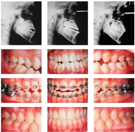
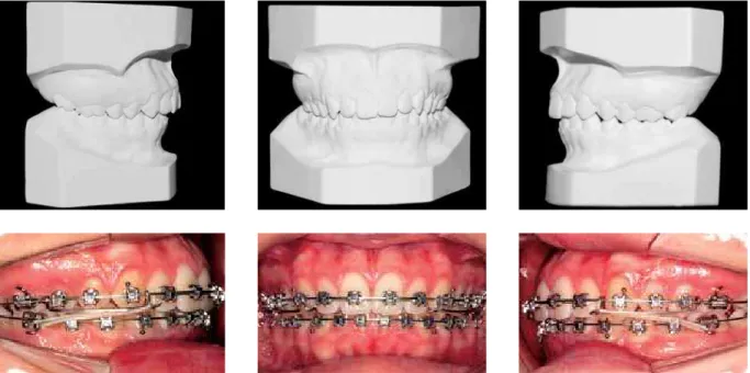
Fortunately, the number of Class III patients in our population is relatively low, around 3.3 to 4.4%,2 and the vast majority’s problems in-volve the maxilla.1 Therefore, the number of Class III patients who require orthognathic sur-gery is negligible (Fig 2). Among patients indi-cated for surgery there are those with a vertical growth pattern, like patients with severe Class II (Fig 3) and Class I with vertical excess (long face syndrome) (Fig 4).
FIGURE 3 - Initial and final lateral radiographs and intraoral photographs - Class II patient with tooth extractions (15, 25, 34, 44) and surgical advancement of the mandible.
In the orthodontic treatment of Class III mal-occlusion in adult patients with surgical indi-cation, the pre-surgical phase tends to “wors-en” patients’ aesthetics and occlusion in order to align the teeth, coordinate the arches and restore the correct axial inclination of the teeth in their supporting bone. What is your opinion about using the Anticipated Beneit Method (ABM) in surgical treatment? Mirian Nakane Matsumoto and Márcio Sobral
With the protocol I used, the number of Class III surgical patients decreased significantly, except for patients with vertical growth pattern and adult pa-tients who would come to me when it was already too late (Fig 5). I have never tried surgical treat-ment with ABM. In my opinion it can and should be used in specific cases, provided that the patient be informed that it is not the conventional procedure used in these cases and that it will entail an extra fi-nancial cost due to the placement of titanium plates
for post-surgical orthodontic movement, in addition to the future need for removing these same plates.
In my view, the main difference between the two techniques is that conventional procedure, after dental decompensation, provides better post-surgical occlusal stability in the short term since the dental arches are perfectly aligned and coordinated. ABM, on the other hand, is likely to develop occlusal instability, hindering the sta-bility of the fragments that remain from the re-cently performed surgery. This could pose future problems involving the movement of fragments. This shortcoming should be carefully assessed in the new technique. It is true, though, that pa-tient comfort is greatly enhanced, firstly because they don’t have to go through that awkward, un-sightly pre-surgical phase and secondly due to a shortened treatment time. I believe it is a prom-ising technique but it still requires further study and improvement before it is properly evaluated.
Orthodontic planning using cone beam to-mography and highly sophisticated, quality software is an undeniable reality in today’s Dentistry. Do you believe that this diagnostic resource is on its way to becoming a routine in orthodontic practice? Luís Antonio Aidar
In the U.S. this routine is already in place, both in clinics and in orthodontics, and oral and maxil-lofacial surgery programs. In Brazil, I have been keeping track of this technology’s expansion and I can tell you that it has advanced dramatically. At conferences, I have noticed that the booths selling this software tend to be always crowded. Numerous professionals are purchasing and dis-seminating this technology in their hometowns. Years ago, I was among the first to try my hand at this software. After many years’ experience and after an initial period of adjustment inherent in any major technological change, I can say that it has done much to raise the level of orthodontics as it is practiced in Brazil today. Cost still stands as the major limiting factor in our country. But I think it’s an investment that has become in-creasingly vital to any professional who wishes to avoid obsolescence. Besides, a few years ago the number of radiological clinics that made cone-beam CT scanning available to orthodontists was extremely small. But fortunately, I see this trend changing, with clinics increasingly acquir-ing these devices and offeracquir-ing this technology, thereby making it more affordable to patients.
Now if you ask me whether it is feasible for a Brazilian orthodontist to purchase a scanner for their “own” use, like Americans are used to do-ing, the answer is no (due to acquisition, main-tenance and infrastructure costs). Therefore, there is no way we can turn our backs on this technology since, above and beyond the many benefits it already offers, it is poised to play an
unprecedented role in the history of orthodon-tics. If we want Brazilian orthodontics to develop, however, the best possible initiative would be to provide this software in specialization and mas-ters programs. Even more so than in private clin-ics, for it would go a long way towards leveraging our already outstanding, worldwide recognized scientific production.
How do you see the gradual replacement of conventional X-rays used in orthodontic diag-nosis by cone beam computed tomography, and what tangible clinical beneits can ortho -dontists derive from this technological innova-tion? Is conventional cephalometry doomed to fall into disuse in the short term? Márcio Sobral and Deocleciano da Silva Carvalho
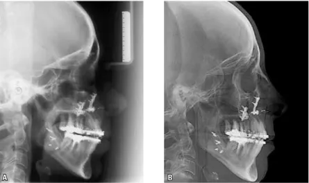
Recent scientific studies have shown that the location of anatomical landmarks on the images obtained through cone beam computed tomog-raphy is much more accurate11,14,20 and, there-fore, better than those obtained from conven-tional cephalometric images. The actual benefit accrued from CBCT is a more reliable cephalom-etry, with reduced measurement error, be it due to image distortion (CT is 1:1) (Fig 6) or to a difficulty in locating anatomical landmarks (CT features better contrast and filters that help more easily identify the landmarks, in both hard and soft tissue) (Fig 7).
A B
FIGURE 6 - Images of the same patient (A = conventional radiograph and B = radiograph taken from CT)
on the same date, showing differences in quality and sharpness between the two images.
FIGURE 7 - Software-generated maximum in-tensification filter.
What are, in your experience, the major indi -cations for cone beam computed tomography in orthodontics? In cases of impacted teeth, are CT scans the only means of diagnosis to establish an orthodontic treatment strategy?
Mirian Nakane Matsumoto
This is a somewhat controversial issue. Some authors recommend CT only in specific cases such as impacted teeth or facial asymmetry cases. After talking to some highly experienced professionals, however, I have come to realize that the trend is to indicate CT for all patients. The reason is sim-ple: cost-effectiveness (not financial, but radioac-tive cost-effecradioac-tiveness). Benefits are so significant in terms of diagnostic tomography, especially with respect to the accuracy of cephalometric measure-ments, that a slightly increased radiation—com-pared to conventional documentation—is fully justified. Furthermore, with the evolution of CT scanners that radiation tends to decrease more and more. With the new generation of CT scanners featuring extended field of view (eFOV, a must for orthodontists), we can acquire a nearly complete
patient skull in one single scan. To say nothing of the fact that, if the patient were to suffer an acci-dent with severe trauma to the face, we would have on file a data set that faithfully reproduces all of the patient’s hard and soft tissue in the face and head, in case a surgical reconstruction is required. And, just as important, we can detect—with greater ease and accuracy—a tumor or lesion that might go un-noticed in conventional panoramic radiography.
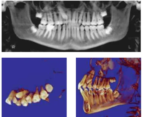
FIGURE 8 - Cone beam tomographic images.
Have you ever made orthodontic preparation of patients for orthognathic surgery (maxil -lomandibular advancement) in patients with severe obstructive sleep apnea, regardless of craniofacial alterations? Luís Antonio Aidar
Until recently, our concern with surgical orth-odontic patients was confined to achieving aes-thetic and functional results without taking into account their breathing condition. Currently, three factors are required to ensure adequate treatment outcome. With the advent of cone beam CT and advances in evaluation software, we are in a com-fortable position to assess pre- and post-treatment conditions and can now determine the volume of air (in mm³) that is moved through a patient’s airway. Moreover, with this type of evaluation we
have noticed very encouraging results in patients with respiratory failure who underwent surgery for maxillary advancement (Fig 9).
FIGURE 9 - Air volume before and after orthognathic surgery combin-ing maxillary and mandibular advancement.
In cases of “en-masse extrusion” of upper posterior teeth, what are your criteria for choosing between intrusion orthodontic procedures and surgical procedures? Luci-ano Castellucci
The emergence of micro-implants and mini titanium plates considerably improved the pre-dictability of orthodontic movements in
muti-lated patients (by reducing the number of indi-cations for this type of surgery).
In most cases I use buccal devices (mini-plates or implants) and palatal micro-implants for en-masse intrusion of posterior teeth and apply closed Nitinol springs or silk threads as elements of force.
Surgical procedures are reserved for patients with a severe vertical pattern, those with ver-tical maxillary excess and who would benefit from maxillary impaction surgery.
How would you advise orthodontists to deal with orthognathic surgeons during the planning of cases that require this type of therapy as well as during treatment de-velopment? What is your view on the fact that, under certain circumstances, a sur-geon’s mistake or inaccuracy can result in a failure for which the orthodontist might eventually take the blame? Deocleciano da Silva Carvalho
We often see patients being referred to sur-geons by orthodontists to assess whether or not it is a surgery case. Actually, it should the other way around. It is up to the orthodontist to de-termine the limitations of orthodontic move-ment. He is the one doing all the planning while the surgeon performs only one treatment phase. The orthodontist is responsible for finishing the case. Therefore, knowing who and how skillful your surgeon is, can prove vital. I usually estab-lish the following protocol for surgical cases:
a) First appointment and request for addi-tional documentation.
b) Develop diagnosis and give patient an idea of costs.
c) Referral to surgeon for further explana-tion of the surgery, risks, and an idea of future costs.
is no looking back, that is, once treat-ment gets started, if he or she decides not to undergo surgery, the case will likely become worse than when he or she start-ed treatment (treatment can only begin with a committed patient, fully aware of his or her responsibility).
e) Once the case is on track, the teeth have been uprighted on the basal bone and dental arches have been coordinated, send the patient back to the surgeon for a general pre-surgical assessment.
f) Request new documentation and plan the surgery with the surgeon to optimize the final aesthetic and functional results. This step is very important because this is where orthodontist and surgeon must see eye to eye to ensure that results are according to plan while minimizing any future problems for those involved in the treatment (orthodontist, surgeon and patient).
g) Placement of surgical hooks by orthodon-tist within the week surgery was sched-uled for. Usually 1 week to 10 days after surgery the patient starts coming to the office on a regular basis for monitoring elastic use, which allows better control and stabilization of surgical fragments. h) Orthodontic treatment is finished. Surely, if we follow those steps carefully, er-rors can be minimized and any minor discrep-ancies that may arise can now be corrected with the use of micro-implants to finish the case in the best possible way.
In your practice, in cases where you need to use as anchorage an implant, with a pro-visional crown, do you usually wait for the osseointegration period of the implant or do you go for immediate loading? Luciano Castellucci
A success rate ranging between 92% and
99% has been reported in the literature—for the maxilla and the mandible, respectively—by studies of short and long term support of fixed partial dentures. These findings have led ortho-dontists to use these implants as orthodontic anchorage. Because of their behavior, which re-sembles an ankylosis, dental implants work as an ideal anchor point for orthodontic accesso-ries, facilitating tooth movement and avoiding the use of headgear.
A prospective study investigated seven adults who used implants as rigid anchorage. After 6 months of osseointegration, all fourteen implants remained stable during treatment, withstanding forces of 150 to 400g. There were no complications. The desired orthodontic re-sults were achieved in all cases. A three-year follow-up has shown that rigid intraoral an-chorages are predictable.9
The horizontal impact of orthodontic forces on dental implants has been examined in sev-eral animal studies, showing no interference with osseointegration. In particular, only small changes can be noted in marginal bone level, pocket depth, bone-implant contact and in-creased bone density.6,18
The literature describes the application of orthodontic force to implants after a 6-month period of osseointegration. Two years after orthodontic treatment, the study found a sur-vival rate of 87.1% in the maxilla and 100% in the mandible. No significant bone loss was ob-served during orthodontic treatment.21
Scientific studies conducted in animals and humans using implants for orthodontic anchor-age suggest, in general, the existence of a heal-ing period rangheal-ing from 12 weeks to 6 months for osseointegration to occur, thus allowing their use for orthodontic anchorage.
chemical surface treatment. The former in-creases initial stability and the latter acceler-ates osseointegration. Efforts have been made to develop protocols for putting the implant in function within a 45-day period.
A 5-year prospective study assessed the ear-ly loading of 104 SLA-treated implants (sand-blasting and acid etching) in 51 patients. The study showed a 99% success rate in the appli-cation of orthodontic force to implants after a period of six weeks of osseointegration. Clinical parameters were similar to other clinical stud-ies and bone crest peri-implant stability was maintained.4 The chemical activation of the implant surface reduced temporary appliance installation time from 6 to 3 weeks.5
Ideally, before starting orthodontic anchor-age with implants, you should consider the type of implant to be used. You should evaluate if the implant has some feature in its geometry and surface that can accelerate osseointegra-tion. It is also advisable to check the place-ment site, if it is in the maxilla or mandible,
and note the different bone densities because if an implant is installed in low density bone it requires a longer osseointegration period than one installed in high density bone. Finally, you should observe the insertion torque and initial implant stability to determine when to activate the implant-supported anchorage.
Ordinarily, I use implants as orthodontic an-chorage with two goals in mind:
1) For orthodontic anchorage.
2) To use the same implant for future oral rehabilitation.
We now know that if we apply forces to im-plants through immediate loading we run the risk of encountering future problems, such as implant tipping, bone loss or even implant loss, which would render our 2nd goal impossible.8
Figure 10 illustrates the use of implants for mesial repositioning of the left lower segment and subsequent rehabilitation of the first mo-lar (36) in a Class II malocclusion patient, on the left side, caused by missing molars in the lower left segment.
In cases of agenesis of upper lateral incisors, when do you distalize canines to place an im-plant in edentulous regions and when do you mesialize canines to close spaces? Luciano Castellucci
The answer to this question depends on an individualized assessment of each case. Several factors have a bearing on the decision: The age of the patient seeking treatment, whether it’s a teenager or an adult, the need for extractions in the lower arch, the patient’s aesthetic require-ments. You should have a very honest, up-front chat with the patient and/or his/her legal guard-ians to discuss the cost-effectiveness of the dif-ferent alternatives, their advantages and disad-vantages in the short and long term.
Let’s try to shed a little more light on the is-sue: Let’s say it’s an adolescent or adult patient who presents with agenesis of a lateral incisor and a skeletal and dental Class I. We will try to con-vince him or her that the best treatment option is the placement of an implant in the missing side to restore symmetry, while explaining the poten-tial future risks, such as discolored gingiva in the implant region or even height differences due to the extrusion of the remaining teeth, especially when gingival exposure is an issue.
In the case of agenesis of lateral incisors given the same skeletal and dental condition, we have to better assess the cost-benefit analysis. In this case, we might also have to convince him or her to have an implant installed, explaining all future risks, as mentioned above.
Should any tooth extractions be required and two upper ageneses be present, we would probably opt for upper space closure and re-placement of laterals with canines, and canines with first premolars. In these cases, I always perform canine extrusion and first premolar intrusion to try and improve the condition of the gingival margins in relation to the upper central incisors.12
As for aesthetics, we know that the sine qua non condition for a successful implant outcome is adequate bone condition,17 which should be in place before implant installation along with prior orthodontic movements or bone grafts whenever necessary.
The truth of the matter is that dental plants had their aesthetic quality greatly im-proved in the late 90’s, so we are talking about nearly 10-years’ experience, which is too short a time period for any conclusive statements. As we speak, I am in the process of putting together a list of my patients who had implants placed to replace the lateral incisors. After I have carried out a thorough evaluation of these cases I will be better equipped to answer this question.
FIGURE 11 - Opening of denture space for implant (12) and clinical crown increase (22).
We constantly hear that self-ligating brack-ets are the future of orthodontics. What are your views on the current scientiic ratio -nale of these appliances and your personal experience with this subject? Deocleciano da Silva Carvalho
I have always been against placing too much emphasis on the role of orthodontic appliances. In my opinion there is no such thing as a smart appliance. It’s the mind behind the pliers that needs to be smart. We witnessed a parade of fad techniques before the emergence of self-ligating brackets. There was the promise of lightning fast results and cases would purportedly finish of their own accord. But this is not what the literature has shown lately. In cases of minor crowding results have been faster. But in cases of severe crowding almost no statistical differences have been found.22
Allow me to comment on our cases treated with self-ligating brackets:
a) The biggest advantage is for patients who live far away in distant cities, who can only come to the office at longer time intervals (up to 6 weeks) and whose treatment is making good headway thanks to heat-activated archwires.
b) In patients with missing teeth requir-ing increased slidrequir-ing mechanics the response is indeed faster (due to reduced friction between bracket and archwire). 13
c) I have also noticed a quicker response when sliding-jigs are used, especially in asym-metric Class II cases (Fig 12).
d) Hygiene is improved thanks to the ab-sence of elastic ligatures on the brackets.
1. Alcan T, Keles A, Erverdi N. The effects of a modiied protraction headgear on maxilla. Am J Orthod Dentofacial Orthop. 2000 Jan;117(1):27-38.
2. Baptista AA, Cury SAA, Motta AFJ, Vilella OV, Mucha JN. A prevalência de más-oclusões em escolares de Niterói. Rev Flum Odontolol. 1998 maio-ago; 2(8):34-41.
3. Bjerklin K, Ericson S. How a computerized tomography examination changed the treatment plans of 80 children with retained and ectopically positioned maxillary canines. Angle Orthod. 2006 Jan;76(1):43-51.
RefeRenCes
on a surgical case hand in hand with a maxil-lofacial surgeon, who gave the appliance a very positive assessment.
f) Retreatment patients who had previously used a conventional appliance also made a favor-able evaluation (less discomfort).
g) My experience shows a gain of approxi-mately 10% in treatment time—though I have
no scientific study to support this claim—espe-cially in cases that require more sliding.
h) I noted a transverse arch development but long-term monitoring is needed to assess stability.
The most critical part is definitely bonding, given the need to reposition brackets during treatment, even if your bonding was perfect.
4. Bornstein MM, Schmid B, Belser UC, Lussi A, Buser D. Early loading of non-submerged titanium implants with a sandblasted and acid etched surface. 5 years results of a prospective study in partially edentulous. Clin Oral Implants Res. 2005 Dec;16(6):631-8.
5. Buser D, Chen ST, Weber HP, Belser UC. Early implant placement following single-tooth extraction in the esthetic zone: biologic rationale and surgical procedures. Int J Periodontics Restorative Dent. 2008 Oct;28(5):441-51.
6. Gotfredsen K, Berglundh T, Lindhe J. Bone reactions adjacent to titanium implants subjected to static load of different duration. Clin Oral Implants Res. 2001 Dec;12(6):552-8.
ACKnOWLeDGeMenTs
I am grateful to Dr. Keila Rodrigues Correia for her assistance in organizing this interview
and Dr. Daniel P. Brunetto for his help and support in the area of tomography and digital documentation.
Márcio sobral
- MSc in Orthodontics, UFRJ.
- Professor, Specialization Course in Orthodontics, UFBA.
Luciano Castellucci
- DDS, UFBA.
- MSc and PhD in Oral Rehabilitation, FOB/USP. - Adjunct Professor, FO/UFBA.
- Scientific Director and Professor, Specialization Courses in Prosthodontics and Implant Dentistry, ABO/BA.
Mirian Aiko nakane Matsumoto
- DDS, FORB/USP, Ribeirão Preto/SP - MSc and PhD in Orthodontics, UFRJ.
- Full Professor, FORB/USP, Ribeirão Preto/São Paulo. - Diplomate of the Brazilian Board of Orthodontics and
Facial Orthopedics (BBO). Deocleciano da silva Carvalho
- DDS, USP, São Paulo.
- MSc in Orthodontics, USP, São Paulo. - PhD in Pediatric Dentistry, USP, São Paulo.
- Director of the Brazilian Board of Orthodontics and Facial Orthopedics.
Luís Antônio de Arruda Aidar
- DDS, UNIMES, Santos, São Paulo State. - Specialist and MSc in Orthodontics, UMESP
(Methodist College/São Paulo).
- PhD (Otolaryngology and Head and Neck Surgery), UNIFESP (EPM/São Paulo).
- Professor, Department of Orthodontics, School of Dentistry, UNISANTA (Santa Cecília/Santos). - Head of the Specialization Course in Orthodontics,
School of Dentistry, UNISANTA (Santa Cecília/ Santos).
Contact address
Ademir Roberto Brunetto Av. 7 de Setembro, 4456 - Batel CEP: 80.250-210 - Curitiba/PR Email: [email protected]
7. Hassel B, Farman AG. Skeletal evaluation using cervical vertebrae. Am J Orthod Dentofacial Orthop. 1995 Jan;107(1):58-66.
8. Higuchi K. Osseointegration and orthodontics. In: Branemark PI, editor. The osseointegration book: from calvarium to calcaneus. 1. Osseointegration. Berlin: Quintessence Books; 2005. p. 251-69.
9. Higuchi KW, Slack JM. The use of titanium ixtures for intraoral anchorage to facilitate orthodontic tooth movement. Int J Oral Maxillofac Implants. 1991 Fall;6(3):338-44.
10. Kapust AJ, Sinclair PM, Turley PK. Cephalometric effects of face mask/expansion therapy in Class III children: a comparison of three age groups. Am J Orthod Dentofacial Orthop. 1998 Feb;113(2):204-12.
11. Kobayashi K, Shimoda S, Nakagawa Y, Yamamoto A. Accuracy
in measurement of distance using limited cone-beam
computerized tomography. Int J Oral Maxillofac Implants. 2004 Mar-Apr;19(2):228-31.
12. Kokich VO Jr, Kinzer GA. Managing congenitally missing lateral incisors. Part I: canine substitution. J Esthet Restor Dent. 2005;17(1):5-10.
13. Krishnan M, Kalathil S, Abraham KM. Comparative evaluation of frictional forces in active and passive self-ligating brackets with various archwires alloys. Am J Orthod Dentofacial Orthop. 2009 Nov;136(5):675-82.
14. Lascala CA, Panella J, Marques MM. Analysis of the accuracy of the linear measurements obtained by cone beam computed tomography (CBCT-NewTom). Dentomaxillofac Radiol. 2004 Sep;33(5):291-4.
15. Li KK, Powell NB, Riley RW, Zonato A, Gervacio L, Guilleminault C. Morbidly obese patients with severe obstructive sleep apnea: is airway reconstructive surgery a viable treatment option? Laryngoscope. 2000 Jun;110(6):982-7.
16. MacDonald KE, Kapust AJ, Turley PK. Cephalometric changes after the correction of Class III malocclusion with maxillary expansion/facemask therapy. Am J Orthod Dentofacial Orthop. 1999 Jul;116(1):13-24.
17. Meirelles JKS, Reis SA, Fornazari RF. Inter-relação ortodontia-implantodontia. Terapia clínica avançada em ortodontia-implantodontia. Säo Paulo: Artes Médica; 2002.
18. Melsen B. Tissue reaction to orthodontic tooth movement – a new paradigm. Eur J Orthod. 2001 Dec;23(6):671-81. 19. Mitani H, Fukazawa H. Effects of chin cup force on the timing
and amount of mandibular growth associated with anterior reversed occlusion (Class III malocclusion) during puberty. Am J Orthod Dentofacial Orthop. 1986 Dec; 90(6):454-63.
20. Misch KA, Yi ES, Sarment DP. Accuracy of cone beam computed tomography for periodontal defect measurements. J Periodontol. 2006 Jul;77(7):1261-6.
21. Molly L. Periodontal parameters around implants anchoring orthodontic appliances: a series of case report. J Periodontol. 2004 Jan;75(1):176-81.
22. Fleming PS, DiBiase AT, Sarri G, Lee RT. Eficiency of mandibular arch alignment with 2 preadjusted Edgewise appliances. Am J Orthod Dentofacial Orthop. 2009 Dec;136(6):756-7.
23. Sugawara J, Asano T, Endo N, Mitani H. Long term effects on chin cup therapy on skeletal proile in mandibular prognathism. Am J Orthod Dentofacial Orthop.1990; 98(2):127-33, 1990. 24. Weissheimer F, Brunetto AR, Petrelli E. Disjunção palatal e