Abstract
Objective: To evaluate if clinical and laboratory parameters could assist in the differential diagnosis of intra and extra-hepatic neonatal cholestasis (NC).
Methods: Retrospective study of NC patients admitted at the Pediatric Hepatology Outpatient Clinic of the teaching hospital of Universidade Estadual de Campinas (UNICAMP), Campinas, Brazil, between December 1980 and March 2005. The approach to the diagnosis of NC was standardized. According to diagnosis, patients were classiied into two groups: I (intra-hepatic neonatal cholestasis) and II (extra-hepatic neonatal cholestasis). In order to verify if there was association with the categorical variable, the chi-square and Mann-Whitney tests were used, with corrections for age for the covariance analysis (ANCOVA). The determination of accuracy of the clinical and laboratory variables for differentiation of the groups was made using the analysis of the ROC curve.
Results: One hundred and sixty-eight patients were evaluated (group I = 54.8% and group II = 45.2%). In the patients with less than 60 days of life there was predominance of intra-hepatic causes, whereas, in those older than 60 days, there was predominance of extra-hepatic etiology (p < 0.001). Median birth weight was lower in group I (p = 0.003), as well as length at birth (p = 0.007). Median values of direct bilirubin were higher in group II (p = 0.006). Values of gamma-glutamyltransferase (GGT) (10 times higher than the limit of normality) presented sensitivity of 56.3%, speciicity of 91.5%, and accuracy of 75.7% for the diagnosis of extra-hepatic cholestasis.
Conclusion: In the present study, extra-hepatic NC presented greater weight and length at birth, fecal hypocholia/ acholia, choluria, hepatomegaly, increase in GGT (10.8 times higher than the limit of normality), and a delay for investigation in the tertiary center.
J Pediatr (Rio J). 2010;86(1):40-44: Neonatal cholestasis, children, hepatitis.
O
RiginAlA
RtiCleCopyright © 2010 by Sociedade Brasileira de Pediatria
40
introduction
Neonatal cholestasis (NC) is a syndrome that poses a major diagnostic challenge to the clinician from an anatomical standpoint. A rapid investigation is always considered to be pathologically necessary.1-3
The frequency of NC in the population is dificult to evaluate. Incidences of 1/2,500,4 1/5,000,5 and 1/9,0006 live births have been recorded in the literature. The focus of the initial approach is to differentiate between intrahepatic
(IHC) and extrahepatic (EHC) cholestasis. Differentiation between IHC and EHC may have a success rate of 90 to 95% when various diagnostic methods are used.2
IHC represents about 2/3 of all cases of NC.4,7-9 Once established that cholestasis is intrahepatic, the new challenge is to determine the etiological diagnosis. In a classiication proposed by Dellert & Balistreri,8 there are around 40 possible causes of IHC. Despite this fact, part of the cases is
Differential diagnosis of neonatal cholestasis:
clinical and laboratory parameters
Maria Angela Bellomo-Brandao,1 luciana tonussi Arnaut,2 Adriana M. A. De tommaso,1 gabriel Hessel1
1. MD, PhD. Department of Pediatrics, Faculty of Medical Sciences, Universidade Estadual de Campinas (UNICAMP), Campinas, SP, Brazil. 2. MD. Department of Pediatrics, Faculty of Medical Sciences, UNICAMP, Campinas, SP, Brazil.
No conflicts of interest declared concerning the publication of this article.
Suggested citation: Bellomo-Brandao MA, Arnaut LT, Tommaso AM, Hessel G. Differential diagnosis of neonatal cholestasis: clinical and laboratory parameters. J Pediatr (Rio J). 2010;86(1):40-44.
Manuscript submitted Oct 19 2009, accepted for publication Dec 8 2009.
of unknown etiology. The causes of EHC are less numerous and include biliary atresia (BA), choledochal cyst, biliary sludge, and biliary duct stricture.1,10
According to the guideline of the North American Society for Pediatric Gastroenterology, Hepatology, and Nutrition (NASPGHAN), any infant noted to be jaundiced at 2 weeks of age (or 3 weeks of age in those who are breastfed) should be investigated for cholestasis by measuring serum bilirubin levels.2
Treatable disorders, such as sepsis, hypothyroidism, panhypopituitarism, and inborn errors of metabolism (for example, galactosemia), must be recognized and treated promptly to prevent signiicant progression of the disease. In BA, urgency in diagnosis is associated with the performance of the Kasai porto-jejunostomy before the infant is 60 days of age and is directly related to successful treatment.2,9,12,13
The most important indings in BA are jaundice and acholic stools starting between ages 4 and 8 weeks in infants who are otherwise apparently in good health.14
For early identiication of BA cases, the use of a stool color card,3,15 ultrasonography for the presence of the triangular cord sign,16,17 and also correlation of clinical, laboratory and histopathological indings have been proposed.3,14,18-20
This study is aimed at evaluating whether clinical and laboratory parameters may help to establish differentiation between IHC and EHC.
Methods
A retrospective study was conducted from December 1980 to March 2005 to review the medical charts of patients referred to a tertiary care center [Pediatric Hepatology Outpatient Facility at Hospital de Clínicas of Universidade Estadual de Campinas (UNICAMP), Campinas, Brazil] for investigation of NC. Inclusion criteria were: cholestatic jaundice starting within the irst 3 months of life and performance of liver biopsy during the irst year of life.
The protocol for data acquisition contained the following information: fetal distress, breastfeeding, gender, age, birth weight, length at birth, dark urine, a history of jaundice at birth or after 1 month of age, pale or acholic stools, vomiting, edema, ascites, hepatomegaly, and splenomegaly.
Laboratory test results were obtained in absolute numbers and number of times the upper limit of normal [alanine aminotransferase (ALT), aspartate aminotransferase (AST), alkaline phosphatase (AP), gamma-glutamyl-transferase (GGT)], in addition to albumin, International Normalized Ratio (INR) and direct bilirubin (DB). To determine the etiology, the following tests were reviewed: liver biopsy, serological test results for cytomegalovirus (CMV), human immunodeiciency virus (HIV), Epstein-Barr virus (EBV),
rubella, toxoplasmosis, and syphilis. Whenever necessary, data were also collected from other tests performed, such as measurement of alpha-1-antitrypsin level, measurement of sweat sodium and chloride concentrations, study of inborn errors of metabolism, hormone levels, study of polymerase chain reaction for CMV (PCR-CMV) in the blood, and antigenemia.
Patients were divided into two groups: group I with IHC and group II with EHC.
Ethical aspects
The study was approved by the Research Ethics Committee of the School of Medicine of UNICAMP.
Statistical analysis
To compare categorical variables between groups of patients with cholestasis, the chi-square test or Fisher’s exact test was used, whenever necessary. To compare numerical variables between groups, the Mann-Whitney test was used. Due to the age difference between the groups, comparisons were repeated and corrected for age, using analysis of covariance (ANCOVA).
Receiver operating characteristic curve (ROC) analysis was carried out to obtain cutoff values for GGT (absolute values and number of times the upper limit of normal) to distinguish between IHC and EHC, using the inal diagnosis as the gold standard.
Results
One hundred and sixty-eight patients were evaluated (group I = 54.8% and group II = 45.2%). A signiicant difference was observed between the groups regarding the following variables: gender, age, birth weight, height at birth, dark urine, acholic stools, hepatomegaly, direct bilirubin, absolute value and number of times the upper limit of normal for GGT.
The predominant gender was male (62.64%) in group I and female in group II (55.26%) with p = 0.026.
In infants investigated before 60 days of age, intrahepatic causes predominated, whereas in those investigated after 60 days of age, extrahepatic causes predominated (p < 0.001).
Weight and length at birth were lower in group I. Number of patients with birth weight lower than 2,500 g was higher in group I. Hepatomegaly, pale/acholic stools, and dark urine predominated in cases of EHC. Results are shown in Table 1. The median serum level of direct bilirubin was higher in group II (p = 0.006). The same occurred with GGT level which was shown as the number of times the upper limit
Variable n group i (n = 92) group ii (n = 76) p
Gender 168 0.026
Male 57 34
Female 35 42
Age (days) 168 56±46 77±36 < 0.001
Birth weight (g) 165 2,630±775.18 2,975±432.81 0.003
Number of patients
with birth weight < 2,500 g 165 31 (n = 90) 5 (n = 75) < 0.001
Length at birth 122 47±4 (n = 60) 48±1.85 (n = 62) 0.007
Pale/acholic stools 167 64 (n = 91) 68 0.002
Dark urine 168 64 67 0.004
Hepatomegaly 157 37 (n = 86) 50 (n = 71) < 0.001
table 1 - Patient clinical characteristic in the irst examination, according to group
Variable n group i (n = 92) group ii (n = 76) p
ALT 159 2.58 (0.10-18.25) 3.63 (0.43-19.45) (n = 67) 0.090
AST 159 4.69 (0.17-44.00) 5.37 (0.02-17.86) (n = 67) 0.546
AP 121 1.16 (0.13-12.10) (n = 77) 1.53 (0.13-10.50) (n = 56) 0.346
GGT 107 3.91 (0.49-8.49) (n = 59) 12.68 (2.10-6.63) (n = 48) < 0.001
Albumin (g/dL) 89 3.77 (1.68-4,84) (n = 51) 3.70 (1.19-5.60) (n = 38) 0.684
INR 59 1.04 ( 1.00-2.77) (n = 15) 1.14 (0.87-2.08) (n = 44) 0.649
DB (mg/dL) 159 6.35 (1.10-19.60) (n = 90) 8.30 (1.68-17.00) (n = 69) 0.006
table 2 - Laboratory test results performed at the beginning of investigation, expressed as the median (interquartile range)
ALT = alanine aminotransferase; AP = alkaline phosphatase; AST = aspartate aminotransferase; DB = direct bilirubin; GGT = gamma-glutamyl-transferase, albumin; INR = International Normalized Ratio.
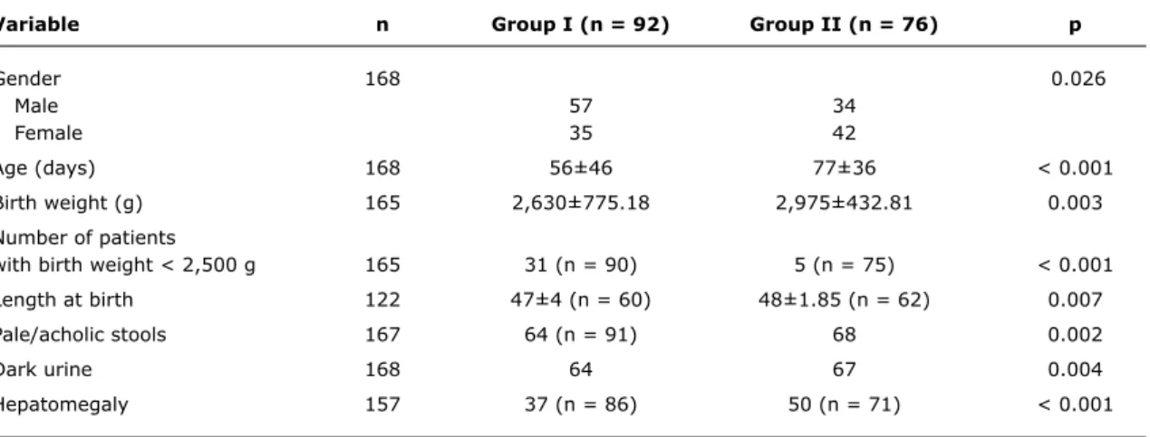
The median level of hepatic enzymes is shown as the number of times the upper limit of normal. For the diagnosis of EHC, serum levels of
gamma-glutamyl-transferase ≥ 429.5 U/L or > 10.8 times the upper limit of normal had a sensitivity of 56.3%, a speciicity of 91.5%, and an accuracy of 75.7% (Figures 1 and 2).
There were no statistical differences between groups I and II concerning history of fetal distress, breastfeeding, history of jaundice at birth or after the irst month of life, vomiting, splenomegaly, presence of edema, and ascites.
Discussion
In the investigation of neonatal cholestasis, the main aim is to make an early diagnosis and promptly implement the most appropriate therapy. For example, cases of sepsis, hypothyroidism, and metabolic disease require early diagnosis and treatment to prevent progression of the disease and its consequent worsening. BA must be diagnosed promptly because early performance of the Kasai procedure (before 2 months of age) is directly related to
successful therapy.2 Liver biopsy is an excellent diagnostic test, as long as it contains ive to seven portal tracts and is performed by an experienced pathologist with expertise in pediatric blood pathology. However, the gold standard of diagnosis is exploratory laparotomy.2,11,21
GGT = gamma-glutamyl-transferase; ROC = receiver operating characteristic. Absolute GGT values area = 0.825; 95%CI 0.749-0.901; p < 0.001; sensitivity = 56.3%; specificity = 91.5%; accuracy = 75.7%; n = 107.
Cutoff value: GGT > 429.5.
GGT = gamma-glutamyl-transferase; ROC = receiver operating characteristic. Number of times the upper limit of gamma-gt: sensitivity = 58.3%; specificity = 91.5%; accuracy = 76.6%; n = 107.
Cutoff: number of times GGT > 10.80.
Figure 2 - Results of ROC curve analysis for absolute GGT values
Figure 1 - Results of ROC curve analysis for number of times the upper limit GGT values
No statistically signiicant differences were found in serum levels of AST, ALT and AP, similar to results obtained in the present study.
Weight and length at birth were lower in the IHC group, possibly relecting prematurity, failure to thrive, neonatal complications, metabolic disorders, and congenital infections.8,14,19,22,23
The median age of infants at the time of referral is a cause for concern. In the EHC group, age at referral was over 60 days (77 days) of life and higher than that in the IHC group. Data published in Brazil show that in the majority of cases (76 to 94.7%), the diagnosis of BA is made after 8 weeks of life.10,24-27
It is likely that infants in the IHC group were more promptly investigated, due to a more severe systemic illness and arrived earlier at a referral center. In the EHC group, referral was delayed, resulting in the performance of the Kasai portoenterostomy at a later date. Perhaps this may have contributed to a more frequent inding of hepatomegaly.
Dark urine and pale/acholic stools predominated in infants with EHC, and the median serum levels of direct bilirubin was higher, relecting a more marked clinical picture of cholestasis.
GGT is an enzyme that transfers the gamma-glutamyl group from glutathione and other peptides. It is present in the biliary epithelium and hepatocytes. GGT levels are elevated in various liver diseases. GGT is also present in the kidney, breast, intestine, brain, pancreas, and spleen. Normal values vary with age, gender, and diagnostic methods. That is why emphasis is placed on results expressed as the number of times the upper limit of normal.28 It is recognized as a useful test for differentiating familial cholestatic syndromes.8
The use of GGT to differentiate IHC from EHC was not recommended by Moyer et al.,2 who classiied it as level C evidence, because a wide variability of tests makes the interpretation of results more dificult.
Wright & Christie29 reported that GGT > 300 UI/L could be suggestive of EHC. Lai et al.17found GGT values > 300 UI/L with a speciicity of 82.8% and accuracy of 72% for the diagnosis of EHC. Wang et al.30 concluded that normal or low levels of serum GGT may indicate a more severe case of idiopathic IHC, albeit reversible.
In the present study, the GGT value expressed as the number of times the upper limit of normal was also higher in cases of EHC.
Correspondence:
Maria Angela Bellomo-Brandao Aristides Lobo, 789
CEP 13083-060 - Campinas, SP - Brazil Tel.: +55 (19) 3579.2998, +55 (19) 9794.3289 E-mail: [email protected]
References
1. Hessel G, Sawamura R. Colestase do lactente. In: Barbiere D, Palma D, editors. Gastroenterologia e Nutrição. Série Atualizações Pediátricas. São Paulo: Atheneu; 2001. p. 143-57.
2. Moyer V, Freese DK, Whitington PF, Olson AD, Brewer F, Colletti RR, et al. Guideline for the evaluation of cholestatic jaundice in infants: recommendations of the North American Society for Pediatric Gastroenterology, Hepatology and Nutrition. J Pediatr Gastroenterol Nutr. 2004;39:115-28.
3. Sokol RJ, Shepherd RW, Superina R, Bezerra JA, Robuck P, Hoofnagle JH. Screening and outcomes in biliary atresia: summary of a National Institutes of Health workshop. Hepatology. 2007;46:566-81.
4. Dick MC, Mowat AP. Hepatitis syndrome in infancy: an epidemiological survey with 10-year follow up. Arch Dis Child. 1985;60:512-6.
5. Danks DM, Campbell PE, Jack I, Rogers J, Smith AL. Studies of the aetiology of neonatal hepatitis and biliary atresia. Arch Dis Child. 1977;52:360-7.
6. Henriksen NT, Drablos PA, Aagenaes O. Cholestatic jaundice in infancy. The importance of familial and genetic factors in aetiology and prognosis. Arch Dis Child. 1981;56:622-7.
7. Eliot N, Odièvre M, Hadchouel M, Hill C, Flamant R. Analyze statistique des donnees cliniques, biologiques et histologiques dans 288 observations de cholestase neonatale. Arch Fr Pediatr. 1977;34:213-20.
8. Dellert SF, Balistreri WF. Neonatal cholestasis. In: Walker WA, Durie PR, Hamilton JR, Walker-Smith JA, Watkins JB, editors. Pediatric gastrointestinal disease: pathophysiology, diagnosis, management. 3rd ed. Ontario: BC Decker; 2000. p. 880-94.
9. Yachha SK, Sharma A. Neonatal cholestasis in India. Indian Pediatr. 2005;42:491-2.
10. de Carvalho E, Ivantes CA, Bezerra JA. Extrahepatic biliary atresia: current concepts and future directions. J Pediatr (Rio J). 2007;83:105-20.
11. Petersen C. Pathogenesis and treatment opportunities for biliary atresia. Clin Liver Dis. 2006;10:73-88, vi.
12. Schreiber RA, Kleinman RE. Biliary atresia. J Pediatr Gastroenterol Nutr. 2002;35 Suppl 1:S11-6.
13. Alagille D. Cholestasis in the irst three months of life. Prog Liver Dis. 1979;6:471-85.
14. Hsiao CH, Chang MH, Chen HL, Lee HC, Wu TC, Lin CC, et al.
Universal screening for biliary atresia using an infant stool color card in Taiwan. Hepatology. 2008;47:1233-40.
15. Park WH, Choi SO, Lee HJ. Technical innovation for noninvasive and early diagnosis of biliary atresia: the ultrasonographic ”triangular cord” sign. J Hepatobiliary Pancreat Surg. 2001;8:337-41.
16. Roquete ML, Ferreira AR, Fagundes ED, Castro LP, Silva RA, Penna FJ. Accuracy of echogenic periportal enlargement image in ultrasonographic exams and histopathology in differential diagnosis of biliary atresia. J Pediatr (Rio J). 2008;84:331-6. 17. Lai MW, Chang MH, Hsu SC, Hsu HC, Su CT, Kao CL, et al.
Differential diagnosis of extrahepatic biliary atresia from neonatal hepatitis: a prospective study. J Pediatr Gastroenterol Nutr. 1994;18:121-7.
18. Mowat AP, Psacharopoulos HT, Williams R. Extrahepatic biliary atresia versus neonatal hepatitis. Review of 137 prospectively investigated infants. Arch Dis Child. 1976;51:763-70.
19. Okçu-Heper A, Erden E, Doganci T, Kuloglu Z, Kansu A, Genc Y.
Nonobstructive neonatal cholestasis: clinical outcome and scoring of the histopathological changes in liver biopsies. Pediatr Dev Pathol. 2006;9:44-51.
20. Rastogi A, Krishnani N, Yachha SK, Khanna V, Poddar U, Lal R.
Histopathological features and accuracy for diagnosing biliary atresia by prelaparotomy liver biopsy in developing countries. J Gastroenterol Hepatol. 2009;24:97-102.
21. Dehghani SM, Haghighat M, Imanieh MH, Geramizadeh B.
Comparison of different diagnostic methods in infants with Cholestasis. World J Gastroenterol. 2006;12:5893-6.
22. Bellomo-Brandão MA, Porta G, Hessel G. Clinical and laboratory evaluation of 101 patients with intrahepatic neonatal cholestasis.
Arq Gastroenterol. 2008;45:152-5.
23. Kieling CO, Santos JL, Vieira SM, Ferreira CT, Linhares AR, Lorentz AL, et al. Biliary atresia: we still operate too late. J Pediatr (Rio J). 2008;84:436-41.
24. Pileggi FO, dos Santos RC, Vicente YA, Machado MI, Zucolotto S. Atresia de vias biliares: estudo de 19 casos. J Pediatr (Rio J). 1996;72:5-8.
25. dos Santos JL, Cerski CT, da Silva VD, da Mello ES, Wagner MB, da Silveira TR. Fatores relacionados ao prognóstico da atresia biliar pós-portoenterostomia. J Pediatr (Rio J). 2002;78:341-6. 26. Suzuki HU, Morais MB, Medeiros EH, Kawakami E, Patrício FR, Wehba
J, et al. Síndrome colestática do lactente: análise retrospectiva de 177 casos. Rev Paul Pediatr. 1991;9:90-4.
27. Tannuri U. Atresia das vias biliares – Evolução nas duas últimas décadas. J Pediatr ( Rio J). 1996;72:1-3
28. Whitield JB. Gamma glutamyl transferase. Crit Rev Clin Lab Sci. 2001;38:263-355.
29. Wright K, Christie DL. Use of gamma-glutamyl transpeptidase in the diagnosis of biliary atresia. Am J Dis Child. 1981;135:134-6. 30. Wang JS, Tan N, Dhawan A. Signiicance of low or normal serum
gamma glutamyl transferase level in infants with idiopathic neonatal hepatitis. Eur J Pediatr. 2006;165:795-801.