Saucerization of osseointegrated
implants and planning of simultaneous
orthodontic clinical cases
Alberto Consolaro*, Renato Savi de Carvalho**, Carlos Eduardo Francischone Jr.***, Maria Fernanda M.O. Consolaro****, Carlos Eduardo Francischone*****
The field for Orthodontics has seen significant expansion with the advent of new diagnostic and therapeutic approaches in all specialties, such as medical and dental implantology, sleep medicine, orthognathic surgery, computed tomography, gerodontology, etc. This requires the mastery of new concepts and technical terms typical of the jargon used by each specific area. Such mastery plays a key role in discussions about diagnosis and planning of clinical cases with professionals from other specialties.
Dental osseointegrated implants, for example, completely changed the practice and scope of dentistry in the last 20 years. Many adult orth-odontic patients have already had one or more osseointegrated implants installed or may be planning, or need to do so. Many young orth-odontic patients have also had osseointegrated implants installed because of tooth loss caused by trauma or partial anodontia.
Osseointegrated implant saucerization is a phenomenon worthy of recognition and con-sideration in orthodontic planning to establish functional and aesthetic prognosis. With this in-sight in mind, we intend to discuss the concept of saucerization, with the specific purpose of answering a few important questions. Given the
occurrence of saucerization, should special care be given to teeth located in the neighborhood of osseointegrated implants when moving teeth and finishing orthodontic cases?
The concept of osseointegration is a peculiar-ity of the teeth and implants in our bodies: The importance of cervical soft tissues
Osseointegration allows the direct anchor-age of an implant through bone tissue forma-tion around the implant without the growth or development of fibrous tissue at the bone-im-plant interface.3,5
Teeth are the only body structures that tra-verse or penetrate an epithelial lining or cover-age (Figs 1, 2 and 3). By extension, dental im-plants also have this feature and the anchorage provided by osseointegration is a prerequisite for implant stability. Long-term implant surviv-al depends on the adhesion of the epithelium and connective tissues to the titanium surface since a complete soft tissue cervical sealing pro-tects the bone from the highly contaminated oral environment.8,10,15,22,23,26
The marginal gingiva and peri-implant mu-cosa share many clinical and microscopic char-acteristics.1,2,19,20,25 The gingival mucosa around
* Full Professor of Pathology, FOB-USP and at FORP-USP Postgraduate courses. ** Professor of Implantology, Sacred Heart University (USC).
*** Professor and MSc in Implantology, USC.
A B
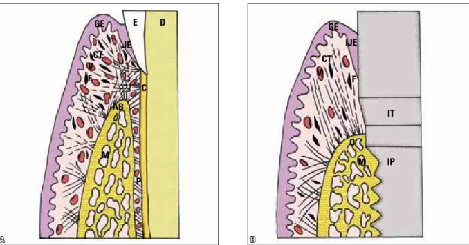
FIGURE 1 - In the normal periodontium, at A, the collagen fibers are highlighted, extending from the gingival alveolar bone (AB) crest to the cementum (C), gingiva and periodontal ligament (P) to form a cross-hatch pattern at the connective attachment. The rich blood vascular (V) and fibroblastic (F) compo-nents can be seen, to a lesser extent in the cervical peri-implant connective tissue (CT). B shows schematically that the bundles of collagen fibers in the peri-implant cervical connective attachment tend to run parallel to the surface of the intermediate prosthesis (IT). GE = gingival epithelium; JE = junctional epithelium, IJE = implant junctional epithelium; D = dentin; M = marrow space; IP = implant.
successful implants usually displays no inflam-matory lesions. When lesions do occur, they are small and located adjacent to the junctional epithelium.1,19 Clinically, a healthy or slightly inflamed gingiva, as well as the peri-implant mu-cosa, if proper oral hygiene is performed, exhibit inflammatory infiltrates at similar locations and with similar extension.20 Several studies have shown similarities between the peri-implant mu-cosa and the gingiva in terms of their epithelial and connective structures.9,16,17,18,24,27 However, the absence of root cementum on the surface of the implants change the orientation plane and the adhesion of the fibers between teeth and im-plants.9 The importance of sealing the soft tissue at implant sites to achieve functional success has not been completely or thoroughly evaluated.
Studies on the topography of periodontal tissue vasculature revealed that the gingiva and
connective tissue above the bone crest of the tooth are nourished by supraperiosteal vessels that originate in the alveolar process and peri-odontal ligament. In the soft and hard peri-im-plant tissues the mucosa region is nourished by terminal branches of wide vessels originating from the periosteum of the bone implant site. In both cases the vessels built a "plexus clevicular" lateral to the junctional epithelium. All natural teeth in the connective portion above the crest showed a rich vasculature, unlike the implant sites as very few vessels were observed in this re-gion.7 This finding reinforces the suspicion that the peri-implant soft tissue may have a slightly decreased ability to defend itself against external aggression compared to the natural periodontal tissues (Fig 1).
The mechanical resistance between the gin-giva and the peri-implant mucosa was tested in
GE GE
IJE JE
E
F
IT
IP O
M C
M
P AB F V
V CT
FIGURE 2 - The tooth is the only structure of the body that crosses the lining epithelium and interacts with the internal environment. Layout of the periodontal structures relative to the biological distances: dentin (D), cementum (C), alveolar bone (B), periodontal ligament (PL), junc-tional epithelium (JE), gingival epithelium (GE) and gingival connec-tive tissue (GCT). The junctional epithelium has 15-30 cell layers and as it proliferates in the apical direction it enables the contact of EGF molecules with bone cells, thereby stimulating bone resorption and maintenance of the biological distances. In the human body, between the epithelium and the bone, there is always connective tissue inter-position due to the presence of EGF in the underlying epithelial and connective tissues. EGF is released by the Epithelial Rests of Malas-sez and keeps the alveolar bone away from the cementum through the same mechanism and thus prevents dentoalveolar ankylosis.
FIGURE 3 - The form of the alveolar bone crest, with its rhomboidal aspect, corresponds to the morphology of the junctional epithelium (JE) which fosters the steady release of EGF, depicted by the arrows. The collagen fibers of the connective attachment (CA) perpendicular to the cementum (C) can help limit the effect of EGF on bone cells. The cementoblasts (Cb) on the root surface have receptors for EGF and other mediators of bone turnover, which ultimately protect teeth from resorption. D = dentin; PL = periodontal ligament; B = alveolar bone, E = enamel; Ob = osteoblasts.
dogs and revealed that probe penetration was greater in implants than in teeth: 2 mm and 0.7 mm, respectively.14 In peri-implant soft tissues, the probe displaced the junctional epithelium and connective tissue on the implant’s adhesion surface interface and stopped at the bone crest. Occasionally, bleeding occurred due to ves-sel rupture. In the teeth, the probe stopped at the apical portion of the junctional epithelium, identifying the bottom of the gingival sulcus. The bleeding was minimal, in contrast with that of the implants.14
The effects of dental bacterial plaque after three weeks and after three months in the gingi-va and peri-implant tissues were comparatively
evaluated.6 Both tissues exhibited inflamma-tory lesions identical in size and composition features. Within three months the bleeding was similar and both inflammatory infiltrates had the same characteristics, but the apical extent was more pronounced in the peri-implant mu-cosa than in the gingiva. This finding implies that the defense mechanisms of the gingiva are more efficient than those of the peri-implant tissues in preventing future spreads of sul-cus microbiota.6 However, the neck of an os-seointegrated dental implant tends to display normal function and aesthetics, provided that adequate oral hygiene is maintained. This also applies to normal teeth.
D
C
Cb
C
Ob
B CA
B
PL E
E
PL D
GE
JE
JE
Saucerization of osseointegrated implants: Concept and Mechanism
Saucerization occurs in all osseointegrated implants, regardless of their design, surface type, platform, connection type, commercial brand or patient conditions (Fig 12). Although the speed with which it occurs can vary, its occurrence seems to be part of the integration of implants with epithelium and gingival connective tissue.
The cervical region of osseointegrated im-plants, when exposed to the oral environment, usually exhibits some degree of bone resorp-tion (Figs 4-11), of approximately 0.2 mm depth.4,5,11 The plane of the resorbed osseoin-tegrated bone surface forms an open angle with the implant’s cervical region on nearly all of its surfaces. Three-dimensionally, this cervical bone resorption—observed in all types of osseointe-grated implants—is in the shape of a saucer, i.e., it is shallow and superficial, hence "sauceriza-tion.” This process can be extended over time,
FIGURE 4 - The gingival stratified squamous epithelium (GE) is juxta-posed with its normal thickness soon after the placement of healing caps or intermediate prosthesis and crown. The ulcerated epithelium has its cell membranes exposed to mediators that interact with their receptors. Under stress the cells increase the production of mediators. The EGF (arrows) of the epithelial cells themselves stimulates implant epithelial proliferation and initiates the formation of the peri-implant junctional epithelium. EGF from saliva (S) probably participates in this process because it is greatly increased during oral surgery.
FIGURE 5 - The peri-implant junctional epithelium (JE) produc-es new cell layers and assumproduc-es a conformation similar to the junctional epithelium of natural teeth. This new conformation of the peri-implant junctional epithelium brings it closer to the osseointegrated surface, increasing the local concentration of EGF and, as a result, accelerating bone resorption and starting saucerization.
FIGURE 6 - The peri-implant junctional epithelium (JE) conforma-tion is similar to the juncconforma-tional epithelium of natural teeth. It de-rives structural balance from the peri-implant connective attach-ment to stabilize its proliferative activity. On the bone surfaces resorption decreases, approaching normal bone turnover. Thus, the peri-implant bone surface undergoes corticalization, indica-tive of process stabilization.
crown
crown crown
GE
Intermediate prosthesis
Intermediate prosthesis Intermediate prosthesis
gingival connective
tissue
gingival connective
tissue
gingival connective
tissue
Bone tissue implants
implants implants
Bone tissue
Bone tissue Gingival
epithelium
Gingival epithelium
S S
JE
A B
FIGURE 7 - During the removal of the healing caps or intermediate prosthesis there occurs the formation of the peri-implant junctional epithelium (JE) that covers the surface interface with the mucosa, including the gingival tissue. When it is still thin and disorganized, the peri-implant junctional epithelium tends to show a reddish appearance and can bleed if touched, given its frailty (A). When organized and mature, the peri-implant junctional epithelium appears pink, resembling the epithelium of the adjacent mucosa. Occasionally, the underlying microcirculation (B) can be seen as the JE becomes transparent.
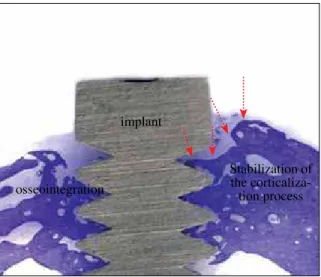
FIGURE 8 - After saucerization, the peri-implant bone surface normal-izes, with corticalization (arrows) indicative of stabilization of the peri-cervical bone remodeling process (toluidine blue, 10X).
consuming on average 0.1 mm of peri-implant cervical bone tissue each year.4,5,11 In a personal communication, Albrektsson reported that this cervical bone loss tends to decrease over the
years to a level even lower than that recorded in previous studies, and that these results would soon be reported in the literature.
Many theories and explanations have been provided to account for saucerization but almost all have had difficulty explaining some of its fea-tures. One of these theories attributes sauceriza-tion to the occlusal masticatory load that im-plants have to sustain. However, when osseoin-tegrated implants are out of occlusion or are fit-ted only with the gingival healing caps for many months or even years, without ever coming into occlusion, saucerization is also present (Fig 13). On the other hand, when implants remain sub-merged for a few months/years, the bone moves toward the more cervical surface and may even grow over the cover screws (Fig 12). This bone gain requires osteotomy maneuvers in order to place healing caps or an intermediate prosthesis.
Shortly after the placement of healing caps, or directly from the intermediate prosthesis and crown, the stratified squamous epithelium Stabilization of
the corticaliza-tion process osseointegration
A B
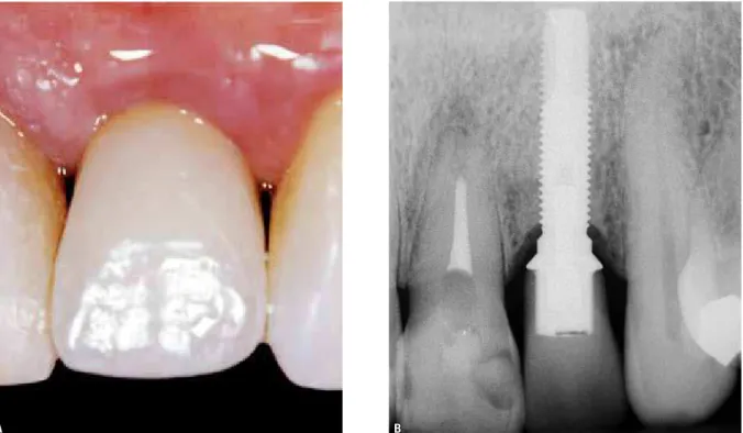
FIGURE 9 - Clinical case of implant in the upper lateral incisor region after six years, highlighting saucerization with regular bone surface and os-seointegration.
of the oral mucosa is juxtaposed to the surface with its normal thickness (Fig 4). When an epi-thelium is ulcerated their cell membranes are exposed to mediators in order to interact with their receptors, in the same manner as in oral ul-cers and surgical wounds, including in the peri-implant region.
The epidermal growth factor (EGF) in the saliva and in the epithelial cells stimulates peri-implant epithelial proliferation, thereby trigger-ing the formation of the peri-implant junctional epithelium. The peri-implant junctional epithe-lium produces new cell layers and assumes a con-formation similar to the junctional epithelium of natural teeth (Fig 5). This new conformation of the peri-implant junctional epithelium brings it closer to the osseointegrated surface, increasing the local concentration of EGF and, as a result, accelerating bone resorption and starting saucer-ization (Fig 5). Two recent papers have reviewed EGF functions and history.12,13
A few weeks or months after the peri-im-plant junctional epithelium and saucerization are formed they start moving away from each other. A stable biological distance is then estab-lished between the implant-integrated cervical bone and the peri-implant junctional epithe-lium, as occurs with natural teeth. From this stage, saucerization balance and stabilization are in place, allowing the bone on the cervical surface to resume corticalization (Figs 6, 8-11). It is probably due to this stabilization over the years that bone loss resulting from cervical sau-cerization diminishes its rhythm,4,5,11 provided that the conditions of hygiene and periodontal health are close to ideal. This situation has been noted in clinical cases that were followed up for many years after placement of osseointegrated implants (Figs 10 and 11).
A B C
A B C
epithelium itself through what is known as the autocrine effect. Although it probably takes place throughout the mucosa, it is particu-larly active in ulcerated areas where this auto-crine effect is compounded by salivary EGF. As a result, a considerable increase occurs in cell layers to the extent that the peri-implant junctional epithelium is formed. Once the
epithelium-implant integration occurs, salivary EGF penetration ceases or is drastically reduced and the process of cell-renewal epithelial prolif-eration goes back to normal.
The thickness of the gingival tissue appears to have a considerable effect on alveolar crest bone loss. When this thickness is 2 mm or smaller, the cervical bone loss tends to be significantly greater.21
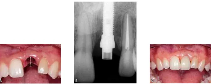
FIGURE 11 - Same clinical case as in the previous figure. A is a five-year control periapical radiograph showing pericervical saucerization and corticalization of peri-implant bone tissue. B shows 15 years of clinical control: Note normality and stability of peri-implant gingival tissue. C shows a 15-year control periapi-cal radiograph: Note the stability of the bone around the implant and increased cortiperiapi-calization.
FIGURE 10 - Implant installed in the region of tooth 21 avulsed in an accident. A shows the abutment installed over the implant. Periapical radiograph at B shows the correct adjustment of the abutment on the implant; the height and shape of the bone tissue around the implant are highlighted. C) Prosthetic
A B
C D
FIGURE 12 - Saucerization invariably occurs in all types of osseointegrated implants. The epithelial tissue has essentially a lining function and it is not very selective as to what it chooses to line. The epithelium will line even root surfaces which, although scraped, still manage to keep LPS (lipopolysaccharide) in its structure. LPS molecules are excessively toxic to our cells, but that does not stop the long junctional epithelium from forming, which is very important for maintaining clinical normality.
These results could probably be explained in light of the EGF. The thickness of the gingival tissue at the time of implant placement is commensurate with the distance from the implant junctional epi-thelium to be formed relative to bone tissue, i.e., EGF molecules rise to the bone surface in lower concentration.
Saucerization timing and orthodontic treatment
In natural teeth, the union of the junctional epithelium to the cervical enamel and surface is performed by means of several kinds of union structures, which effect an efficient sealing for salivary EGF (Figs 1, 2 and 3) in the peri-implant O
EG EJI
TCG
crown crown
Intermediate prosthesis Intermediate prosthesis
implant
en bloc implant
cone morse intermediate
A B
C
E
D
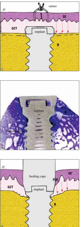
FIGURE 13 - Osseointegrated Implants submerged from A to D. In this situation saucerization does not occur. Bone repair fosters partial over-lap of implant coverage (as at B, C and D) because there is no formation of peri-implant junctional epithelium that would provide EGF molecules (arrows) in the vicinity of the bone surface. As soon as the healing caps are fitted, the formation of the peri-implant junctional epithelium (PJE) begins and so does saucerization (E). GE = gingival epithelium; GCT = gingival connective tissue; B = alveolar bone. (C: toluidine blue, 10X). healing caps
Stabilization of the
corticaliza-tion process
GCT GCT
GE GE
osseointegration
B B
B
implant implant
implant
implant suture
GCT
GE
PJE
15A
15B
15C 14A
14B
FIGURE 15 - The same clinical case of the previous figure with abutment mounted on the implant (A). Periapical radiograph (B) showing adequate interradicular space between 11 and 13, which allowed the installation of the implant in the correct position. C shows the prosthetic crown ce-mented onto the abutment.
nomena related to cell and tissue saucerization, the more we will be able to learn about the care, and the aesthetic and functional nuances involved. Additional refinement and details con-cerning the evolution of the operative and restor-ative procedures of dentistry as a whole come to light every day, dissolving boundaries or obstacles between the most diverse specialties.
Final considerations
Orthodontists should increasingly familiar-ize themselves with the jargon of other clini-cal specialties, including implantology, as well as their concepts and more specific issues. This need stems from increased transdisciplinary ac-tions undertaken by professionals in the joint planning of clinical cases involving multiple specialties, and whose ultimate goal is to reha-bilitate the patient's mouth.
Bone saucerization around osseointegrated implants is one such concept that forms a spe-cific part of the implantology jargon. Orthodon-tists should consider the occurrence of this peri-implant bone phenomenon while simultaneous-ly placing osseointegrated implants and moving the other teeth, realigning or relocating them harmoniously, many a time with such proximity to the cervical region that the condition should be carefully evaluated for its risks and aesthetic and functional benefits.
Further research is probably needed to answer the following question: Given the occurrence of saucerization, what are the special needs and care required by teeth located in the neighbor-hood of osseointegrated implants when moving teeth and finishing orthodontic cases?
junctional epithelium. This sealing, however— provided by the epithelium-implant junction—is less efficient and supposedly allows a constant salivary EGF input which, in conjunction with the EGF of the junctional epithelium and mu-cosa, sets in motion a process of slow and steady approach to the cervical bone (Figs 1, 4, 5, 6, 9).
After an osseointegrated implant has been placed, peri-implant saucerization can normally be expected to occur, regardless of implant type (Figs 14 and 15). So what is the average distance that should be maintained by orthodontists be-tween the cervical regions of neighboring natural teeth—when using osseointegrated implants—so that the cervical bone level of these implants is not affected by neighboring saucerization?
This concern may be even greater in upper anterior teeth such as, for example, lateral incisor implants (Figs 10, 11, 14, 15) in cases of par-tial unilateral or bilateral anodontia. Or, again, in cases of incisors and canines lost by accidental in-jury. The aesthetic and functional implications of the gingiva should be considered in planning and installing implants, such as the shape and size of the papillae, as well as the maintenance of a har-monious smile line.
Can saucerization, eventually, adversely affect the cervical hard and soft tissues of teeth locat-ed in the neighborhood of implants in patients treated orthodontically and whose teeth were harmoniously aligned with the implants? What special orthodontic care would be required to avoid or reduce the undesirable long-term conse-quences of osseointegrated implant saucerization occurring in the neighborhood of natural teeth?
phe-1. Adell R, Lekholm U, Rockler B, Branemark PI, Lindhe J, Eriksson B, et al. Marginal tissue reactions at osseointegrated titanium ixtures (I). A 3-year longitudinal prospective study. Int J Oral Maxillofac Surg. 1986;15:39-52.
2. Akagawa Y, Takata T, Matsumoto T, Nikai H, Tsuru H. Correlation between clinical and histological evaluations of the peri-implant gingiva around single cristal sapphire endosseous implant. J Oral Rehabil. 1989;16:581-7. 3. Albrektsson T. On long-term maintenance of the
osseointegrated response. Aust Prosthodont J. 1993;7:15-24. 4. Albrektsson T, Brånemark PI, Hansson HA, Lindström J.
Osseointegrated titanium implants: requirements for ensuring a long-lasting, direct bone to implant anchorage in man. Acta Orthop Scand. 1981;52(2):155-70.
5. Albrektsson T, Zarb G, Worthington P, Eriksson RA. The long-term eficacy of currently used dental implants: a review and proposed criteria of success. Int J Oral Maxillofac Implants. 1986;1(1):11-25.
6. Berglundh T, Lindhe J, Marinello CP, Ericsson I, Liljenberg B. Soft tissue reactions to de novo plaque formation at implants and teeth. An experimental study in the dog. Clin Oral Implants Res. 1992 Mar;3(1):1-8.
7. Berglundh T, Lindhe J, Jonsson K, Ericsson I. The topography of the vascular systems in the periodontal and peri-implant tissues in the dog. J Clin Periodontol. 1994 Mar;21(3):189-93.
8. Branemark PI. Introduction to osseointegration. In: Branemark PI, Zarb GA, Albrektsson T, editors. Tissue-integrated prostheses: osseointegration in clinical dentistry. Chicago: Quintessence; 1985. p. 11-76
9. Buser D, Stich H, Krekeler G, Schroeder A. Faserstrukturen der periimplantaren mukosa bei titanimlantaten. Eine experimentelle studie am beagle-hund. Zeitschrift fur Zahnarztliche Implantologie. 1989;5:15-23.
10. Carmichael RP, Apse P, Zarg GA, McCulloch CAG. Biological, microbiological and clinical aspects of the peri-implant mucosa. In: Albrektsson T, Zarb GA, editors. The Branemark osseointegrated implant. Chicago: Quintessence; 1989. p. 39-78. 11. Cochran DL, Nummikoski PV, Schoolield JD, Jones AA, Oates
TW. A prospective multicenter 5-year radiographic evaluation of crestal bone levels over time in 596 dental implants placed in 192 patients. J Periodontol. 2009 May;80(5):725-33. 12. Consolaro A, Consolaro MFMO. ERM functions, EGF and
orthodontic movement or why doesn't orthodontic movement cause alveolodental ankylosis? Dental Press J Orthod. 2010 Mar-Abr;15(2);24-32.
13. Consolaro A, Carvalho RS, Francischone CE Jr, Francischone CE. Mecanismo da saucerização nos implantes
osseointegrados. Rev Dental Press Periodontia Implantol. 2009 out-dez;3(4):25-39.
ReFeRenCeS
14. Ericsson I, Lindhe J. Probing depth at implants and teeth. An experimental study in the dog. J Clin Periodontol. 1993;20:623-7.
15. Gould TRL. Clinical implications of the attachment of oral tissues to perimucosal implants. Exerpta Medica. 1985;19:253-70.
16. Gould TRL, Brunette DM, Westbury L. The attachment mechanism of epithelial cells to titanium in vitro. J Periodontal Res. 1981;16(6):611-6.
17. Hashimoto M, Akagawa Y, Nikai H, Tsuru H. Single-cristal sapphire endosseous dental implant loaded with functional stress: clinical and histological evaluation of peri-implant tissues. J Oral Rehabil. 1988;15:65-76.
18. Jansen JA, Wijn JR, Wolters-Lutgerhorst JML, van Mullem PJ. Ultrastructural study of epithelial cell attachment to implant material. J Dental Res. 1985;64:891-6. 19. Lekholm U, Adell R, Lindhe J, Branemark PI, Eriksson
B, Rockler B, et al. Marginal tissue reactions at osseointegrated titanium fixtures. A cross-sectional retrospective study. Int J Oral Maxillofacial Surg. 1986;15:53-61.
20. Lekholm U, Eriksson B, Adell R, Slots J. The condition of the soft tissues at tooth and fixture abutments supporting fixed bridges. A microbiological and histological study. J Clin Periodontol. 1986;13:558-62.
21. Linkevicius T, Apse P, Grybauskas S, Puisys A. The influence of soft tissue thickness on crestal bone changes around implants: a 1-year prospective controlled clinical trial. Int J Oral Maxillofac Implants. 2009 Jul-Aug;24(4):712-9. 22. McKinney RV, Steflik DE, Koth DL. Evidence for junctional
epithelial attachment to ceramic dental implants, a transmission electron microscope study. J Periodontol. 1985;6:425-36.
23. McKinney RV, Steflik DE, Koth DL. The epithelium-dental implant interface. J Oral Implantol. 1988;13:622-41. 24. Schroeder A, van der Zypen E, Stich H, Sutter F. The
reaction of bone, connective tissue and epithelium to endosteal implants with sprayed titanium surfaces. J Maxillofacial Surg. 1981;4:191-7.
25. Seymour GJ, Gemmel E, Lenz LJ, Henry P, Bower R, Yamazaki K. Immunohistologic analysis of the inflammatory infiltrates associated with osseointegrated implants. Int J Oral Maxillofac Implants. 1989;4(3):191-7.
26. Ten Cate AR. The gingival junction. In: Branemark PI, Zarb GA, Albrektsson T, editors. Tissue-integrated prostheses: osseointegration in clinical dentistry. Chicago: Quintessence; 1985. p. 145-53.
27. Van Drie HJY, Beertsen W, Grevers A. Healing of the gingiva following installment of Biotes implants in beagle dogs. Adv Biomater. 1988;8:485-90.
Contact address Alberto Consolaro