Dental and skeletal changes in patients with mandibular
retrognathism following treatment with Herbst and
pre-adjusted fixed appliance
Fabio de Abreu Vigorito1, Gladys Cristina Dominguez2, Luís Antônio de Arruda Aidar3
How to cite this article: Vigorito FA, Dominguez GC, Aidar LAA. Dental and skeletal changes in patients with mandibular retrognathism following treat-ment with Herbst and pre-adjusted fixed appliance. Dental Press J Orthod. 2014 Jan-Feb;19(1):46-54. doi: http://dx.doi.org/10.1590/2176-9451.19.1.046-054.oar
Submitted: April 03, 2012 - Revised and accepted: March 03, 2013
Contact address: Gladys Cristina Dominguez
Rua Dr. Alceu de Campos Rodrigues, 247 – Vila Nova Conceição São Paulo/SP — Brazil
CEP: 04.544-000 – E-mail: [email protected]
1 PhD in Orthodontics, School of Dentistry, University of São Paulo (USP).
2 Full professor, Department of Orthodontics, University of São Paulo (USP).
3 Full professor, Department of Orthodontics, University of Santa Cecília
(UNISANTA).
» The authors report no commercial, proprietary or financial interest in the products or companies described in this article.
» Patients displayed in this article previously approved the use of their facial and in-traoral photographs.
Objective:To assess the dentoskeletal changes observed in treatment of Class II, division 1 malocclusion patients with mandibular retrognathism. Treatment was performed with the Herbst orthopedic appliance during 13 months (phase I) and pre-adjusted orthodontic fixed appliance (phase II). Methods: Lateral cephalograms of 17 adolescents were taken in phase I onset (T1) and completion (T2); in the first thirteen months of phase II (T3) and in phase II completion (T4). Differences among the cephalometric variables were statistically analyzed (Bonferroni variance and multiple comparisons). Results: From T1 to T4, 42% of overall maxillary growth was observed between T1 and T2 (P < 0.01), 40.3% between T2 and T3 (P < 0.05) and 17.7% between T3 and T4 (n.s.). As for overall mandibular move-ment, 48.2% was observed between T1 and T2 (P < 0.001) and 51.8% between T2 and T4 (P < 0.01) of which 15.1% was observed between T2 and T3 (n.s.) and 36.7% between T3 and T4 (P < 0.01). Class II molar relationship and overjet were properly corrected. The occlusal plane which rotated clockwise between T1 and T2, returned to its initial position between T2 and T3 remaining stable until T4. The mandibular plane inclination did not change at any time during treat-ment. Conclusion: Mandibular growth was significantly greater in comparison to maxillary, allowing sagittal maxil-lomandibular adjustment. The dentoalveolar changes (upper molar) that overcorrected the malocclusion in phase I, partially recurred in phase II, but did not hinder correction of the malocclusion. Facial type was preserved.
Keywords:Angle Class II malocclusion. Orthopedics. Orthodontics.
Objetivo:avaliar as alterações dentoesqueléticas observadas no tratamento da má oclusão de Classe II com retrognatismo mandibular, realizado com aparelho ortopédico de Herbst durante 13 meses (Fase I) e aparelho ortodôntico fixo pré-ajustado (Fase II). Métodos: foram obtidas telerradiografias laterais de 17 adolescentes, ao início (T1), final da Fase I (T2), primeiros 13 meses da Fase II (T3) e término da Fase II (T4). As diferenças entre as variáveis cefalométricas foram analisadas estatisti-camente (variância e comparações múltiplas de Bonferroni). Resultados: de T1 a T4, do total da projeção da maxila, 42% foram observados de T1 a T2 (p < 0,01); 40,3% de T2 a T3 (p < 0,05); e 17,7% de T3 a T4 (n.s.). Do total da projeção da man-díbula, foi notado 48,2% de T1 a T2 (p < 0,001) e 51,8% de T2 a T4 (p < 0,01), sendo 15,1% (n.s.) de T2 a T3, e 36,7% de T3 a T4 (p < 0,01). A relação molar e a sobressaliência foram corrigidas idealmente. Em T4, todos apresentavam características de oclusão normal. O plano oclusal que de T1 a T2 rotacionou no sentido horário, de T2 a T3 retornou aos valores iniciais, mantendo-se estável até T4. A inclinação do plano mandibular, responsável pela caracterização do tipo facial, não alterou em nenhum tempo. Conclusão: a mandíbula cresceu significativamente mais que a maxila, favorecendo o ajuste sagital maxilomandibular. As mudanças dentárias (molares superiores), que sobrecorrigiram a má oclusão na Fase I, recidivaram parcialmente na Fase II, sem comprometer a correção da má oclusão. O tipo facial foi preservado.
INTRODUCTION
Growing patients with Class II malocclusion and mandibular retrognathism may be treated with a variety of techniques, as described in the literature. Some of the techniques include treatment performed with an orthope-dic phase employing appliances such as the Herbst. This
treatment has been widely studied by Pancherz1-4 and other
researchers5-22 who took several aspects into consideration
and revealed that this type of treatment not only represents an alternative to the correction of Class II malocclusion, but also preserves the stomatognathic system. However, with regard to Brazilian individuals, these results are ques-tioned: Are treatment efects skeletal or dentoalveolar? Is the mandibular growth curve modiied when stimulated by the Herbst appliance? Are the obtained results lost ater the appliance is removed? The complexity of clarifying the referred doubts lays in the diiculty of performing longi-tudinal studies in homogeneous casuistries. With a view to eliminating the tendency towards including only suc-cessful cases and, thus, confuse the results, the ideal would be that prospective studies were conducted with groups of consecutive patients. From this point of view, in 2007, a
study23 was carried out to assess and compare, in patients
treated during growth spurt, the dentoskeletal changes observed in the Herbst active phase and during a period of same duration ater the appliance had been removed. The obtained results were the motivation to perform the present study which aims at assessing full treatment per-formed in adolescents in two phases: phase I – orthope-dic with Herbst appliance and phase II – orthodontic with pre-adjusted ixed appliance.
MATERIAL AND METHODS
The sample comprised 17 Brazilian adolescent patients (12 men and 5 women), with mean age of 12 years and 4 months ± 1 year and 2 months, and bone age correspond-ing to the growth spurt, as revealed by a hand-wrist radio-graph. The patients were selected according to the follow-ing inclusion criteria: individuals with mandibular retrog-nathism and Angle Class II, division 1 malocclusion greater than half-cusp (> 3 mm); individuals with overjet > 5 mm (permanent dentition); with model discrepancy under 4 mm; with clinical recommendation for mandibular ad-vancement to be performed with functional orthopedic ap-pliance. Individuals with absence of teeth, dental fractures and dental caries were excluded. Treatment was carried out in two phases. Initially, the orthopedic phase (phase I)
performed with Herbst functional orthopedic appliance placed onto acrylic splints associated with maxillary
expan-sion screw.24 The objective was to correct the transversal
discrepancy,25 activating the expansion screw during the
irst month of treatment. The appliance was made accord-ing to a wax bite registration obtained with 6 mm of ini-tial advancement, and progressive advancements of 2 mm every 2 months, according to individual needs. This phase lasted for an average of 13.9 ± 2.1 months. Thereater, the orthodontic phase (phase II) was performed with pre-ad-justed ixed appliance and aimed at leveling and aligning the upper and lower teeth as well as at obtaining functional occlusion with adequate overjet and overbite. This phase lasted for 46 months.
Complete orthodontic documentation (panoramic and hand-wrist radiographs, lateral and frontal cephalo-grams; intra and extraoral photographs; study casts) was
prepared for all patients at four stages: T1, immediately
before treatment onset; T2, ater 13 months using the
Herbst appliance, which represented the end of phase I;
T3, 13 months ater phase II or orthodontic phase had
begun; and T4, phase II completion, totalizing a period
of 33 months. All 68 lateral cephalograms were manu-ally traced by the same operator at monthly intervals. They were analyzed with regard to the cephalometric variables of sagittal changes analysis (SO-analysis)
sug-gested by Pancherz4 (Figs 1 and 2).
Patients’ guardians signed an informed consent form, agreeing with all stages of the study and the posterior disclosure of results. The project was approved by the Institutional Review Board of the School of Dentistry/ USP and registered under protocol 109/06.
STATISTICAL ANALYSIS
Method error assessment (Dahlberg26) was
per-formed in 11.8% of the sample.
RESULTS
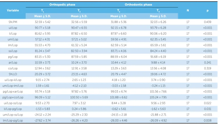
For a better understanding of the characteristics of each moment of growth (Fig 2) and the diferences be-tween them, the results are presented in three tables. Table 1 presents the measures, the relation between measurements at each moment of assessment and the result of the analysis of variance.
Figure 1 - Analysis of sagittal changes (SO-analysis) of Pancherz. Figure 2 - Superimposition of tracings (according to analysis of Pancherz4),
of one of the patients from the sample, in all four observation stages: T1 = yellow, T2 = blue, T3 = red and T4 = green.
Variable
Orthopedic phase Orthodontic phase
N p
T1 T2 T3 T4
Mean ± S.D. Mean ± S.D. Mean ± S.D. Mean ± S.D.
SN.PM 32.59 ± 5.42 32.56 ± 5.59 31.88 ± 5.36 32.03 ± 6.26 17 0.439 ui/Lop 90.77 ± 5.18 90.47 ± 6.55 92.15 ± 6.78 93.79 ± 6.28 17 <0.001 li/Lop 81.62 ± 5.95 87.82 ± 6.50 87.97 ± 6.60 90.06 ± 6.20 17 <0.001 um/Lop 57.12 ± 4.31 57.21 ± 5.02 59.56 ± 4.91 62.35 ± 5.45 17 <0.001 lm/Lop 55.53 ± 4.70 61.32 ± 5.24 62.59 ± 5.14 65.59 ± 5.61 17 <0.001 ss/Lop 81.24 ± 3.47 82.50 ± 3.94 83.71 ± 4.06 84.24 ± 4.40 17 <0.001 pg/Lop 83.15 ± 5.30 87.59 ± 5.85 88.59 ± 6.04 91.68 ± 6.19 17 <0.001 ar/Lop 10.59 ± 3.73 10.24 ± 3.70 10.44 ± 4.12 9.88 ± 4.14 17 0.241 co/Lop 12.94 ± 3.62 12.91 ± 3.58 13.29 ± 3.63 13.56 ± 4.08 17 0.319 SN.LO 20.29 ± 3.72 23.15 ± 4.63 20.79 ± 4.47 19.06 ± 4.72 17 <0.001 ui/Lop-li/Lop 9.15 ± 2.74 2.65 ± 1.23 4.18 ± 1.20 3.74 ± 0.90 17 <0.001 um/Lop-lm/Lop 1.59 ± 1.61 -4.12 ± 2.10 -3.03 ± 1.58 -3.24 ± 1.15 17 <0.001 pg/Lop+ar/Lop 93.74 ± 5.18 97.82 ± 5.76 99.03 ± 6.74 101.56 ± 7.66 17 <0.001 pg/Lop+co/Lop 96.09 ± 5.12 100.50 ± 5.69 101.88 ± 6.62 105.24 ± 7.95 17 <0.001 ui/Lop-ss/Lop 9.53 ± 2.70 7.97 ± 3.12 8.44 ± 3.28 9.56 ± 2.93 17 0.022 li/Lop-pg/Lop -1.53 ± 5.83 0.24 ± 5.86 -0.62 ± 5.61 -1.62 ± 5.63 17 0.031 um/Lop-ss/Lop -24.12 ± 2.24 -25.29 ± 2.30 -24.15 ± 2.18 -21.88 ± 2.71 17 <0.001 lm/Lop-pg/Lop -27.62 ± 3.74 -26.26 ± 4.20 -26.00 ± 4.46 -26.09 ± 4.92 17 0.008 Table 1 - Measures and relations between values obtained at each moment of assessment and result of the analysis of variance.
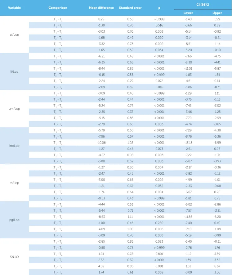
Variable Comparison Mean diference Standard error p CI (95%)
Lower Upper
ui/Lop
T 1 - T2 0.29 0.56 > 0.999 -1.40 1.99
T 1 - T3 -1.38 0.76 0.516 -3.66 0.89
T 1 - T4 -3.03 0.70 0.003 -5.14 -0.92
T2 - T3 -1.68 0.49 0.020 -3.14 -0.21
T2 - T4 -3.32 0.73 0.002 -5.51 -1.14
T3 - T4 -1.65 0.52 0.034 -3.20 -0.10
li/Lop
T 1 - T2 -6.21 0.48 < 0.001 -7.66 -4.75
T 1 - T3 -6.35 0.65 < 0.001 -8.30 -4.41
T 1 - T4 -8.44 0.86 < 0.001 -11.01 -5.87
T2 - T3 -0.15 0.56 > 0.999 -1.83 1.54
T2 - T4 -2.24 0.79 0.072 -4.61 0.14
T3 - T4 -2.09 0.59 0.016 -3.86 -0.31
um/Lop
T 1 - T2 -0.09 0.40 > 0.999 -1.29 1.11
T 1 - T3 -2.44 0.44 < 0.001 -3.75 -1.13
T 1 - T4 -5.24 0.74 < 0.001 -7.45 -3.02
T2 - T3 -2.35 0.37 < 0.001 -3.46 -1.25
T2 - T4 -5.15 0.85 < 0.001 -7.70 -2.59
T3 - T4 -2.79 0.65 0.003 -4.74 -0.85
lm/Lop
T 1 - T2 -5.79 0.50 < 0.001 -7.29 -4.30
T 1 - T3 -7.06 0.57 < 0.001 -8.76 -5.36
T 1 - T4 -10.06 1.02 < 0.001 -13.13 -6.99
T2 - T3 -1.27 0.45 0.073 -2.61 0.08
T2 - T4 -4.27 0.98 0.003 -7.22 -1.31
T3 - T4 -3.00 0.69 0.003 -5.07 -0.93
ss/Lop
T 1 - T2 -1.27 0.30 0.004 -2.17 -0.36
T 1 - T3 -2.47 0.45 < 0.001 -3.82 -1.12
T 1 - T4 -3.00 0.66 0.002 -4.99 -1.01
T2 - T3 -1.21 0.37 0.032 -2.33 -0.08
T2 - T4 -1.74 0.64 0.094 -3.67 0.20
T3 - T4 -0.53 0.43 > 0.999 -1.81 0.75
pg/Lop
T 1 - T2 -4.44 0.53 < 0.001 -6.02 -2.86
T 1 - T3 -5.44 0.71 < 0.001 -7.57 -3.31
T 1 - T4 -8.53 1.11 < 0.001 -11.86 -5.20
T2 - T3 -1.00 0.46 0.280 -2.40 0.40
T2 - T4 -4.09 1.00 0.005 -7.10 -1.08
T3 - T4 -3.09 0.70 0.003 -5.19 -0.99
SN.LO
T 1 - T2 -2.85 0.85 0.023 -5.40 -0.31
T 1 - T3 -0.50 0.75 > 0.999 -2.76 1.76
T 1 - T4 1.24 0.78 0.801 -1.12 3.59
T2 - T3 2.35 0.32 < 0.001 1.39 3.32
T2 - T4 4.09 0.86 0.001 1.51 6.67
T3 - T4 1.74 0.61 0.068 -0.09 3.56
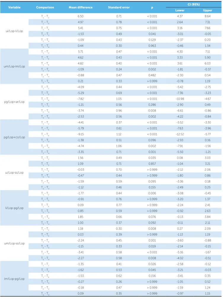
Variable Comparison Mean diference Standard error p CI (95%)
Lower Upper
ui/Lop-li/Lop
T1 - T2 6.50 0.71 < 0.001 4.37 8.64
T1 - T3 4.97 0.78 < 0.001 2.64 7.31
T1 - T4 5.41 0.75 < 0.001 3.16 7.66
T2 - T3 -1.53 0.49 0.041 -3.01 -0.05
T2 - T4 -1.09 0.43 0.129 -2.37 0.20
T3 - T4 0.44 0.30 0.963 -0.46 1.34
um/Lop-lm/Lop
T1 - T2 5.71 0.47 < 0.001 4.30 7.11
T1 - T3 4.62 0.43 < 0.001 3.33 5.90
T1 - T4 4.82 0.40 < 0.001 3.61 6.03
T2 - T3 -1.09 0.24 0.002 -1.81 -0.37
T2 - T4 -0.88 0.47 0.482 -2.30 0.54
T3 - T4 0.21 0.33 > 0.999 -0.78 1.19
pg/Lop+ar/Lop
T1 - T2 -4.09 0.44 < 0.001 -5.42 -2.75
T1 - T3 -5.29 0.69 < 0.001 -7.36 -3.23
T1 - T4 -7.82 1.05 < 0.001 -10.98 -4.67
T2 - T3 -1.21 0.56 0.286 -2.90 0.49
T2 - T4 -3.74 0.96 0.008 -6.61 -0.86
T3 - T4 -2.53 0.56 0.002 -4.22 -0.84
pg/Lop+co/Lop
T1 - T2 -4.41 0.37 < 0.001 -5.52 -3.30
T1 - T3 -5.79 0.61 < 0.001 -7.63 -3.96
T1 - T4 -9.15 1.12 < 0.001 -12.52 -5.77
T2 - T3 -1.38 0.51 0.096 -2.93 0.16
T2 - T4 -4.74 1.06 0.002 -7.91 -1.56
T3 - T4 -3.35 0.71 0.001 -5.50 -1.21
ui/Lop-ss/Lop
T1 - T2 1.56 0.49 0.035 0.08 3.03
T1 - T3 1.09 0.71 0.857 -1.04 3.21
T1 - T4 -0.03 0.70 > 0.999 -2.12 2.06
T2 - T3 -0.47 0.44 > 0.999 -1.80 0.86
T2 - T4 -1.59 0.59 0.095 -3.36 0.18
T3 - T4 -1.12 0.46 0.155 -2.49 0.25
li/Lop-pg/Lop
T1 - T2 -1.77 0.44 0.006 -3.08 -0.45
T1 - T3 -0.91 0.76 > 0.999 -3.20 1.37
T1 - T4 0.09 0.77 > 0.999 -2.24 2.41
T2 - T3 0.85 0.59 > 0.999 -0.92 2.63
T2 - T4 1.85 0.66 0.076 -0.13 3.84
T3 - T4 1.00 0.37 0.092 -0.11 2.11
um/Lop-ss/Lop
T1 - T2 1.18 0.30 0.008 0.27 2.09
T1 - T3 0.03 0.39 > 0.999 -1.13 1.19
T1 - T4 -2.24 0.45 0.001 -3.60 -0.88
T2 - T3 -1.15 0.33 0.019 -2.14 -0.15
T2 - T4 -3.41 0.58 < 0.001 -5.16 -1.67
T3 - T4 -2.27 0.58 0.008 -4.02 -0.51
lm/Lop-pg/Lop
T1 - T2 -1.35 0.41 0.026 -2.58 -0.12
T1 - T3 -1.62 0.53 0.045 -3.21 -0.03
T1 - T4 -1.53 0.62 0.156 -3.41 0.35
T2 - T3 -0.27 0.26 > 0.999 -1.05 0.52
T2 - T4 -0.18 0.47 > 0.999 -1.59 1.24
T3 - T4 0.09 0.35 > 0.999 -0.97 1.15
DISCUSSION
All patients that comprised this study
present-ed, in T1, typical characteristics of Class II division 1
malocclusion, as conirmed by the initial cephalo-metric variables that describe the molar relationship (um/Lop - lm/Lop: 1.59 ± 1.61 mm) and the overjet (ui/Lop - li/Lop: 9.15 ± 2.74 mm). According to the inclusion criteria, all patients clinically presented man-dibular retrognathism and accepted treatment that in-cluded mandibular advancement.
The results yielded by the present study are in agreement with previous studies that used similar
methods.9,12,23,27 Both the maxilla (SS/Lop) and
man-dible (PPg/Lop) were anteriorly projected, but since mandibular growth increment was 3.5 times greater, there was a favorable sagittal maxillomandibular adjust-ment. In order to identify the contribution of mandibu-lar growth, measurements of the absolute mandibumandibu-lar length (pg/Lop+co/Lop and pg/Lop+ar/Lop) were as-sessed and signiicant growth increment was observed, although the condylar (co/Lop) and articular (ar/Lop) points did not present any alterations.
The registered amount of skeletal growth allowed better a understanding of how the teeth varied in their sagittal spatial position. Overcorrection of the observed molar relationship (um/Lop-lm/Lop: 5.71 mm) was due to the association between maintenance of upper molars position (um/Lop: -0.09) while the maxilla was anteriorly projected (SS/Lop: -1.27 mm), and mesial-ization of lower molars (lm/Lop: -5.79 mm) along with mandibular anterior projection (pg/Lop: -4.44 mm). Overjet was signiicantly reduced from 9.5 mm to 2.65 mm, as a result of mandibular anterior projection (pg/Lop: -4.44 mm) and buccal inclination of lower in-cisors in their bone base (li/Lop: -6.21 mm). The me-chanical efect observed in the inclination of lower inci-sors restricts the recommendation of this type of therapy to individuals who do not present increased inclination at treatment onset.
The occlusal plane (SN.LO), which in the beginning presented a mean value that is typical of a mesofacial
pattern (32.59± 5.42o), was rotated clockwise (2.85o) by
the presence of interocclusal acrylic splints. This might have caused the efect of molar intrusion, since, when the appliance was removed, an important posterior dis-occlusion was observed in all patients. This speculation can be done because, diferently from the occlusal plane,
the inclination of the mandibular plane (SN.PM) did not undergo any alterations, thus conirming that it was just a dentoalveolar efect and not a skeletal one, there-fore, the facial type did not change.
In the following 13 months ater the Herbst ap-pliance had been removed, which corresponded to
orthodontic treatment onset (T2-T3), the maxilla
con-tinued to be anteriorly projected (ss/Lop: -1.21 mm), whereas mandibular projection was little signiicant (pg/Lop: -1 mm). It was observed that partial
re-currence of molar relationship (um/Lop - lm/Lop:
-1.09 mm) occurred as a result of mesialization of up-per molars (um/Lop-ss/Lop: -1.15 mm) along with non-signiicant mesialization of lower molars (lm/Lop-pg/Lop: -027). However, considering that a relation of overcorrection of molar relationship was observed
in T2 (um/Lop - lm/Lop: -4.12 mm), this recurrence
was favorable to adjust the molars in Class I relation (um/Lop - lm/Lop: -3.03 mm). Additionally, there was a partial recurrence of 1.53 mm in overjet (ui/Lop-li/Lop) as a result of diferential growth of the maxilla, which led the upper incisors to occupy a more anterior spatial position (is/Lop: -1.68 mm). This could not have been due to the insigniicant uprighting of lower incisors (li/Lop: -0.15 mm) because, in this case, they did not change their position (li/Lop-pg/Lop: 0.85 mm).
The occlusal plane (SL.LO) rotated
counterclock-wise, since, from T2 to T3, with the removal of the
Herbst appliance, the molars were free from the in-terocclusal splints and, additionally, were actively lev-eled to the orthodontic appliance, restoring the vertical spatial position that they presented at treatment onset.
These data corroborate data found in the literature,9,10
thus conirming that this movement happened without afecting the inclination of the mandibular plane (SN-PM), therefore, with preservation of facial type.
The complementary assessment carried out in this study, between the thirteen-month interval ater re-moval of the Herbst appliance and the end of the
ac-tive orthodontic treatment (T3-T4), showed that,
Nevertheless, when analyzing the maintenance of dental stability, during a period in which there was sig-niicant expression of mandibular growth and absence of signiicant maxillary growth, it could be observed that tooth movement was compensatory, maintain-ing both molar and overjet relations. While the up-per incisors (ui/Lop: -1.65 mm) and the upup-per mo-lars (um/Lop: -2.79 mm) were anteriorly projected in the absence of signiicant maxillary growth (ss/Lop: -053 mm), the lower incisors (li/Lop: -2.09 mm) and lower molars (lm/Lop:-3 mm) were also spatially ante-riorly projected, however, in association with signii-cant mandibular growth (pg/Lop: -3.09 mm). Thus, it can be concluded by means of the diferential calcu-lus (dental movement minus skeletal movement) that only the upper molars had a signiicant movement of mesialization, regardless of the growth of its bone base (um/Lop-ss/Lop: -2.27 mm). This movement was nec-essary to maintain Class I molar relationship. The
oc-clusal plane remained stable (SN.LO: 1.74o). This fact
can be explained because in T3, the molars already
pre-sented interocclusal contact and there were no
addition-al verticaddition-al movements until T4. The mandibular plane
remained unchanged, revealing a uniform behavior during the entire treatment, thus, preserving facial type.
When considering the series of changes observed
from the beginning to the end of treatment (T1-T4), it
is veriied that out of the total of maxillary anterior pro-jection (3 mm), 42% happened during the orthopedic
phase (T1-T2) and 58% during the orthodontic phase
(T2-T4), of which the most part (40.3%) happened
dur-ing the irst 13 months (T2-T3) and the rest (17.7%),
an insigniicant increase, between T3 and T4. As shown
in Tables 1 to 3, the mandibular anterior displacements (Pg/Lop) were compatible to the corresponding incre-ment of the mandibular absolute growth (Pg/Lop + ar/ Lop and Pg/Lop + co/Lop). When analyzing the variable Pg/Lop + co/Lop, it is veriied that 48.2% of mandibular growth happened during the 13 months of the
orthope-dic phase (T1-T2) as a response to the stimulus provided
by the Herbst appliance, in a period when the potential growth was intense; whereas 51.8% happened during
the orthodontic phase (T2-T4). However, it must be
emphasized that during the 13 months ater the Herbst
appliance was removed (T2-T3), there was growth
de-celeration, with slight, non-signiicant growth incre-ment (15.1%) and, therefore, without anterior
projec-tion. Signiicant growth was soon resumed, expressing
the remaining 36.7% in the following months until T4.
This type of response agrees with previous studies.4,9
It was very important to assess the amount of growth
during the orthodontic phase (T2-T4) as proposed in
this study. Moreover, dividing observation into two
pe-riods, T2-T3 (13 months) and T3-T4 (33 months), was
important to understand whether or not the curve of mandibular growth could modify its usual course before the stimulus given by the use of the Herbst appliance.
Franchi et al28 claim thatmandibular growth follows the
physical growth spurt and it is characterized by a gradu-al increase in the amount of increments until it reaches its maximum, when the greatest amount of growth is expressed. Aterwards, it gradually decelerates again, however, linearly, until growth is complete. In the pres-ent study, it was observed that during the 13 months
of stimulus (T1-T2) provided by the Herbst appliance,
the increments were intense. Nevertheless, a
decelera-tion in the following 13 months (T2-T3), and then, a
resumption of growth (T3-T4), explain that the growth
veriied between T1 and T2 represents the favorable
expression of the present growth potential, for being in its maximum (as revealed by the hand-wrist
radio-graph in T1), which is summed up to the anticipation
of growth in the subsequent 13 months, which, with-out the use of the appliance, would not have manifest-ed at that moment, thus, modifying the behavior of the descendant curve of growth spurt in adolescence.
As for growth complexity and mandibular spatial projection in the face, our results can be explained by
those observed by Pancherz et al29 who assessed the
“efective condylar growth” and its inluence over the spatial position of the symphysis in the face. Their ind-ings reveal that condylar growth triplicated during the active phase of six months in which the Herbst appli-ance was used, decelerated in a similar period ater the removal of the appliance, and soon resumed its nor-mal growth in the subsequent 30 months. Comparison between total mandibular and maxillary projection,
from T1 to T4, revealed that the mandible (pg/Lop:
8.47 mm) was projected 2.8 times more than the max-illa (ss/Lop: 3 mm), a fact that favored sagittal maxil-lomandibular adjustment.
With regard to dentoalveolar correction of Class II malocclusion, a favorable response was observed
increase in overjet that patients presented at treatment
onset were ideally corrected. In T4, all of them showed
characteristics of normal occlusion, with good molar re-lationship and adequate overjet, thus, achieving the pur-pose of the treatment. In order to produce such results, treatment evolved from sagittal overcorrection of molar relationship, which was associated with great reduction in overjet during the 13-month orthopedic phase; par-tially relapsed at the beginning of the orthodontic phase and became stable in the following 33 months until the end of the treatment.
Based on the aforementioned observations, it is im-portant to emphasize that: First, the recurrence of the
overcorrected molar relationship between T2 and T3 was
necessary for molars to obtain cusp-to-fossa relationship instead of cusp-to-cusp, which probably contributed to ofer the stability observed in the subsequent period. Ad-ditionally, despite being signiicant, the degree of overjet
relapse registered between T2 and T3, did not prevent the
values from being within the clinical parameters of nor-mality by the end of the treatment. The second aspect
is with regard to the stability observed in T3 and T4, a
period of 33 months. The advantage of lasting nearly two times longer than each previous period allowed the stabil-ity of results to be assessed.
Clockwise rotation of the occlusal plane was
signii-cant during the orthopedic phase (T1-T2) and it
hap-pened as a result of the presence of interocclusal splints.
In the subsequent phase (T2-T3), it rotated
counter-clockwise, therefore, relapsing by the removal of the splints and active orthodontic leveling, thus, restoring intermaxillary occlusal contacts. This pattern of coun-terclockwise rotation continued in the following 33 months, however, insigniicantly. As for the changes that occurred in opposite directions, the comparison between orthopedic and orthodontic phases reveal that they did not present any adverse clinical efect, since the
changes occurred without inluencing the inclination of the mandibular plane. On the other hand, the occlusal
plane restored its initial inclination in T3 and remained
stable until T4. The mandibular plane (SN.PM), which
deines the facial type, was maintained in all periods of assessment, a fact that is favorable to the stability achieved in the long term, all of which agreed with
oth-er authors in the litoth-erature.9,30
The size of the sample is a limitation of this study. However, it is of great value considering that it is a pro-spective study carried out with consecutive patients and that had never been performed with Brazilian patients. The results obtained from assessing these patients by means of the treatment protocol allowed us to visualize not only that the therapy applied was eicient, but also that the series of skeletal and dental changes observed did not cause a temporary impact, but an impact that is com-patible with the conditions of stability in the long term. However, further studies are necessary to longitudinally assess the post-treatment phase. Finally, it is important to emphasize the undesirable efect that the use of the Herbst appliance can cause to individuals with increased buccal inclination of the lower incisors at treatment onset.
CONCLUSIONS
1. Pancherz H. Treatment of Class II malocclusions by jumping the bite with the Herbst appliance. A cephalometric investigation. Am J Orthod.1979;76(4):423-42.
2. Pancherz H, Hansen K. Occlusal changes during and after Herbst treatment: a cephalometric investigation. Eur J Orthod. 1986;8(4):215-28. 3. Pancherz H, Fackel U. The skeletofacial growth pattern pre and
post-dentofacial orthopaedics. A long-term study of Class II malocclusions treated with the Herbst appliance. Eur J Orthod. 1990;12(2):209-18. 4. Pancherz H, Ruf S. The Herbst appliance. Research based clinical
management. Quintessence; 2008. cap. 6, p. 43-6. 5. Hägg U, Pancherz H. Dentofacial orthopaedics in relation to
chronological age, growth period and skeletal development. An analysis of 72 male patients with Class II division 1 malocclusion treated with Herbst appliance. Eur J Orthod. 1988;10(3):169-76.
6. McNamara JA Jr, Howe RP, Dischinger TG. A comparison of the Herbst and Fränkel appliances in the treatment of Class II malocclusion. Am J Orthod Dentofacial Orthop. 1990;98(2):134-44.
7. Schiavoni R, Grenga V, Macri V. Treatment of Class II high angle malocclusions with the Herbst appliance: a cephalometric investigation. Am J Orthod Dentofacial Orthop. 1992;102(5):393-409.
8. Windmiller EC. The acrylic-splint Herbst appliance: a cephalometric evaluation. Am J Orthod Dentofacial Orthop. 1993;104(1):73-84. 9. Lai M, McNamara JA Jr. An evaluation of two-phase treatment with the
Herbst appliance and preadjusted edgewise therapy. Semin Orthod. 1998;4(1):46-58.
10. Franchi L, Baccetti T, McNamara JA Jr. Treatment and posttreatment efects of acrylic splint Herbst appliance therapy. Am J Orthod Dentofacial Orthop. 1999;115(4):429-38.
11. Manfredi C, Cimino R, Trani A, Pancherz H. Skeletal changes of Herbst appliance therapy investigated with more conventional cephalometrics and European norms. Angle Orthod. 2001;71(3):170-6.
12. Schütz TCB, Vigorito JW, Rodrigues CRMD, Domínguez-Rodríguez GC. Avaliação cefalométrico-radiográica das modiicações dentoalveolares decorrentes do tratamento com o aparelho Herbst em adolescentes com maloclusão de Classe II, divisão 1ª de Angle – Parte I. Ortodontia. 2002;35(4):22-34.
13. Schütz TCB, Vigorito JW, Domínguez-Rodríguez GC. Avaliação cefalométrico-radiográica das modiicações esqueléticas e do peril facial decorrentes do tratamento com o aparelho Herbst em adolescentes com maloclusão de Classe II, divisão 1ª de Angle – Parte II. Ortodontia. 2003;36(1):44-61.
14. Ruf S. Short and long-term efects of the Herbst appliance on temporomandibular joint function. Semin Orthod. 2003;9(1):74-86. 15. Schaefer AT, McNamara JA Jr, Franchi L, Baccetti T. A cephalometric
comparison of treatment with the Twin-block and stainless steel crown Herbst appliances followed by ixed appliance therapy. Am J Orthod Dentofacial Orthop. 2004;126(1):7-15.
REFERENCES
16. VanLaecken R, Martin CA, Dischinger T, Razmus T, Ngan P. Treatment efects of the edgewise Herbst appliance: a cephalometric and tomographic investigation. Am J Orthod Dentofacial Orthop. 2006;130(5):582-93.
17. Ruf S, Pancherz H. Herbst/multibracket appliance treatment of Class II division 1 malocclusions in early and late adulthood. A prospective cephalometric study of consecutively treated subjects. Eur J Orthod. 2006;76(2):352-60.
18. Bock N, Pancherz H. Herbst treatment of Class II division 1 malocclusions in retrognathic and prognathic facial types. Angle Orthod. 2006;76(6):930-41. 19. Aidar LA, Dominguez GC, Abrahão M, Yamashita HK, Vigorito JW. Efects of Herbst appliance treatment on temporomandibular joint disc position and morphology: a prospective magnetic resonance imaging study. Am J Orthod Dentofacial Orthop. 2009;136(3):412-24.
20. Aidar LA, Dominguez GC, Yamashita HK, Abrahão M.Changes in
temporomandibular joint disc position and form following Herbst and ixed orthodontic treatment. Angle Orthod. 2010;80(5):843-52.
21. Siara-Olds NJ, Pangrazio-Kulbersh V, Berger J, Bayirli B. Long-term dentoskeletal changes with the Bionator, Herbst, Twin Block, and MARA Functional Appliances. Angle Orthod. 2010;80(1):18-29.
22. Schütz TC, Dominguez GC, Hallinan MP, Cunha TC, Tuik S. Class II correction improves nocturnal breathing in adolescents. Angle Orthod. 2011;81(2):222-8.
23. Vigorito FA, Domínguez GC. Comparação dos efeitos dento-esqueléticos decorrentes do tratamento realizado em duas fases (com aparelho de Herbst e aparelho ixo pré-ajustado) em adolescentes com retrognatismo mandibular. Ortodontia. 2007;40(4):263-70.
24. Howe RP, McNamara JA Jr. Clinical management of the bonded Herbst appliance. J Clin Orthod. 1983;17(7):456-63.
25. Tollero I, Baccetti T, Franchi L, Tanasescu CD. Role of the posterior transverse interarch discrepancy in Class II, division 1 malocclusion during the mixed dentition phase. Am J Orthod Dentofacial Orthop. 1996;110(4):417-22.
26. Houston WJB. The analysis of errors in orthodontic measurements. Am J Orthod. 1983;83(5):383-90.
27. Flores-Mir C, Ayeh, Goswani A, Charkhandeh S. Skeletal and dental changes in class II division 1 malocclusions treated with Splint-Type Herbst appliances: a systematic review. Angle Orthod. 2007;77(2):376-81. 28. Franchi L, Baccetti T, McNamara JA. Mandibular growth as related to
cervical vertebral maturation and body height. Am J Orthod Dentofacial Orthop. 2000;118(3):335-40.
29. Pancherz H, Ruf S, Kohlhas P. Efective condylar growth and chin position changes in Herbst treatment: a cephalometric roentgenographic long-term study. Am J Orthod Dentofacial Orthop. 1998;114(4):437-46. 30. Ruf S, Pancherz H. The efect of Herbst appliance on the mandibular