Anti-inflammatory and antispasmodic
activity of
Ipomoea imperati
(Vahl)
Griseb (Convolvulaceae)
Departamentos de 1Farmacologia, Instituto de Ciências Biomédicas, and 2Fisiologia,Instituto de Biociências, Universidade de São Paulo, São Paulo,
SP, Brasil A.C.B. Paula1,
L.S.S. Hayashi1
and J.C. Freitas1,2
Abstract
Ipomoea imperati (Convolvulaceae) lives on the sandy shores of the Brazilian coast and in other areas of the world. The anti-inflammatory activity of a methanol-water extract of the leaves of I. imperati was investigated in experimental models of acute and subchronic inflam-mation. Topical application of the extract (10 mg/ear) inhibited mouse ear edema induced by croton oil (89.0 ± 1.3% by the lipid fraction with an IC50 of 3.97 mg/ear and 57.0 ± 1.3% by the aqueous fraction with
an IC50 of 3.5 mg/ear) and arachidonic acid (42.0 ± 2.0% with an IC50
of 4.98 mg/ear and 31.0 ± 2.0% with an IC50 of 4.72 mg/ear).
Phospholipase A2,purified from Apis mellifera bee venom, was also
inhibited by the extract (5.0 mg/ml lipid and aqueous fraction) in vitro
in a dose-dependent manner (85% by the lipid fraction with an IC50 of
3.22 mg/ml and 25% by the aqueous fraction with an IC50 of 3.43 mg/
ml). The methanol-water extract of I. imperati (1000 mg/kg) adminis-tered by the oral route also inhibited the formation of cotton pellet-induced granulomas (73.2 ± 1.2% by the lipid fraction and 56.14 ± 2.7% by the aqueous fraction) and did not cause gastric mucosal lesions. I. imperati extracts (10 mg/ml) also inhibited in a dose-dependent manner the muscle contractions of guinea pig ileum in-duced by acetylcholine and histamine (IC50 of 1.60 mg/ml for the lipid
fraction and 4.12 mg/ml for the aqueous fraction). These results suggest the use of I. imperati as an anti-inflammatory and antispas-modic agent in traditional medicine.
Correspondence J.C. Freitas
Departamento de Fisiologia Instituto de Biociências, USP Rua do Matão, Travessa 14, 101 05508-900 São Paulo, SP Brasil
Fax: +55-11-818-7416 E-mail: [email protected]
Research supported by FAPESP.
Received July 6, 2001 Accepted October 2, 2002
Key words
·Anti-inflammatory activity ·Antispasmodic activity ·Arachidonic acid ·Croton oil ·Medicinal plants ·Phospholipase A2
Introduction
Natural products have served as a source of drugs for centuries, and about half of the pharmaceuticals in use today are derived from natural products (1). Dependence on plants as the source of medicines is prevalent in developing countries where traditional medicine plays a major role in health care (2).
Ipomoea littoralis (Convolvulaceae),
cur-rently known as I. imperati (Vahl) Griseb, is a member of the Ipomoea section Batatas, and is the only species in the section that is native and endemic to the old world (3). I.
imperati blooms from December to April
treatment of inflammation (the leaves being employed to treat furunculosis), swelling and wounds, as well as a diuretic (5). The extract
of I. imperati is usually obtained from its
leaves, which are also used to treat pains after childbirth and for stomach problems. A clinical study has reported an analgesic ef-fect of this extract (5). In addition, other species of the family Convolvulaceae such
as I. pes-caprae also have been reported to
have anti-inflammatory, antispasmodic and antihemolytic properties (6).
In the present study, we examined the anti-inflammatory and antispasmodic activ-ity of an alcohol-water extract of I. imperati
and of its aqueous and lipid subfractions following oral and topical administration in several animal models in vivo, and also in
vitro using the enzyme phospholipase A2
(PLA2).
Material and Methods
Animals
Fasted male Wistar rats (180 ± 10 g), male Swiss mice (25 ± 5 g) and male guinea pigs (200-350 g) obtained from the breeding colony of the Institute of Biology, University of São Paulo (IB/USP) were used. The ani-mals had free access to water. All experi-ments were approved by the Ethics Commit-tee of the Physiology Department, IB/USP, São Paulo.
Plant material
The leaves of I. imperati were collected by one of the authors (A.C.B. Paula) along the seashore at Boracéia, São Paulo State, Brazil, in January and February from 1995 to 1998. The specimens were identified by Dr. José Rubens Pirani (University of São Paulo, São Paulo, Brazil) and Dr. Rosângela Simão Bianchini (Botanical Institute, São Paulo, Brazil) and were deposited in the Botanical Institute under voucher number SP 351848.
Extraction and fractionation procedures
The leaves of I. imperati (Vahl) Griseb (2.65 kg) were extracted with 40% metha-nol. After filtration, solvents were evapo-rated under vacuum (40ºC). Partition using water:dichlorometane (1:1, v/v) separated polar (80 g) and apolar (65 g) compounds. Lyophilization was used for the aqueous fraction and evaporation in vacuo at 40ºC (lipid fraction) was used to obtain the two fractions used for the pharmacological tests.
Acute toxicity in animals
Acute toxicity was studied in groups (N = 10) of male albino mice (18-22 g). I. imperati
(lipid and aqueous fraction) was injected intraperitoneally and mortality was recorded for 24 h (7).
Mouse ear edema induced by arachidonic acid and croton oil
The in vivo anti-inflammatory activity of
I. imperati was assessed in the mouse ear
edema model using arachidonic acid and croton oil to induce inflammation (8). Con-trol mice received only the irritant agents, whereas experimental mice also received the extract (2.5, 5.0, 7.5 and 10.0 mg/ear of both fractions) applied topically together with the irritant agent. Arachidonic acid and croton oil were dissolved in acetone to concentra-tions of 100 and 10 mg/ml, respectively. Each mouse received 0.5 mg of arachidonic acid/ear or 200 µg of croton oil/ear in the left ear.
The drugs were applied topically to the inner surface of the ear with an automatic pipette in a volume of 5 µl (arachidonic acid) or 20 µl (croton oil). The right ear (control) received 20 µl of acetone alone (vehicle).
6-mm diameter disc) and edema was assessed by subtracting the weight of the disc from the right control ear from the weight of the disc from the left treated ear. Hydrocortisone (217.0 µg/ear) was used as the positive control.
Drug effects were calculated as percent inhibition using the following equation:
(weight of left minus right control ears) - (weight of left minus right treated ears) x 100
(weight of left control ear)
Cotton pellet granuloma
Dental cotton rolls (Johnson and Johnson, New Brunswick, NJ, USA) were cut into 5-mm pieces and sterilized in groups of four pellets (160 mg). Rats were anesthetized and the pellets were implanted subcutaneously at four symmetrical positions in the abdo-men (9,10).
Two hours after implantation of the cot-ton pellets, the animal groups (N = 10 each) were treated orally with carboxymethylcel-lulose (0.2 mg/kg), mineral water (1 ml), dexamethasone (0.2 mg/kg), or the lipid and aqueous fractions of the extract of I. imperati
at doses of 10, 30, 100, 300 or 1000 mg/kg. Administration was then continued daily for 6 days. On the seventh day, the animals were killed by cervical dislocation, and the granu-lomas were removed, dried (60ºC), and weighed. The difference between the initial and final dry weight corresponded to the weight of the granulomatous tissue formed. The animals sacrificed after subchronic treat-ment had their stomachs excised and opened along the greater curvature to determine the number of lesions.
Phospholipase activity
The inhibition of PLA2 activity (purified
from Apis mellifera bee venom) by the I.
imperati extract was assayed by measuring
the decrease in the pH of the incubation mixture using a pH electrode in a closed
stirring chamber. The assay medium con-tained 4 mM sodium taurocholate, 12 mM calcium chloride and 7 mM phosphatidyl-choline dipalmitate (Sigma, St. Louis, MO, USA). According to Ref. 11, this technique is reliable down to pH 5.0. In the present study, the mean initial pH of the phospho-lipid mixture was 8.0. Positive controls were set up using purified PLA2 (0.33 µg/ml)
from bee venom. The total incubation vol-ume was 2.5 ml. The four different concen-trations used in these in vitro experiments were 2.5, 3.0, 3.5 and 5.0 mg/ml of both fractions. In the case of the lipid fraction we used propylene glycol as an apolar solvent and mineral water as a polar solvent.
Isolated ileum
Guinea pigs were sacrificed by cervical dislocation and exsanguinated. Ileum seg-ments (2 cm each) were prepared and mounted to record isometric contractions using force transducers (Grass FTO3) con-nected to a Narco Bio Systems polygraph. A 30-min period for stabilization was followed by a 10-min period during which basal activ-ity was recorded. The test substances (lipid and aqueous fractions of the extract dis-solved in carboxymethylcellulose at doses of 1.0, 3.0, 5.0 and 10.0 mg/ml) were added to the bath in a volume of 50 µl; one concen-tration was tested per ileum segment.
In the same experiments, the prepara-tions were electrically stimulated with su-pramaximal rectangular pulses (20 V, 1.0 ms, 0.1 Hz) and the effects of non-cumula-tive extract concentrations were evaluated as described above.
The influence of the extract on the con-tractions induced by acetylcholine (1 µM) and histamine (1 µM) was studied in prepa-rations preincubated for 10 min with the fraction. The fractions of the I. imperati
extract were tested at their corresponding IC50 for the inhibition of electrically induced
was assessed by comparing the contractile responses to acetylcholine and histamine ob-tained in the absence and in the presence of extract components (1.0, 3.0 and 10.0 mg/ ml) (12).
Statistical analysis
Results are reported as means ± SEM and were analyzed statistically by analysis of variance (ANOVA) followed by the Tukey test. P values of less than 0.05 were consid-ered significant.
Results
Acute toxicity study in animals
The methanol-water extract of I. imperati
at doses up to 2000 mg/kg did not produce any mortality in male albino mice (N = 10) up to 24 h after intraperitoneal injection.
Effect of Ipomoea imperati on croton oil- and arachidonic acid-induced mouse ear edema
The mouse ear edema reached a maxi-mum at 6 h after croton oil application and 1 h after arachidonic acid application. The lipid and aqueous fraction, 10 mg/ear of I.
imperati extract, significantly inhibited
swell-ing, considerably reducing the vascular per-meability response to croton oil and arachi-donic acid application. I. imperati inhibited the inflammation induced by croton oil in a concentration-dependent manner. The maxi-mum inhibition induced by the lipid and aqueous fractions was 89.0 ± 1.3% with an IC50 of 3.97 mg/ear and 57.0 ± 1.3% with an
IC50 of 3.5 mg/ear, respectively (P<0.05),
with a confidence interval (CI) of 2.51-6.27 and 2.98-6.09 mg/ear, respectively. The posi-tive control dexamethasone (217 µg/ear) in-duced a maximum inhibition of 52.0 ± 1.0% with an IC50 of 28.3 µg/ear and a CI of
26.2-30.45 µg/ear (Figure 1A).
Similarly, I. imperati extracts (10 mg of lipid or aqueous fractions/ear) dose depend-ently inhibited the inflammation induced by arachidonic acid (0.5 mg/ear) by 42.3 ± 2.0 and 31.0 ± 2.0%, respectively. The IC50 was
4.98 mg/ear, with a CI of 2.94-5.08 mg/ear, and 4.72 mg/ear with a CI of 3.94-6.08 mg/ ear, respectively (P<0.05) (Figure 1B).
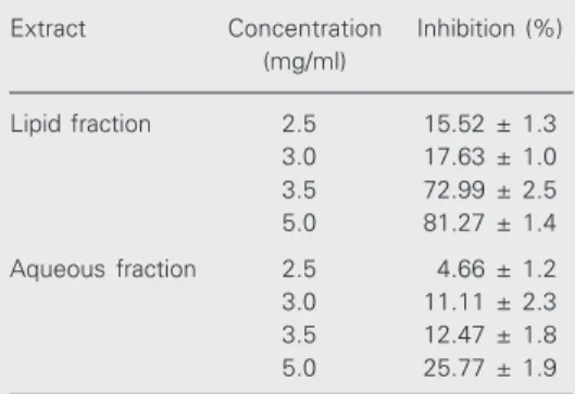
Both fractions of I. imperati extract (5.0 mg/ml) had a significant inhibitory activity against PLA2 (0.33 µg/ml bee venom) (Table
1). The lipid fraction inhibited PLA2 activity
by 85%, but the aqueous fraction only inhib-ited it by 25% (P<0.05). The IC50 for both
fractions against PLA2 activity was 3.22 and
Figure 1. Effect of topical appli-cation of Ipomoea imperati lipid and aqueous fractions on croton oil- (200 µg/ear) (A) and arachi-donic acid-induced (0.5 mg/ear) (B) mouse ear edema. The posi-tive control dexamethasone (217 µg/ear) induced a maximum inhi-bition of 52.0 ± 1.0% and ac-etone was the negative control. Each column is the mean of 5 mice, and the bars indicate the SEM. *P<0.05 compared to con-trol (Tukey test).
Edema inhibition (%)
120 100 80 60 40 20 0
2.5 3.5 5.0 7.5 10.0 12.5 Extract (mg/ear) 12 12 12 12 12 12 12 12 12 12 12 12 12 12 12 12 12 12 12 123 123 123 123 123 123 123 123 12 12 12 12 123 123 123 123 123 123 123 123 123 123 123 12 12 12 12 12 12 12 12 12 12 12 12 12 12 12 12 12 12 12 12 12 12 12 12 12 12 12 12 12 12 12 12 12 12 12 50 40 30 20 10 0
2.5 3.5 5.0 7.5 10.0 12.5
Extract (mg/ear) * * * * * * 12 12
Lipid fraction Aqueous fraction
A
B
Table 1. Inhibitory action of the lipid and aqueous fractions of Ipomoea imperati leaf extract on phos-pholipase A2 activity.
Extract Concentration Inhibition (%) (mg/ml)
Lipid fraction 2.5 15.52 ± 1.3 3.0 17.63 ± 1.0 3.5 72.99 ± 2.5 5.0 81.27 ± 1.4
Aqueous fraction 2.5 4.66 ± 1.2 3.0 11.11 ± 2.3 3.5 12.47 ± 1.8 5.0 25.77 ± 1.9
Data are reported as the mean % inhibition ± SEM for 5 rats. Lipid and aqueous fractions of I. imperati
3.43 mg/ml, respectively, and the CI was 2.47-3.98 and 2.83-4.04 mg/ml, respectively, which are not proportional to the weight of the extract.
The extract exhibited significant inhibi-tory activity on the formation of granulation tissue in the cotton pellet test. The lipid and aqueous fractions of I. imperati (at the dose of 1000 mg/kg) inhibited the inflammatory process by 73.2 ± 1.2% (IC50 of 93.7 mg/ear
and CI of 68.1-128.95 mg/ear) and by 56.14 ± 2.7% (IC50 of 102.5 mg/ear with a CI of
72.10-145.59 mg/ear), respectively, 6 days after implantation of the cotton pellet (Table 2). The lipid fraction (1000 mg/kg) was as effective as dexamethasone (0.20 mg/kg) in inhibiting this inflammation by 73%. No gastric mucosal lesions were observed after 6 days in the rats used in these experiments. Other doses used in this experiment proved to be effective, but maximum inhibition was obtained with 1000 mg/kg (Table 2).
The methanol-water extract of I. imperati
dose dependently inhibited the tone and am-plitude of the contractions of guinea pig ileum in electrically stimulated preparations. The lipid fraction had an IC50 of 1.60 mg/ml and
caused a maximum inhibition of 85.0 ± 1.0% (CI of 0.72-3.58 mg/ml), while the aqueous fractions had an IC50 of 4.12 mg/ml and caused
a maximum inhibition of 81.0 ± 1.53% (P<0.05; Figure 2) with a CI of 2.13-7.97 mg/ml.
The contractions in response to acetyl-choline and histamine (1 µM, each) were antagonized by the I. imperati methanol-water extract (10 mg/ml). The IC50 for the
lipid and aqueous fractions against acetyl-choline were 3.48 mg/ml (CI of 1.23-5.68) and 1.84 mg/ml (CI of 0.28-2.34 mg/ml), respectively, and maximum inhibition was 68.0 ± 0.9% for both fractions. With hista-mine, the IC50 was 4.03 mg/ml (CI of
0.67-6.10 mg/ml) and 1.69 mg/ml (CI of 0.9-3.45 mg/ml) for the lipid and aqueous fractions, respectively. The maximum inhibition for both fractions was 57 ± 0.86% (P<0.05; Figure 3).
Inhibition of
contraction (%)
100
80
60
40
20
0
0.0 2.5 5.0 7.5 10.0 12.5
Concentration (mg/ml)
*
Lipid fraction Aqueous fraction
* *
Table 2. Effect of the oral administration of Ipomoea imperati leaf lipid and aqueous extracts on rat granuloma tissue formation.
Treatment Dose Dry granuloma weight (mg) Inhibition (%) (mg/kg)
Initial Final
Carboxymethylcellulose 0.2 298.0 ± 3.1 492.0 ± 2.0
-Ipomoea imperati 10 240.0 ± 0.8 220.0 ± 1.3 20.3 ± 1.3 (lipid fraction) 30 239.0 ± 1.2 204.0 ± 2.0 35.1 ± 1.8*
100 245.0 ± 1.9 194.2 ± 1.0 51.2 ± 1.9* 300 243.0 ± 0.9 181.1 ± 0.89 62.2 ± 1.8* 1000 241.7 ± 1.9 168.4 ± 1.8 73.2 ± 1.2*
Ipomoea imperati 10 239.0 ± 1.2 225.9 ± 1.6 13.1 ± 0.98 (aqueous fraction) 30 242.0 ± 1.5 223.8 ± 1.2 18.2 ± 1.4
100 250.0 ± 1.2 220.0 ± 1.4 30.5 ± 1.6* 300 243.0 ± 0.9 200.5 ± 1.0 42.5 ± 1.8* 1000 241.9 ± 1.8 185.8 ± 1.8 56.1 ± 2.7*
Dexamethasone 0.2 240.3 ± 2.3 166.3 ± 1.5 74.0 ± 3.2*
Data are reported as the means ± SEM for 10 rats. Lipid fractions 30, 100, 300 and 1000 mg/kg of both fractions and the positive control (dexamethasone) were signifi-cantly different (*P<0.05, Tukey test).
Inhibition of
contraction (%)
100
80
60
40
20
0
0.0 2.5 5.0 7.5 10.0 12.5 Concentration (mg/ml)
Acetylcholine + lipid fraction Acetylcholine + aqueous fraction Histamine + lipid fraction Histamine + aqueous fraction
Figure 2. Dose-dependent inhi-bition of contractions by Ipo-moea imperati extracts in elec-trically stimulated isolated guin-ea pig ileum. Each point repre-sents the mean ± SEM of 5 experiments. Lipid fractions (1.0, 3.0 and 5.0 mg/ml) were significantly different from the aqueous fraction (*P<0.05, Tukey test).
Figure 3. Dose-dependent inhi-bition of 1 µM acetylcholine- and 1 µM histamine-induced con-tractions in guinea pig ileum by
Ipomoea imperati extracts. Data are reported as means ± SEM for 5 experiments. *P<0.05 compared to the aqueous frac-tion (Tukey test).
Discussion
A large number of herbal drugs are re-puted to be of excellent medicinal value, and are used for the treatment of several ail-ments. In folk medicine, various indigenous drugs are used in single and/or in combined form to treat different types of inflammatory and arthritic conditions, with considerable success (13).
In the present study we showed that the lipid fraction of the methanol-water I.
imperati leaf extract contains the active agent
against topical inflammation induced by cro-ton oil and arachidonic acid. However, the positive control dexamethasone was effec-tive only in inhibiting croton oil-induced topical inflammation. It is known that the anti-inflammatory effect of dexamethasone depends on the model used. Lee et al. (14) reported that dexamethasone (0.05 mg/ear dissolved in acetone) applied topically 16 h before the induction of mouse ear edema inhibited by 92% the inflammation induced by 12-O tetradecanoyl phorbol acetate (TPA). It has been established that inflammation induced by TPA/croton oil is related to the activation of PLA2, which releases
arachi-donic acid from the cell membrane. Arachi-donic acid, in turn, is metabolized to prosta-glandins and leukotrienes. Substances able to inhibit edema could be inhibitors of cyclo-oxygenase (COX) and/or 5-lipcyclo-oxygenase (15). However, other agents are involved in edema generation such as serotonin and bradykinin, which act directly through spe-cific receptors (5HT2, B2) present in the
endothelial cells of postcapillary venules. On the other hand, pretreatment with an-tagonists of protein kinase C may suppress the inflammation induced in guinea pig illeum. However, the inflammation observed in this model is related to the activation of PLA2. Bresnick et al. (16) stated that PLA2
catalyzes the sn-2 hydrolysis of phospho-lipids liberating free fatty acids, predomi-nantly arachidonic acid, and
lysophospho-lipids. These products can have biological actions or be further metabolized to form a variety of proinflammatory lipid mediators including prostaglandins, leukotrienes, or platelet-activating factor and thus the inhibi-tion of PLA2 by pharmacological agents
should have led to an anti-inflammatory ef-fect.
The mechanism of croton oil-induced inflammation involves an increase in PLA2
activity (17,18), which in turn leads to the release of arachidonic acid and subsequent biosynthesis of leukotrienes and prostaglan-dins (19,20), thus also involving the lipoxy-genase pathway. It has been firmly estab-lished that arachidonic acid metabolites act as mediators of the inflammatory response via COX and lipoxygenase activity, and have therefore been a target for the development of therapeutic agents (21).
Enzyme inhibition assays are important tools in the search for new drugs. Our exper-imental work also concerns the effects of I.
imperati extracts on several in vivo and in
vitro eicosanoid-releasing systems, to
inves-tigate whether they contribute to the anti-inflammatory activity of these compounds. The anti-inflammatory action of I. imperati
extracts on arachidonic acid-induced inflam-mation involved inhibition of the PLA2
en-zyme and probably of COX II (COX-2), the key enzyme involved in prostaglandin bio-synthesis. So PLA2 and probably the COX-2
enzyme of arachidonate metabolism can be inhibited by I. imperati, suggesting that this plant can provisionally be classified as a dual inhibitor, with greater PLA2 than
COX-2 activity. The step preceding arachi-donic acid liberation, which is catalyzed by PLA2, can be a target of the drugs (22).
Since endothelial cell contraction in-creases vascular permeability, and since this permeability is inhibited by vasodilating agents, the antispasmodic activity of I.
pes-caprae can be assumed to contribute to the
hypo-thesis on the tentative theory that endothelial cell contraction causes increased vascular permeability and on the fact that agents such as ß-adrenoceptor agonists and calcium an-tagonists inhibit this permeability with gen-eral relaxant properties.
During the process of acute inflamma-tion vascular leakage is a cardinal effect believed to result from the actions of certain inflammatory mediators, such as histamine, 5-hydroxytryptamine, bradykinin, LTE4 and
platelet-activating factor, on the postcapillary venules. The mechanism of this leakage was proposed to be due to mediator-induced con-traction of endothelial cells. The cells are thereby pulled apart from each other and create gaps, allowing blood components to leak into the interstitial compartment creat-ing edema (23). Pongprayoon et al. (24) suggested that endothelial cells and smooth muscle cells share a similar contractile mech-anism. Vasoconstriction has been implicated in the dermatitis caused by jellyfish stings, leading to local vascular insufficiency and gangrene. Agents with a direct vasodilatory action, e.g., papaverine (IC50 values of 30
µg/ml) have been recommended for the treat-ment of such toxin-induced dermatitis be-cause of their nonspecific antispasmodic ac-tion.
I. imperati extracts have local and
sys-temic anti-inflammatory actions in mice and rats, respectively. The pharmacological mechanism involved in this anti-inflamma-tory effect may be related to the inhibition of PLA2 and COX-2. Additional experiments
are necessary to demonstrate the inhibition of COX-2 by I. imperati. The extract also presented a nonspecific antispasmodic ac-tivity on the isolated ileum, inhibiting hista-mine and acetylcholine. In the acute toxicity assay, 1 mg/kg of I. imperati methanol-water extract caused no mortality in mice after 24 h.
Acknowledgments
The authors thank Dr. José Rubens Pirani, Institute of Biology, University of São Paulo, and Dr. Rosângela Simões Bianchini, Bo-tanical Institute of São Paulo, Brazil, for the identification of Ipomoea imperati.
References
1. Clark AM (1996). Natural products as a resource for new drugs.
Pharmaceutical Research, 13: 1133-1141.
2. Austin DF (1991). Ipomoea littoralis (Convolvulaceae) - Taxonomy, distribution and ethnobotany. Economic Botany, 45: 251-256. 3. Austin DF (1975). Flora of Panama. Annals of the Missouri Botanical
Garden, 62: 198-201.
4. Sastri BN (1965). The Wealth of India, Raw Materials. Council of Scientific and Industrial Research, New Delhi, India.
5. Fosberg FR & Sachet MH (1977). Convolvulaceae. In: Flora of Micronesia 3. Smithsonian Contribution Botanic 36, Caracas, Ven-ezuela.
6. Pongprayoon U, Baeckstrom P, Jacobsson U, Lindstrom M & Bohlin L (1992). Antispasmodic activity of ß-damascenona and E-phytol isolated from Ipomoea pes-caprae. Planta Medica, 58: 19-21. 7. Litchfield JT & Wilcoxon F (1949). A simplified method of evaluating
a dose effect experiments. Journal of Pharmacology and Experimen-tal Therapeutics, 96: 99-103.
8. Van Arman GC (1974). Anti-inflammatory drugs. Clinical Pharmacolo-gy and Therapeutics, 16: 900-904.
9. Meier R, Schuler W & Desaulles P (1950). Zur Frage des Mechanismus der Hemmung des Bindegewebewachstums durch
Cortisone. Experientia, 6: 469-471.
10. Niemegeers CJE, Van Bruggen W, Awouters F, Outer F & Janssen PAJ (1975). The effects of suprofen in rats with implanted cotton pellets. Arzneimittel-Forschung, 25: 1524-1526.
11. Roberts MF, Deems RA & Dennis EA (1977). Spectral perturbations of the histidine and tryptophan in cobra venom phospholipase A2 upon metal ion and mixed binding. Journal of Biological Chemistry, 252: 6011-6017.
12. Rang HP (1964). Stimulant actions of volatile anaesthetics on smooth muscle. Journal of Pharmaceutical Chemotherapy, 22: 356-365. 13. Abad MJ, Bermejo E, Carretero E, Martinez-Acitores C, Noguera B &
Villar A (1996). Antiinflammatory activity of some medicinal plant extracts from Venezuela. Journal ofEthnopharmacology, 55: 63-68. 14. Lee D, Marshall LA, Bolognese B & Adams JL (1997). Tetrazole is an effective Sn-3 phosphate replacement in substrate analog inhibitors of 14 kDa phospholipase A2. Prostaglandins, 13: 183-187. 15. Benito PB, Abad Martinez MJ, Silván Sem AM, San Gómez A,
Fernández Matellano L, Sánchez Contreras S & Diaz Lanza AM (1998). In vivo and in vitro anti-inflammatory activity of saikosaponins.
Life Sciences, 63: 1147-1156.
Phospholi-pase activity in skin after application of phorbol esters and 3-methyl-cholanthrene. Carcinogenesis, 2: 1119-1122.
17. Kondoh H, Sato Y & Kanoh H (1985). Arachidonic acid metabolism in cultured mouse keratinocytes. Journal of Investigative Dermatol-ogy, 85: 64-69.
18. McColl SR, Hurst NP & Cleland LG (1986). Modulation by phorbol myristate acetate of arachidonic acid release and leukotriene syn-thesis by human polymorphonuclear leukocytes stimulated with A23187. Biochemical and Biophysical Research Communications, 141: 399-404.
19. Ashendel GL & Boutwell RK (1979). Prostaglandin E and F levels in mouse epidermis are increased by tumor promoting phorbol esters.
Biochemical and Biophysical Research Communications, 90: 623-627.
20. Furstenberger G & Marks F (1980). Early prostaglandin E synthesis is an obligatory event in the induction of cell proliferation in mouse
epidermis in vivo by phorbol ester TPA. Biochemical and Biophysical Research Communications, 92: 749-756.
21. Inoue H, Mori T, Shibata S & Koshihara V (1989). Modulation by glycyrrhetenic acid derivatives of TPA-induced mouse ear edema.
British Journal of Pharmacology, 96: 204-210.
22. Opas EE, Bonney RJ & Humes JL (1985). Prostaglandin and leukotri-ene synthesis in mouse ears inflamed by arachidonic acid. Journal of Investigative Dermatology, 84: 253-256.
23. Williamson JA, Burnett JW, Fenner PJ, Hach-Wunderle V, Hoe LY & Adiga KM (1988). Papaverine recommended for the treatment of toxin-induced dermatitis non-specific antispasmodic action. Medical Journal of Australia, 149: 698-700.