Printed version ISSN 0001-3765 / Online version ISSN 1678-2690 http://dx.doi.org/10.1590/0001-3765201520150056
www.scielo.br/aabc
Antioxidant, analgesic and anti-inflammatory effects of lavender essential oil
GABRIELA L. DA SILVA1
, CAROLINA LUFT1
, ADROALDO LUNARDELLI1
, ROBSON H. AMARAL2 , DENIZAR A. DA SILVA MELO1
, MÁRCIO V.F. DONADIO1
, FERNANDA B. NUNES1
, MARCOS S. DE AZAMBUJA1
, JOÃO C. SANTANA 1
, CRISTINA M.B. MORAES1
, RICARDO O. MELLO1
, EDUARDO CASSEL3, MARCOS AURÉLIO DE ALMEIDA PEREIRA3 and JARBAS R. DE OLIVEIRA1
1Laboratório de Pesquisa em Biofísica Celular e Inflamação, Pontifícia Universidade Católica do Rio Grande do Sul, Avenida Ipiranga, 6681, Partenon, 90619-900 Porto Alegre, RS, Brasil 2Laboratório de Análises Clínicas, Instituto de Cardiologia do Rio Grande do Sul, Avenida
Princesa Isabel, 395, Santana, 90620-001 Porto Alegre, RS, Brasil
3Laboratório de Operações Unitárias, Pontifícia Universidade Católica do Rio Grande do Sul, Faculdade de Engenharia, Departamento de Engenharia Química,
Avenida Ipiranga, 6681, Prédio 30, Sala 208, Partenon, 90619-900 Porto Alegre, RS, Brasil
Manuscript received on January 26, 2015; accepted for publication on March 18, 2015
ABSTRACT
Several studies have investigated the antinociceptive, immunomodulatory and anti-inflammatory properties of compounds found in the lavender essential oil (LEO), however to date, there is still lack of substantial data.The objective of this study was to assess the antioxidant, anti-inflammatory and antinociceptive effects of lavender essential oil. The 1,1-diphenyl-2-picrylhydrazyl radical decolorization assay was used for antioxidant activity evaluation. The anti-inflammatory activity was tested using two models of acute inflammation: carrageenan-induced pleurisy and croton oil-induced ear edema. The antinociceptive activity was tested using the pain model induced by formalin. LEO has antioxidant activity, which is dose-dependent response. The inflammatory response evoked by carrageenan and by croton oil was reduced through the pre-treatment of animals with LEO. In the pleurisy model, the drug used as positive control, dexamethasone, was more efficacious. However, in the ear swelling, the antiedematogenic effect of the oil was similar to that observed for dexamethasone. In the formalin test, LEO consistently inhibited spontaneous nociception and presented a similar effect to that of tramadol. The results of this study reveal (in vivo)the analgesic and anti-inflammatory activities of LEO and demonstrates its important therapeutic potential.
Key words: antioxidants, inflammation, lavender, nociception.
Correspondence to: Jarbas Rodrigues de Oliveira E-mail: [email protected]
INTRODUCTION
Lavender genus is an important member of the Lamiaceae family. Lavandula species are widely distributed in the Mediterranean region and
cultivated in France, Italy and Spain. The Lavandula augustifolia Mill. specie is well known among
disorders (Hajhashemi et al. 2003, Gören et al. 2002). Lavandula augustifolia essential oil (LEO) and its major components, (-)-linalool and linalyl acetate, also presented anti-inflammatory properties in rats (Peana et al. 2002). In an in vitro study, (-)-linalool decreased the production and the release of nitric oxide (NO) without interference in the prostaglandins pathway (Peana et al. 2006).
Several studies have investigated the antinoci-ceptive, immunomodulatory and anti-inflammatory properties of compounds found in the lavender es-sential oil (Peana et al. 2002, Kim and Cho 1999). These studies investigated the effects of different constituents of essential oils, such as α–pipine, α-terpinene, terpin-4-ol, α-terpineol, linalyl acetate and linalool. Taken together, these studies conclud -ed that the compounds present in the lavender es-sential oil may have direct or indirect anti-inflam -matory or antinociceptive activities.
Although a growing number of investigations have been conducted in these last years, there is a lack of more substantial data on the effects and mechanisms of action of lavender essential oil. In this work, the in vitro antioxidant activity of lavender essential oil was investigated. Furthermore, the anti-inflammatory effects were evaluated by using different models of acute inflammation and the antinociceptive activity was tested by using the formalin-induced pain model. In all of them, the lavender essential oil effect was compared to well-known anti-inflammatory and analgesic drugs. Attempts have also been made in order to investigate some mechanisms of action. Considering the potential pharmacological effects of the lavender essential oil, we also investigated the possible toxic effects in rats using histopathological, biochemical and hematological parameters.
MATERIALS AND METHODS
Plant Materialand GC/MS analySeS
L a v e n d e r o i l w a s p u r c h a s e d f r o m t h e Bioessencia (Brazil). The oil was analyzed by
gas chromatography-mass spectrometry (GC/ MS). The equipment used was the Brand Agilent Technologies, model 7890A GC system, equipped with a data processor. Capillary column HP - 5 MS (30 m x 0.250 mm; film thickness 0.25 μm), 60 to 325/350°C temperature. Helium was the carrier gas used. The mass spectrometry (MS) analysis was performed in equipment from Agilent Technologies Brand, model 5975C VL MSD, operating at 70 eV, and temperature of the ion source was maintained at 250ºC. The injected volume was 5 μL of diluted sample (1:1) in n-hexane for each analysis. Identification of components in the oil was based on GC retention indexes relative by comparison of the fragmentation patterns of mass spectra with those reported in the literature (Adams 2001).
aniMalS
The animals were cared for and used in accordance with the Guiding Principles in the Care and Use of Animals approved by the Council of the American Physiologic Society. Male Swiss mice (30 ± 5 g), female Wistar rats (200 ± 20 g) and male Wistar rats (200 ± 20 g) were obtained from the Fundação Estadual de Pesquisa e Produção em Saúde (Porto Alegre, Brazil). Animals were housed under conditions of optimum light (12 hours light-dark cycle), temperature (22 ± 1°C) and humidity, with food and water provided ad libitum. All experiments were carried out between 8:00 a.m. and 7:00 p.m. The Ethics Committee of the Pontifícia Universidade Católica do Rio Grande do Sul approved all experimental procedures. All efforts were made in order to minimize animal suffering.
BleaChinGofthe free-radiCal 1,1-diPhenyl -2-PiCrylhydrazyl (dPPh teSt)
nm, which disappears upon reduction of an anti-radical compound. An aliquot containing different amounts of the oil was added to freshly prepared DPPH solution (150 μM in methanol); the concentrations of lavender essential oil employed were 0.1 mg/mL; 1.0 mg/mL; 10 mg/mL; 20 mg/ mL; 40 mg/mL; 60 mg/mL; 80 mg/mL; 100 mg/ mL; 120 mg/mL and 150 mg/mL. An equal volume of methanol was added to control tubes. After starting the reaction, and a 60 minutes incubation period at room temperature, the absorbance was read against a blank at 515 nm. All experiments were carried out in triplicate. We expressed the radical scavenger activity in terms of the amount of antioxidants necessary to decrease the initial DPPH absorbance by 50% (IC50). The IC50 value was determined graphically by plotting the percentage disappearance of DPPH as a function of the sample concentration. The antioxidant activity in percent (I %) was calculated as follows:
I (%) = (Ablank −Asample)/Ablank × 100
Where Ablank is the absorbance of the control reaction (containing all reagents except the test compound), and Asample is the absorbance of the test compound.
oral aCute toxiCity
Female Wistar rats were used. The animals were randomly divided into five groups (n=6). The first group (control group) received saline solution per oral route (p.o.). Groups 2 to 5 were treated with lavender essential oil at the doses of 0.6, 1.5, 3.0 and 5.0 g/kg, respectively. Animals were observed for 14 days after treatment. During these 14 days, the appearance of general toxic signs were observed. In this period of observation, the following parameters were also measured: weight changes, food consumption and mortality recording. The surviving animals were euthanized by decapitation and then blood collection for hematological and biochemical analysis was carried out. The animals that died during the 14 days had their lungs, liver
and kidneys collected for histological analysis. The lethal dose 50 (LD50) was calculated by linear
regression analysis.
heMatoloGiCaland BioCheMiCal analySiS
Leukocyte counting was performed using an automatic hematological analyzer (Cell Dyn 1700, Abbott). The differential leukocyte counting was performed with optical microscope after staining and, in each case, 100 cells were counted. Biochemical analyses were made in an automatic biochemical analyzer. The blood was centrifuged at 1000 x g for 10 minutes to obtain the serum. The following parameters were analyzed: alanine aminotransferase (ALT), aspartate aminotransferase (AST), urea, creatinine, sodium, potassium and total protein.
hiStoloGiCal exaMinationofthe tiSSueS
Liver, kidneys and lungs were carefully dissected. Small slices of these freshly harvested tissues were fixed in buffered formaldehyde solution (10%), dehydrated by serial ethanol solution, diaphanized with ethanol–benzene and enclosed with paraffin. Micrometer sections, cut by a microtome (Leica Leitz 1512), were stained with hematoxylin–eosin and examined under a light microscope.
PleuriSy
of carrageenan/saline, the animals were euthanized in a CO2 chamber and the pleural exudate was collected by pleural cavity lavage with 2 mL of saline solution (NaCl 0.9%) containing EDTA 1%. The samples of the pleural lavage were collected to determine exudation, total proteins, total and differential leukocyte counts. Exudate volumes were measured and the results were calculated by subtracting the volume injected into the pleural cavity (2 mL) from the total volume recovered (Lunardelli et al. 2006). The total cell count in each sample was estimated after dilution with Türk solution (1:20), using a Neubauer cell counting chamber (Boeco, Germany). Cytospin preparations of the samples were stained with May–Grunwald– Giemsa for the differential leukocyte count, which was performed under the immersion objective of a light microscope. After the cell count, the samples of the pleural lavage obtained from control and treated animals were collected, separated and stored at −20°C. Protein concentration was measured by the biuret technique.
ear edeMa
To estimate the topical and oral anti-inflammatory activity of the LEO, the mouse ear edema model was used (n=6). Briefly, 65 μL of acetone solution containing 2% croton oil was applied topically to the right ear of male mice. The left ear received an equal volume of acetone. Six hours after the application of croton oil, the mice were euthanized and a plug (6 mm diameter) was removed from both, treated (right) and untreated (left) ears. The edematous response was measured by the weight difference between the two plugs. LEO was applied topically to the right ear (50 μL/ear) 60 minutes before the croton oil treatment (LEO topic). To evaluate the oral anti-inflammatory activity, the other group was treated orally with the lavender essential oil 0.6 g/kg p.o. (LEO p.o.) diluted in DMSO. Another group was treated only with diluent (DMSO) and was used as vehicle control (VC, DMSO p.o.). Dexamethasone (DEX) was
used as a reference drug (0.5 mg/kg s.c. - 60 min before). The control group (C) had no pre-treated.
forMalin teSt
To assess the antinociceptive activity of the LEO, the formalin-induced nociception model was employed. In this test, a diluted formalin solution (in which formaldehyde is the active ingredient) was injected into the paw of a rodent, and pain-related behaviors were observed. Male Wistar rats were used in these experiments. The animals were divided into three groups (n=6) and pre-treated p.o. with lavender, indomethacin or tramadol, 1 hour before formalin injection, and placed in individual cages (22.0 x 15.0 x 12.5 cm) which served as the observation chamber after the injection of formalin. In order to reduce variability, animals were placed in open cages observation chamber for 30 minutes to allow them to get used to the environment and then were removed in order to receive to the formalin administration. Nociceptive behavior was induced by injecting 50 μL 2% formalin (s.c.) into the surface of the right hind paw (a control group received an injection of 50 μL of saline). Animals were then returned to the observation chamber. A mirror was placed behind the chamber to enable observation of the formalin-injected paw. Rats were observed for nociceptive behavior immediately after formalin injection every 5 minutes until 60 minutes after injection. Nociceptive flinching behavior was quantified as the number of flinches of the injected paw during the observation period. Phase I was defined as the first 10 minutes and the second phase was defined as the remaining time. At the end of the experiments, the rats were euthanized in a CO2 chamber.
aCute toxiCity Study
The acute treatment with LEO by oral administration at doses up to 1.5 g/kg did not produce any sign of toxicity or death in rats the 14 days of observation.
There were no changes in the behavior of animals chosen based on data reported in literature
(García-Hernández et al. 2007, Mohajer et al. 2005).
StatiStiCal analySiS
The results were evaluated statistically using SPSS (Statistical Package for Social Sciences) 18.0 software. The Shapiro-Wilk test was used for analysis of the data distribution. After confirmation of normal data, differences among control and experimental groups were compared by analysis of variance (ANOVA) followed by Bonferroni post hoc test. In the formalin test, the differences were evaluated by repeated measures ANOVA. The results were expressed as the mean ± standard error of the mean (SEM). The level of statistical significance was defined as p < 0.05.
RESULTS
GC and GC/MS analySeS
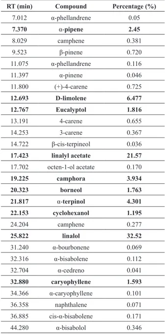
The LEO showed a diverse composition with 28 constituents, comprising approximately 82% of the total oil composition, reported in Table I. The components found are in agreement with the British Pharmacopoeia and, as reported in the literature, the oil is predominantly constituted by linalol (32.52%) and linalyl acetate (21.57%), demonstrating the authenticity sample.
dPPh-radiCal-SCavenGinG aCtivity
Figure 1 shows the absorbance reduction when DPPH solution was tested against various concentrations of the LEO. The greatest inhibitory activity was observed at the concentrations of 150, 120 and 100 mg/mL. No significant differences were found between these doses (p > 0.05). The lavender essential oil concentration resulting in a 50% inhibition of the free radical (IC50) was 51.05 mg/mL. This value was calculated by linear regression using geometric means.
TABLE I
The percentage composition of the total oil from Lavandula angustifolia.
RT (min) Compound Percentage (%)
7.012 α-phellandrene 0.05
7.370 α-pipene 2.45
8.029 camphene 0.381
9.523 β-pinene 0.720
11.075 α-phellandrene 0.116
11.397 α-pinene 0.046
11.800 (+)-4-carene 0.725
12.693 D-limolene 6.477
12.767 Eucalyptol 1.816
13.191 4-carene 0.655
14.253 3-carene 0.367
14.722 β-cis-terpineol 0.036
17.423 linalyl acetate 21.57
17.702 octen-1-ol acetate 0.170
19.225 camphora 3.934
20.323 borneol 1.763
21.817 α-terpinol 4.301
22.153 cyclohexanol 1.195
24.204 camphene 0.277
25.822 linalol 32.52
31.240 α-bourbonene 0.069
32.316 α-bisabolene 0.112
32.704 α-cedreno 0.041
32.880 caryophyllene 1.593
34.366 α-caryophyllene 0.101
36.358 naphthalene 0.071
36.885 cis-α-bisabolene 0.171
44.280 α-bisabolol 0.346
TABLE II
Effects of the Lavandula augustifolia essential oil (LEO) administered orally on serum biochemical and hematological parameters in female Wistar rats.
Control LEO
0.6 mg/kg 1.5 mg/kgLEO 3.0 mg/kgLEO 5.0 mg/kgLEO Urea (mg/dL) 56.75 ± 3.60 47.75 ± 5.56 49.8 ± 4.27 51.5 ± 9.19 48.00 ± 7.07 Creatinine (mg/dL) 0.94 ± 0.15 0.79 ± 0.07 0.74 ± 0.11 0.94 ± 0.37 0.82 ± 0.06 Total protein (g/dL) 7.05 ± 0.24 6.3 ± 0,42 7.28 ± 0.76 8.15 ± 0.64 6.65 ± 0.21 AST (U/L) 114.50 ± 17.54 117.00 ± 15.93 225.40 ± 62.28* 195.50 ± 68.29* 223.50 ± 34.65* ALT (U/L) 60.50 ± 9.88 68.40 ± 12.50 134.20 ± 33.15* 117.00 ± 32.53* 81.50 ± 4.95*
Na+ (mEq/L) 146.75 ± 8.10 146.50 ± 12.8 142.40 ± 3.65 141.00 ± 2.83 142.00 ± 1.41
K+ (mEq/L)
6.28 ± 1.28 5.53 ± 0.52 6.34 ± 0.76 6.00 ± 0.14 5.75 ± 0.07
Leukocytes (x103/µL)
6400 ± 916.52 6133 ± 736.36 3000 ± 400.00* 2200 ± 282.84* 2900 ± 141.42*
ALT = alanine aminotransferase, AST = aspartate aminotransferase, Na+ = sodium, K+
= potassium. The results are expressed as
the mean ± SDM. * p < 0.05.
Figure 1 - Linear regression of radical scavenging effect of lavender essential oil (LEO)
concentration on 2,2-diphenyl-1-picrylhydrazyl radical test.
treated with doses of 0.6 g/kg and 1.5 g/kg. In higher doses (3.0 g/kg and 5.0 g/kg) the main signs of toxicity observed were: atypical locomotion, anorexia, ataxia, piloerection, hypo activity and respiratory depression. The lethal 50 dose (LD50)
found was 3.55 g/kg. The hematological and biochemical profile of control and treated groups
hiStoloGiCal analySiS
Histological analyses of control rats showed normal structures. Leo did not cause any histological changes to the kidneys or the liver. However, the animals treated with the higher doses (3.0 g/kg and 5.0 g/kg) showed inflammatory infiltration in the lungs. The essential oil caused enlargement of lobules due to inflammatory process and lesion of bronchiolar mucosa.
PleuriSy
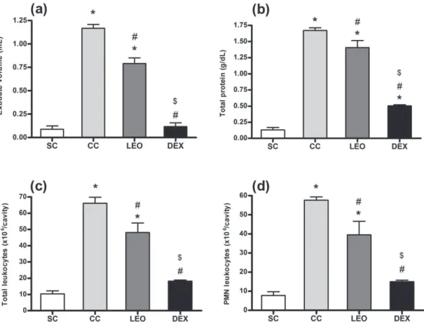
The results shown in Figure 2 demonstrate that edematogenic response evoked by i.pl. injection of carrageenan in rats was significantly reduced through the pre-treatment of animals with the Lavandula angustifolia oil (0.6 g/kg, p.o.),
administered 1 hour prior to the pleurisy induction. The essential oil caused a marked reduction in the volume (Fig. 2a) and total protein concentration
(Fig. 2b) in the collected exudates. There was also a reduction in the total leukocytes counting (Fig. 2c) and in the number of polymorphonuclear leukocytes (Fig. 2d), that migrated into the pleural cavity. Dexamethasone, the drug used as reference, produced similar effects, but it has been significantly more effective than the essential oil.
ear edeMa
edema. The inhibitory effect of the oil was similar to the inhibition caused by dexamethasone (Fig. 3).
forMalin teSt
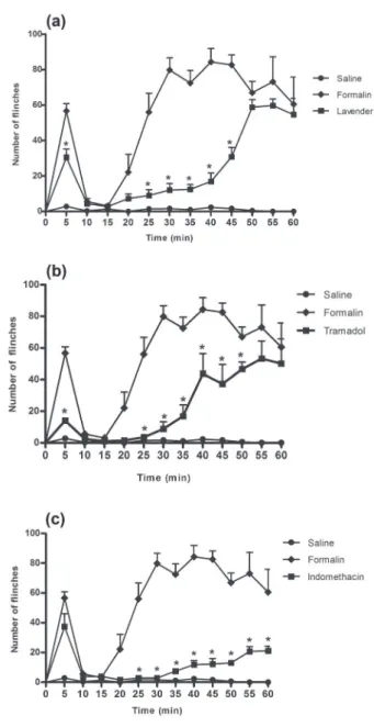
Formalin injection produced a typical pattern of flinching behavior. Two phases of spontaneous licking behavior were observed after the formalin injection. The first phase started immediately after administration of formalin and then diminished gradually after about 10 minutes. The second phase started about 15 minutes and lasted for a period of 1 hour.
We demonstrated that, when administered prior to the 2% formalin injection, the lavender essential oil consistently inhibited the spontaneous nociception and presented a similar effect to tramadol. The administration of lavender essential oil (Fig. 4a) or tramadol (Fig. 4b) significantly reduced the number of flinches in the first phase. In contrast, the administration of indomethacin was not able to reduce the flinching behavior in this stage (Fig. 4c).
In the second phase, all treatments inhibited the nociceptive behavior, but only indomethacin was effective throughout the entire observation period.
Figure 3 - Comparison of topical and oral anti-inflammatory
effects of lavender essential oil in croton oil-induced ear
edema. C = control group (mice not treated); VC = vehicle control group (mice treated with DMSO p.o.); LEO p.o. = mice treated with lavender essential oil 0.6 g/kg p.o.; LEO topic = mice treated with lavender essential oil topically 50 μL/ ear; DEXA = mice treated with dexamethasone 0.5 mg/kg s.c. Values represent the mean ± SEM. * p < 0.05 vs. control group
(C) and vehicle control group (VC).
Figure 4 - Dose- and time-response curves of flinches
induced by injection of formalin 2% s.c. into the surface of the right hind paw. Effect of treatments on the nociceptive behavior. (a) Lavender essential oil 0.6 g/kg, p.o., 1 hour prior. (b) Tramadol 30 mg/kg, p.o., 1 hour prior. (c)
Indomethacin 5 mg/kg, p.o., 1 hour prior. Values represent
DISCUSSION
Many compounds of plant origin have potent antioxidant activities, thus in the present study a screening of antioxidant activity was carried out prior to performing in vivo tests. The free radical scavenging capacity of the lavender essential oil was determined using the stable radical, DPPH. As expected, the lavender essential oil presented antioxidant activity and this ability was concentration-dependent.
Despite the potential pharmacological activities attributed to LEO, there is insufficient data about its toxicity. We performed a study of acute toxicity that showed that lavender essential oil has some toxic effects in doses up to 1.5 g/kg, although the dose of 0.6 g/kg appears to be well tolerated orally. Histopathological examination revealed some alterations in lungs of animals treated with high doses (3.0 g/kg and 5.0 g/kg). A lesion of bronchiolar mucosa was observed, associated with inflammatory infiltration. These data are consistent with clinical signs of toxicity observed before death (hyperventilation followed by respiratory depression). However, considering that such changes were found only in animals treated with high doses, this data can be taken as a result of an overload of the evaluated product. Furthermore, the treatment with lavender essential oil caused a decrease in the total leukocytes and an increase in transaminases (AST and ALT) at doses higher than 0.6 g/kg. For this reason, a dose of 0.6 g/kg was considered safe and can be used in further studies.
Carrageenan-induced pleurisy is a well characterized experimental model of inflammation that permits the quantification and correlation of both exudate and cellular migration with changes of other inflammatory parameters (Santos et al. 2010). This model has been widely used to investigate the pathophysiology of acute inflammation and also to evaluate the efficacy of drugs in inflammation (Paul et al. 2009). Our results revealed that treatment with lavender essential oil was effective in reducing the
responses induced by carrageenan. However, the drug used as reference, dexamethasone, was more effective in preventing these effects. It has been recently shown that carrageenan activates the toll-like receptor 4 (TLR4) (Bhattacharyya et al. 2008). TLR4 is an important receptor in the initiation of the inflammatory response in human cells (Lin and Yeh 2005). The identification of TLR4 as the receptor membrane that interacts with carrageenan strengthens the hypothesis that the effects of carrageenan are attributable to the activation of NF-kB.
The anti-inflammatory activity of lavender oil was further evaluated by the inhibition of croton oil-induced ear edema. The fact that carrageenan and croton oil have different inflammatory mechanisms, led us to suggest a possible mechanism of action for LEO. The croton oil is a highly irritating substance that stimulated an inflammatory response in the epidermis. Tetradecanoyl-phorbol acetate is a phorbol ester derivative of croton oil (Pitot 1979). TPA-induced inflammation is associated with alterations in cytokine production and increased production of prostaglandins and leukotrienes. These effects are thought to be mediated by protein kinase C (Passos et al. 2013). Many protein kinases C are activated by diacylglycerol and by high levels of intracellular calcium, both produced by activation of receptors coupled to G protein. Protein kinase C mediates a number of intracellular signal transduction pathways implicated in the pathogenesis of inflammation, including phospholipase A2-dependent arachidonic acid release and eicosanoid production.
The receptors involved in pain are also activated in inflammation. Pain is not a single entity, but differs in its underlying causes, symptoms and neurobiological mechanisms. On a coarse scale, three major forms of pain can be distinguished. Nociceptive or acute pain originates from the acute activation of primary nociceptive nerve fibers, and inflammatory pain originates from all forms of inflammation. The neuropathic pain originates from the damage of peripheral or central nerves and neurons. Inflammatory and neuropathic pain can turn into chronic pain syndromes, in which plastic changes in nociceptive processing have occurred that are no longer readily reversible by pharmacological treatment (Zeilhofer 2007).
Acute and chronic pain can be relieved effectively by non-steroidal anti-inflammatory drugs that inhibit cyclooxygenases (COX-1 and COX-2) and, consequently, are widely used (Hoogstraate et al. 2003). The opioid analgesic drugs remain the most effective therapy available for the treatment of moderate to severe pain; however, the problems arising from unwanted side-effects persist. A range of new therapies has been developed in recent decades to treat inflammation and chronic inflammatory diseases. Nonetheless, the number of such drugs remains small and in addition to the fact that they all display side effects that limit their use. Thus, the development of new pain management strategies can allow clinicians to have additional options for the management pain.
The antinociceptive properties of lavender species have been demonstrated by many authors (Hajhashemi et al. 2003, Barocelli et al. 2004). In this study, we evaluated the antinociceptive effect of Lavandula angustifolia Mill. essential oil using the formalin-induced pain model. There is a correlation between nociceptive and inflammatory effects in this model. This relationship is dependent on the concentration of formalin injected into the rat’s hind paw. Thus, only high concentrations of formalin, which produce significant plasma extravasation, are suitable for demonstrating the antinociceptive effects of anti-inflammatory agents
(Yashpal and Coderre 1998). In a pilot test in our laboratory, we found that this concentration would be 2%, since the antinociceptive effects of indomethacin parallel their anti-inflammatory action were observed throughout the second phase.
Subcutaneous injection of formalin, a chemical irritant, into the rat’s paw results in a reproducible biphasic response (Rocha-González et al. 2005). The reaction in the first phase (phase 1, 0–5 minutes) is caused by the initial tissue injury and a direct activation of peripheral nociceptors by formalin. The second phase (phase 2, 15–60 minutes) is mediated by a low level of peripheral nerve activity, whose effects are then enhanced in the spinal level by central sensitization (Yashpal and Coderre 1998). It has been well documented that inflammatory mediators such as neuropeptides, kinins, protons, prostanoids, serotonin and other substances are released during the inflammation produced by tissue damage. These mediators cause a painful inflammatory response.
In this work, we demonstrated that, in addition to anti-inflammatory activity, the oral treatment with lavender oil produces significant antinociception. In the first phase, lavender essential oil, as well as tramadol, presented antinociceptive effects. Indeed, indomethacin, a non-selective COX inhibitor, was able to inhibit the antinociceptive behavior only in the second phase. This data supports a relevant role for cyclooxygenases in this model. With this rationale one would think that, if lavender treatment caused a similar behavior to tramadol and different from indomethacin, then possibly the mechanism of action of lavender essential oil is not involved with inactivation of COX. However, it could be related to opioidergic neurotransmission. These findings are in agreement with previous study (Barocelli et al. 2004) which showed that treatment with naloxone, an opioid antagonist, prevents the analgesic action of lavender essential oil.
via G protein. Thus, the opioids or its agonists promotes the opening of potassium channels and inhibit the opening of calcium channels. The fact that lavender oil presented similar effect to tramadol and have been able to inhibit the action of croton oil (a protein kinase C activator), suggesting that the mechanism of action can be related to the activation of channels via protein G.
In conclusion, the results of the present study show the analgesic and anti-inflammatory activities of the lavender oil. Furthermore, the effectiveness of the oil without evidence of significant toxic effects supports the interest for application of the lavender essential oil as a therapeutic agent. Further studies should be conducted to evaluate and characterize the receptors involved in the antinociceptive and anti-inflammatory effects.
RESUMO
Diversos estudos têm investigado as propriedades antinociceptivas, imunomoduladoras e anti-inflamatórias dos compostos encontrados no óleo essencial de lavanda (OEL), contudo, até o momento, ainda faltam dados mais substanciais. O objetivo deste estudo foi avaliar os efeitos antioxidantes, anti-inflamatórios e antinociceptivos do óleo essencial de lavanda. Para avaliação da atividade antioxidante foi realizado o ensaio de descoloração do radical 1,1-difenil-2-picrilhidrazil. A atividade anti-inflamatória foi testada utilizando dois modelos de inflamação aguda: indução de pleurisia por carragenina e edema de orelha induzido pela aplicação de óleo de cróton. A atividade antinociceptiva foi avaliada utilizando o modelo de dor provocada por formalina. OEL apresenta atividade antioxidante, sendo esta resposta dose-dependente. A resposta inflamatória provocada pela carragenina e pelo óleo de cróton foi reduzida com o pré-tratamento dos animais com OEL. No modelo de pleurisia, a droga usada como controle positivo, dexametasona, foi mais eficaz. Entretanto, no edema de orelha, o efeito antiedematogênico do óleo foi similar ao observado na dexametasona. No teste da formalina, o OEL inibiu consistentemente a nocicepção espontânea e apresentou efeito similar ao do tramadol. Os resultados deste estudo revelam a atividade (in vivo)
analgésica e anti-inflamatória do OEL, demonstrando seu importante potencial terapêutico.
Palavras-chave: antioxidante, inflamação, lavanda, nocicepção.
REFERENCES
ADAMS RP. 2001. Identification of essential oils by gas chromatography quadrupole mass spectrometry. JASMS 16: 1902-1903.
AQUINOR, MORELLIS, LAURO MR, ABDOS, SAIJA A AND
TOMAINO A. 2001. Phenolic constituents and antioxidant activity of an extract of Anthurium versicolor leaves. J Nat Prod 64: 1019-1023.
BAROCELLIE, CALCINA F, CHIAVARINI M, IMPICCIATORE
M, BRUNI R, BIANCHI A AND BALLABENI V. 2004. Anti-nociceptive and gastroprotective effects of inhaled and orally administered Lavandula hybrida Reverchon “Grosso” essential oil. Life Sci 76: 213-223.
BHATTACHARYYAS, GILLR, CHEN ML, ZHANGF, LINHARDT
RJ, DUDEJA PK AND TOBACMAN JK. 2008. Toll-like
receptor 4 mediates induction of Bcl10-NFkB-IL-8 inflammatory pathway by carrageenan in human intestinal epithelial cells. J Biol Chem 283: 10550-10558.
GARCÍA-HERNÁNDEZ L, DÉCIGA-CAMPOS M, GUEVARA
-LÓPEZUAND LÓPEZ-MUÑOZ FJ. 2007. Co-administration of rofecoxib and tramadol results in additive or sub-additive interaction during arthritic nociception in rat. Pharmacol Biochem Behav 87: 331-340.
GÖRENA, TOPÇUG, BILSEL G, BILSEL M, AYDOĞMUŞ Z
AND PEZZUTO JM. 2002. The chemical contituents and
biological activity of Lavandula stocheas ssp. stocheas. Z Naturforsch 57: 797-800.
HAJHASHEMIV, GHANNADI A AND SHARIF B. 2003. Anti-inflammatory properties of the leaf extracts and essential oil of Lavandula angustifolia Mill. J Ethnopharmacol 89: 67-71.
HOOGSTRAATE J, ANDERSSON LI, BERGE OG, JONZON B
AND OJTEG G. 2003. Cox-inhibiting nitric oxide donators (CINODs) - a new paradigm in the treatment of pain and inflammation. Inflammopharmacology 11: 423-428.
KIMHM AND CHO SH. 1999. Lavender oil inhibits immediate-type allergic reaction in mice and rats. J Pharm Pharmacol 51: 221-226.
LINWJ AND YEHWC. 2005. Implication of Toll-like receptor
and tumor necrosis factor alpha signaling in septic shock.
Shock 24: 206-209.
LUNARDELLIA, LEITE CE, PIRES MG ANDDE OLIVEIRA JR.
2006. Extract of the bristles of Dirphia sp. increases nitric oxide in a rat pleurisy model. Inflamm Res 55:129-135.
MOHAJERM, SARKHAILP, HAJAROLASVADIN, ZAMANI MJ,
lanceolata Boiss. and Hojen. Extracts and examination of their components. Int J Pharm 2: 50-54.
PASSOS GF, MEDEIROS R, MARCON R, NASCIMENTO AF,
CALIXTO JB AND PIANOWSKI LF. 2013. The role of
PKC/ERK1/2 signaling in the anti-inflammatory effect of tetracyclic triterpene euphol on TPA-induced skin inflammation in mice. Eur J Pharmacol 698: 413-420. PAUL EL ET AL. 2009. Anti-inflammatory and
immunomod-ulatory effects of Baccharis trimera aqueous extract on induced pleurisy in rats and lymphoproliferation in vitro. Inflammation 32: 419-425.
PEANA AT, D’AQUILA PS, PANIN F, SERRA G, PIPPIA P
AND MORETTI MD. 2002. Anti-inflammatory activity of linalool and linalyl acetate constituents of essential oils. Phytomedicine 9: 721-726.
PEANA AT, MARZOCCOS, POPOLO A AND PINTO A. 2006. (-)-Linalol inhibits in vitro NO formation: Probable involvement in the anti-nociceptive activity of this monoterprne coumpond. Life Sci 78: 719-723.
PINHEIRO RM AND CALIXTO JB. 2002. Effects of the selective
COX-2 inhibitors, celecoxib and rofecoxib in a rat acute
model of inflammation. Inflamm Res 51: 603-610. PITOTHC. 1979. Biological and enzymatic events in chemical
carcinogenisis. Annu Rev Med 30: 25-39.
ROCHA-GONZÁLEZ HI, MENESES A, CARLTON SM AND
GRANADOS-SOTO V. 2005. Pronociceptive role of
peripheral and spinal 5-HT7 receptors in the formalin test. Pain 117: 182-192.
SANTOS RC ET AL. 2010. Anti-inflammatory and immuno-modulatory effects of Ulomoides dermestoides on induced pleurisy in rats and lymphoproliferation in vitro. Inflam-mation 33: 173-179.
YASHPALKAND CODERRE TJ. 1998. Influence of formalin
concentration on the antinociceptive effects of anti-inflammatory drugs in the formalin test in rats: separate mechanisms underlying the nociceptive effects of low- and high-concentration formalin. Eur J Pain 2: 63-68.
ZEILHOFERHU. 2007. Prostanoids in nociception and pain.