Artigo
*e-mail: mhsarragiotto@uem.br
CHEMICAL CONSTITUENTS, ANTI-INFLAMMATORY, AND FREE-RADICAL SCAVENGING ACTIVITIES OF
Guettardaviburnoides CHAM. & SCHLTDL. (RUBIACEAE)
Maria Augusta Naressia, Daniele Domingos Manholera, Franciele Queiroz Amesb, Ciomar Aparecida Bersani-Amadob, Anelise Samara Nazari Formagioc, Zefa Valdivina Pereirad, Willian Ferreira da Costaa, Debora Cristina Baldoquia and Maria Helena Sarragiottoa,*
aDepartamento de Química, Universidade Estadual de Maringá, Avenida Colombo, 5790, 87020-900 Maringá – PR, Brasil bDepartamento de Farmacologia e Terapêutica, Universidade Estadual de Maringá, Avenida Colombo, 5790, 87020-900 Maringá – PR, Brasil
cFaculdade de Ciências Agrárias, Universidade Federal da Grande Dourados, Rodovia Dourados-Itahum, Km 12, 79804-970 Dourados – MS, Brasil
dFaculdade de Ciências Biológicas e Ambientais, Universidade Federal da Grande Dourados, Rodovia Dourados-Itahum, Km 12, 79804-970 Dourados – MS, Brasil
Recebido em 04/02/2015; aceito em 29/04/2015; publicado na web em 29/05/2015
Chemical investigation of Guettarda viburnoides (leaves) led to the isolation of ursolic acid, uncaric acid, secoxyloganin, and grandifloroside, along with a mixture of quercetin-3-O-β-D-galactopyranoside and quercetin-3-O-β-D-glucopyranoside, and of β-sitosterol and stigmasterol. The structures of the isolated compounds were elucidated on the basis of their NMR data. The crude extract, ethyl acetate fraction, aqueous-methanol fraction, and grandifloroside showed significant DPPH free-radical scavenging activities with IC50 ranging from 18.92 to 26.47 µg mL−1. The topical administration of the crude extract and fractions markedly reduced the croton oil–induced mice ear edemain 67.0%–99.0%. Inhibition of tissue MPO activity was also observed, which demonstrated an anti-inflammatory effect of the G. viburnoides species.
Keywords: Guettarda viburnoides; chemical constituents; anti-inflammatory activity; antioxidant activity.
INTRODUCTION
The genus Guettarda belongs to the Guettardeae tribe (Rubiaceae family), and comprises approximately 150 species, ranging from eastern Africa through the islands of the Indian and Pacific Oceans to the Neotropics.1
Plants of Guettarda genusare popularly used in South America for wounds and inflammation treatments.2 Studies concerning to the biological activity have demonstrated anti-inflammatory,2-4 antioxi-dant and antiviral properties for Guettarda species.5,6
Chemical studies on this genus reported the isolation of quinine and quinicine-derived alkaloids from the bark of Guettarda noumeana and G. trimera;7 indole alkaloids from the leaves of G. eximia and G. heterosepala,8 and from the roots of G. ovalifolia,8 G. platypoda and G. acreana;2,9 triterpenoids from various plant parts of G. gra-zielae and G. platypoda, and from root barks of G. angelica and G. acreana.2,3,5,9,10 Triterpene saponins and quinic acid derivatives were identified in the roots and leaves of G. pohliana.4,6 A glycerol
α-D-glucuronide and a megastigmane glycoside were isolated from the leaves of G. speciosa L..11 Iridoids and secoiridoids were found in the roots of G. platypoda, stem bark of G. grazielae, and leaves of G. speciosa and of G. pohliana.4,9,12
Guettarda viburnoides Cham. & Schltdl. is a semideciduous shrub or tree distributed in Brazil and Paraguay,13 and no chemical or biological investigation have been reported for this species.
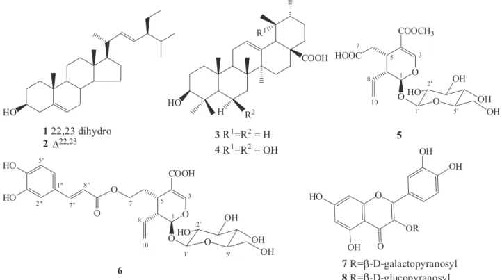
In the present work we describes the isolation and identification of β-sitosterol (1) and stigmasterol (2) as a mixture, ursolic acid (3), uncaric acid (4), secoxyloganin (5), and grandifloroside (6), along with a mixture of quercetin-3-O-β-D-galactopyranoside (7) and quercetin-3-O-β-D-glucopyranoside (8) from Guettarda viburnoides
(leaves). The structures of the isolated compounds were assigned on the basis of their NMR data, including two-dimensional NMR methods. The antiradical properties of crude extract, fractions, and of the secoiridoid 6, were evaluated by the DPPH method. Additionally, topical anti-inflammatory effects of the crude extract and fractions were investigated by using the croton oil-induced ear edema in mice model.
EXPERIMENTAL
General methods
1H and 13C-NMR spectra were recorded on a Varian Mercury Plus spectrometer operating at 300 MHz and 75.5 MHz, respectively, using D2O, CD3OD and CDCl3 as solvent, and tetramethylsilane (TMS) as internal reference. Chromatography columns (CC) were performed on silica gel 60 (0.063-0.200 mm, Merck) or Sephadex LH-20 (Sigma). TLC was performed on normal phase pre-coated silica gel 60 G or 60 GF254 (Merck) plates. Visualization of the compounds on TLC was accomplished by UV irradiation at 254 and 366 nm, and/or by spraying with a H2SO4/MeOH (1:1) or H2SO4/anisaldehyde/ acetic acid (1:0.5:50 mL) solutions followed by heating at 100 °C.
Plant material
Isolation of chemical constituents
The dried and ground leaves of G. viburnoides (960 g) were extracted with MeOH, at room temperature, and the solvent evapo-rated under vacuum to afford the crude extract (EGV; 51.59 g). Part of EGV (36.39 g) was dissolved in H2O:MeOH 1:1 and partitioned into n-hexane, chloroform and ethyl acetate. Evaporation of the solvents resulted in the n-hexane (HF; 3.56 g), chloroform (CF; 2.76 g), EtOAc (EAF; 3.44 g) and aqueous-methanol (AMF; 23.61 g) fractions. Part of HF fraction (1.69 g) was fractionated on silica gel CC using a mixture of n-hexane/ EtOAc in increasing polarity as eluent, to afford the sub-fractions HF.1 to HF.20. Purification of the sub-fraction eluted with n-hexane/EtOAc 10% (HF.9; 0.09 g), on CC (silica gel, n-hexane-AcOEt 10 to 70%) afforded a mixture of 1 and 2 (6.30 mg). The CF fraction (1.28 g) was fractionated by CC on silica gel (n-hexane/EtOAc 10 to 80% and EtOAc/MeOH 10 to 50%) to give compound 3 (65.1 mg).
Fractionation of part of EAF (1.6 g) on CC in silica gel using a mixture of n-hexane/EtOAc and EtOAc/MeOH, in increasing polarity, yielded the sub-fractions EAF.1 to EAF.12. Sub-fraction eluted with n-hexane/EtOAc 30% (EAF.3) provided compound 4 (16.1 mg). The sub-fraction EAF.6 (0.50 g), eluted in EtOAc/MeOH 10%, was purified by successive CC on Sephadex LH-20 using H2O, H2O/MeOH 8:2 to 2:8, and MeOH as eluent, to give compounds 5 (25.2 mg) and 6 (9.6 mg). Another part of the EAF fraction (0.80 g) was purified on Sephadex LH-20 eluted with H2O, H2O-MeOH 8:2 to 2:8, and MeOH to afford a mixture of compounds 7 and 8 (25.2 mg).
Purification of AMF fraction (0.50 g) on Sephadex LH-20 (H2O, H2O-MeOH 8:2, 6:4, 4:6, and 2:8, and MeOH, afforded the previously isolated compounds 5 (3.2 mg) and 6 (6.7 mg).
Secoxyloganin (5) 1H NMR (δ
H D2O,300 MHz): 5.51 (1H, d, J = 5.1 Hz, H-1), 7.53 (1H, d, J = 1.2 Hz, H-3), 3.23 (1H, m, H-5), 2.45 (1H, dd, J = 16.2, 7.3 Hz, H-6a), 2.65 (1H, dd, J = 16.2, 6.1 Hz, H-6b), 5.66 (1H, ddd, J = 16.8, 10.2, 9.9 Hz, H-8), 2.77 (1H, m, H-9); 5.34 (1H, d, J = 16.8; 1.8 Hz, H-10a), 5.28 (1H, dd, J= 10.2; 1.8 Hz, H-10b), 4.85 (1H, d, J = 7.3 Hz, H-1’), 3.21-3.75 (4 H, m, H-2’-H-5’), 3.91 (1H, dd, J = 12.6; 1.8, H-6’a), 3.70 (1H, m, H-6’b), 3,72 (3H, s, OCH3). 13C NMR (δ
C D2O, 75.5 MHz): 99.8 (C-1); 155.8 (C-3), 111.7 (C-4), 31.1 (C-5), 37.8 (C-6), 180.0 (C-7), 135.2 (C-8), 46.6 (C-9), 123.6 (C-10), 172.2 (C-11), 101.5 (C-1’), 75.4 (C-2’), 78.5 (C-3’), 72.4 (C-4’), 79.2 (C-5’), 63.6 (C-6’), 54.6 (OCH3).
Grandifloroside (6) 1H NMR ( δ
H CD3OD,300 MHz): 5.42 (1H, d, J = 5.7 Hz, H-1), 7.25 (1H, brs, H-3), 2.85 (1H, m, H-5), 1.99 (1H, td, J = 14.0; 6.8, H-6a), 1.78 (1H, td, J = 14.0; 6.8, H-6b), 4.15-4.28 (2H, m, H-7), 5.75 (1H, ddd, J = 16.9, 10.5, 6.9 Hz, H-8), 2.65 (1H, m, H-9), 5.25 (1H, d, J = 10.5 Hz, H-10), 5.30 (1H, dd, J = 10.5; 1.8, H-10a), 5.35 (1H, dd, J = 16.9; 1.8, H-10b), 4.72 (1H, d, J = 8.1 Hz, H-1’), 3.20 (1H, t, J = 9.0 Hz, H-2’), 3.39 (1H, t, J = 9.0 Hz, H-3’), 3.31 (1H, t, J = 9.3 Hz, H-4’), 3.36 (1H, m, H-5’), 3.65 (1H, dd, J = 12.0, 5.5 Hz, H-6’a), 3.84 (1H, dd, J = 12.0; 2.0 Hz, H-6’b), 7.07 (1H, brs, H-2”), 6.83 (1H, d, J = 8.1 Hz, H-5”), 7.00 (1H, dd, J = 8.1; 1.2 Hz, H-6”), 7.50 (1H, d, J = 15.9 Hz, H-7”), 6.25 (1H, d, J = 15.9, H-8”). 13C NMR (δ
C CD3OD, 75.5 MHz): 99.6 (C-1), 152.5 (C-3), 115.8 (C-4), 32.6 (C-5), 30.9 (C-6), 66.2 (C-7), 136.8 (C-8), 46.4 (C-9), 122.2 (C-10), 175.2 (C-11), 101.4 (C-1’), 75.6 (C-2’), 78.6 (C-3’), 72.5 (C-4’), 79.2 (C-5’), 63.6 (C-6’), 129.6 (C-1”), 117.4 (C-2”), 147.6 (C-3”), 150.2 (C-4”), 118.7 (C-5”), 125.3 (C-6”), 148.7 (C-7”), 117.1 (C-8”), 172.1 (C-9”).
Anti-inflammatory assays
Animals
Male Swiss mice weighting 25–30 g were used. The animals were maintained under controlled temperature of 22 °C and at 12 h light/ dark cycle, with water and food ad libitum. Animal care and the ex-perimental protocol followed the principles and guidelines suggested by the Brazilian College of Animal Experimentation (COBEA), and were approved by the local ethical committee.
Croton oil–induced mice ear edema
Ear edema was induced by applying 20 µL of croton oil solu-tion (200 µg diluted in 10 mL of the acetone/water 7:3 v/v) on the inner surface of the left ear of mice.14 The right ear received only the vehicle (20 µL). Indomethacin (1.0 mg per ear) was used as reference anti-inflammatory drug (positive control). Immediately after the application of croton oil, the treated groups received 20 µL of the EGV and fractions (CF, EAF and AMF) at the doses of 2.5 and 5.0 mg and the same volume of the reference drug on the inner ear surface; the control group received 20 µL of the vehicle. After 6 hours, the animals were killed, and each ear was perforated with a metal punch to provide a 6-mm-diameter disc. Edema was assessed by the difference in weight (mg) between the left and right ears. The percentage of edema was determined by Equation 1:
(%) inhibition = weight of left earcontrol- weight of left eartreated × 100
weight of left earcontrol- weight of right earvehicle
(1)
Myeloperoxidase (MPO) activity
Myeloperoxidase (MPO) activity was assayed using homogenate supernatants of the ear sections (i.e., untreated controls and animals treated with crude extract, fractions and indomethacin). The ear tis-sue was placed in potassium phosphate buffer 50 mmol L-1 (pH 6.0), containing 0.5% hexadecyltrimethyl-ammonium bromide (1 mL/50 mg tissue) in a Potter homogenizer. The homogenate was shaken in a vortex mixer and centrifuged for 5 min at 2500 rpm, and 10 µL of the supernatant was added to each well of a 96-well microplate in triplicate. A buffer solution (200 µL) containing o-dianisidine dihydrochloride (16.7 mg), redistilled water (90 mL), potassium phosphate buffer (10 mL), and 1% H2O2 (50 µL), were added. The enzyme reaction was stopped by the addition of a 1.46 mol L-1 sodium acetate solution (30 µL). Enzyme activity was determined by absorbance measured at 450 nm using a microplate spectrophotometer (Spectra Max Plus).15 DPPH free-radical scavenging activity
The free-radical scavenging activity of the crude extract EGV and of the HF, CF, EAF and AMF fractions, and of the compound 6, were determined by the DPPH method.16 Solutions of different concentrations of the samples in MeOH were added to 2 mL of daily-prepared DPPH solution (4.7 mg in 75 mL of MeOH). The solutions were left to stand at room temperature in the dark, and the absorbance was measured at 515.5 nm after 30 min. A DPPH solution without addition of the samples was used as control. The BHT was used as positive control. The assays were carried out in triplicate. The free-radical scavenging capacity was determined using linear regression analysis on confidence interval of 95% (P<0.05). The results were expressed as IC50 that represents the sample concentration required to decrease the initial DPPH concentration by 50%.
RESULTS AND DISCUSSION
isolation of β-sitosterol (1) and stigmasterol (2) as a mixture,17 ursolic acid (3), uncaric acid (4),18 secoxyloganin (5),19 grandifloroside (6),20 and a mixture of the flavonoids quercetin-3-O-β-D-galactopyranoside (hyperin) (7) and quercetin-3-O-β-D-glucopyranoside (isoquercitrin) (8)21 (Figure 1).
The 1H and 13C NMR data of compound 5 were characteristic of an iridoid skeleton, mainly by the signals for a hemiacetal group at δH 5.51 (d, J = 5.1 Hz, H-1)/δC 99.8 (C-1), and for the α,β-unsaturated carbonyl system at δH 7.53 (d, J = 1.2 Hz, H-3)/δC 155.8 (C-3), δC 111.7 (C-4) and δC 172.2 (C-11). The carboxymethylene unit attached to C-5 was evidenced by the signals at δH 2.45 (dd, J = 16.2, 7.3 Hz, H-6a) and δH 2.65 (dd, J = 16.2, 6.1 Hz, H-6b), which showed cor-relation with H-5 (δH 3.23, m) in the COSY spectra, and with the methylene carbon at δC 37.8 (C-6) in HSQC spectra. The signals for H-8 at δH 5.66 (ddd, J = 16.8, 10.2, 9.9 Hz), and for H-10 at δH 5.34 (dd, J = 16.8; 1.8 Hz, H-10a) and δH 5.28 (dd, J = 10.2; 1.8 Hz, H-10b), together with the methylene carbon at δC 123.6 (C-10), confirmed the terminal vinyl unit. The signals for the β-glucopyranosyl moiety were observed at δH 3.21-3.91 and δH 4.85 (H-1’) in the
1H NMR spectra. These data were consistent with of those described in literature for secoxyloganin (5).19 This secoiridoid was earlier reported in Guettarda platypoda and G. pohliana.4,12
Compound 6 showed the same basic skeleton of secoiridoid 5, differing in the nature of the substituent attached to C-5. The pres-ence of a trans-caffeoyl moiety in compound 6 was evidenced by the signals for the aromatic system at δH 7.07 (brs, H-2”)/δC 117.4 (C-2”),
δH 6.83 (d, J = 8.1 Hz, H-5”)/δC 118.7 (C-5”), and δH 7.00 (dd, J = 8.1; 1.2 Hz, H-6”)/δC 125.3 (C-6”), and for trans olefinic hydrogens atδH 7.50 (d, J = 15.9 Hz, H-7”) and δH 6.25 (d, J = 15.9 Hz, H-8”). The signals for the oxyethylene moiety were observed at δH 1.99 (1H, td, J = 14.0; 6.8, H-6a); δH 1.78 (1H, td, J = 14.0, 6.8, H-6b)/δC 30.9 (C-6) and δH 4.15-4.28 (2H, m, H-7)/δC 66.2 (C-7). The positioning of the trans-O-caffeoyl moiety at C-7 was confirmed by comparison of the chemical shift for C-7 of 6 (δC 66.2) with the reported for the demethylsecologanol (δC 59.8 ppm).
22 The structure of compound 6 was elucidated as grandifloroside based on the similarity of their NMR
data with those of literature for its 11-methyl ester derivative.20 These secoiridoids were reported in Adina racemosa,20 and in Neonauclea sessilifolia,23 both species belonging to the Rubiaceae family
The DPPH assay results (Table 1) showed that the crude extract (EGV), ethyl acetate (EAF) and aqueous-methanol (AMF) fractions, and grandifloroside (6) presented the highest free-radical scavenging activities, with IC50 values of 24.69, 18.92, 26.47 and 20.52 µg mL
-1, respectively. The EAF and AMF fractions afforded compounds 4-8, and 5 and 6, respectively.The antioxidant activities of compounds 5, 7 and 8 is well known.24 The DPPH free-radical scavenging activity of compound 6 is being reported here for the first time.
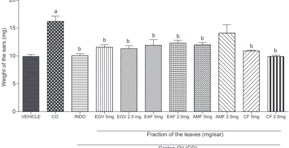
The anti-inflammatory effects of the crude extract (EGV) and of CF, EAF and AMF fractions were evaluated by using the ear edema induced by croton oil model.14 The determination of ear swelling and MPO enzyme activity are an apparently simple, sensitive and a quick procedure for evaluating the degree of inflammation, and the thera-peutic efficacy of drugs. The HF fraction was not tested due its low solubility in the vehicle (acetone/water 7:3 v/v) used for the assays. The data (Figure 2) showed that topical administration of EGV, and of CF, EAF and AMF fractions, at a dose of 2.5 mg/ear, markedly reduced the ear edema (77.6, 99.0, 61.0 and 67.0%, respectively). Among the sample tested, the chloroform fraction (CF) was the most
Figure 1. Chemical structures of compounds (1-8) isolated of G. viburnoides
Table 1. DPPH free-radical scavenging activity of G. viburnoides
Sample IC50µg mL
-1 (95% confidence limit) Crude Extract (EGV) 24.69 (19.80 – 29.58) Hexxane Fraction (HF) 100.52 (96.65 – 104.39) Chloroform Fraction (CF) 123.98 (121.72 – 126.24) Ethyl Acetate Fraction (EAF) 18.92 (14.20 – 23.64) Aqueous-Methanol Fraction (AMF) 26.47 (24.92 – 28.02) Grandifloroside (6) 20.52 (18.59 – 22.45)
active, displaying anti-inflammatory activity comparable to the posi-tive control indomethacin, which showed 90.4% inhibition (Figure 2). From the fraction CF was isolated ursolic acid (3), whose anti- and pro-inflammatory activities has been reported in the literature.25 Also, the crude extract and fractions were effective in inhibiting the elevated tissue MPO activity. MPO is a marker of the presence of leukocyte in inflamed site, and its activity is directly related to the amount of neutrophil infiltration, indicating an inflammatory process.15 The administration of EGV, CF, EAF and AMF, at dosage of 2.5 mg /ear, provoked an enzyme activity inhibition of 84.5, 80.8, 67.4 and 66.0% for EGV, CF, EAF and AMF, respectively, suggesting a decrease in cell migration to the inflamed site (Figure 3). These effects were similar to that of indomethacin that reduced the enzyme activity in 75.7%. These results indicate an anti-inflammatory effect of the crude extract and its fractions in this experimental model.
CONCLUSION
Compounds 4, 7 and 8 are being described for the first time in Guettarda genus. Ursolic acid (3) and secoxyloganin (5) were previ-ously isolated from Guettarda. Grandifloroside (6) is being described for the first time in Guettarda; however, the presence of secoiridoid 6, and of its 11-methyl ester derivative was reported in species belonging to the Rubiaceae family. Iridoids have been used as chemotaxonomic markers and the isolation of 5 and 6 from Guettarda viburnoides may contribute to chemotaxonomic studies of the Guettarda genus and Rubiaceae family. The assays results demonstrated the antioxidant and anti-inflammatory effects of G. viburnoides in the experimental models tested.
Figure 2. Effect of the EGV, CF, EAF and AMF fractions (2.5 and 5.0 mg) of Guettarda viburnoides on edema of the ear induced by CO in male Swiss mice (25-30 g). Notes: The animals (n= 6) were treated topically with the EGV or fractions, at the indicated concentrations, immediately after application of CO (200 mg) to the left ear. Indomethacin (Indo, 1 mg) was administered topically and used as reference anti-inflammatory (positive control). Each column represents the mean weight of the ears ± SEM 6 h after the application of CO. a p<0,001 compared to the control group vehicle (acetone/water 70%); b p<0,001 compared to the control group (CO); c p<0,01 compared to the control group (CO), (ANOVA, Tukey’s test)
SUPPLEMENTARY MATERIAL
The 1H and 13C NMR, COSY, HSQC and HMBC spectra for compounds 3-6 are available at http://quimicanova.sbq.org.br as a free-access PDF file.
ACKNOWLEDGEMENTS
The authors are grateful to CAPES and CNPq for fellowships (M. A. Naressi, D. D. Manholer).
REFERENCES
1. Achille F.; Motley T. J.; Lowry II, P. P.; Jérémie, J.; Ann. Mo. Bot. Gard.
2006, 93, 103.
2. Capasso, A.; Balderrama, L.; Sivila, S. C.; de Tommasi, N.; Sorrentino, L.; Pizza, C.; Planta Med. 1998, 64, 348.
3. Bhattacharyya, J.; de Almeida, M. Z.; J. Nat. Prod. 1985, 48, 148. 4. Testa, G.; de Oliveira, P. R. N.; da Silva, C. C.; Schuquel, I. T. A.;
Santin, S. M. O.; Kato, L.; de Oliverira, C. M. A.; de Arruda, L. L. M; Bersani-Amado, C. A.; Quim. Nova 2012, 35, 527.
5. Aquino, R.; de Simone, F.; Pizza, C.; Conti, C.; Stein, M. L.; J. Nat. Prod. 1989, 52, 679.
6. De Oliveira, P. R. N.; Testa, G.; de Sena, S. B.; da Costa, W. F.; Sarragiotto, M. H.; Santin, S. M. O.; de Souza, M. C.; Quim. Nova 2008, 31, 755.
7. Montagnac, A.; Litaudon, M.; Pais, M.; Phytochemistry 1997, 46, 973; Kan-Fan, C.; Brillanceau, M. H.; Pusset, J.; Chauviere, G.; Husson, H. P.; Phytochemistry 1985, 24, 2773.
8. Kan-Fan, C.; Husson, H.; J. Chem. Soc., Chem. Commun. 1979, 22, 1015; Brillanceae, M. H.; Kan-Fan, C.; Kan, S. K.; Husson, H. P.; Tet-rahedron Lett. 1984, 25, 2767; Kan-Fan, C.; Brillanceau, M. H.; Husson, H. P.; J. Nat. Prod. 1986, 49, 1130; Jiang, Y.; Weniger, B.; Quirion, J.; Muller, C.; Anton, R.; Planta Med. 1994, 60, 294.
9. Ferrari, F.; Messana, I.; Botta, B.; de Mello, J. F.; J. Nat. Prod. 1986,49, 1150.
10. Lima, G. S.; Moura, F. S.; Lemos, R. P. L.; Conserva, L. M.; Braz. J. Pharmacogn. 2009, 19, 284; Souza, M. P.; Matos, M. E. O.; Machado, M. I. L.; Braz Filho, R.; Vencato, I.; Mascarenhas, Y. P.; Phytochemistry
1984, 23, 2589; Matos, M. E. O.; Sousa, M. P.; Machado, M. I. L.; Braz Filho, R.; Phytochemistry 1986, 25, 1419.
11. Cai, W. H.; Matsunami, K.; Otsuka, H.; Shinzato, T.; Takeda, Y.; J. Nat. Med. 2011, 65, 364.
12. Aquino, R.; de Simone, F.; Senatore, F., Pizza, C.; Pharmacol. Res. Commun. 1988, 20, 105; Moura, F. S.; Lima, G. S.; Meneghetti, M. R.; Lemos, R. P. L.; Conserva, L. M.; Nat. Prod. Res. 2011, 25, 1614; Inouye, H.; Takeda, Y.; Nishimura, H.; Kanomi, A.; Okuda, T.; Puff, C.; Phytochemistry 1988, 27, 2591.
13. Taylor, C.; Delprete, P. G.; Vincentini, A.; Cortés, R.; Zappi D.; Persson C.; Costa, C. B.; da Anunciação, E. A. In Flora of the Venezuelan Guay-ana, vol. 8; Steyermark, J. A.; Berry, P. E.; Yatskievych, K.; Holst, B. K., eds; Missouri Botanical Garden Press: St. Louis, 2004, p. 497–847; Lorenzi, H.: Brazilian Trees – vol. 2, 4th ed., Instituto Plantarum: Nova Odessa, 2002.
14. Van Arman, G. C.; Clin. Pharmacol. Ther. 1974, 16, 900.
15. Bradley, P. P.; Priebat, D. A.; Chiristensen, R. D.; Rothstein, G.; J. In-vest. Dermatol. 1982, 78, 206.
16. Nazari, A. S.; Dias, S. A.; da Costa, W. F.; Bersani-Amado, C. A.; Vidotti, G. J.; de Souza, M. C.; Sarragiotto, M. H.; Pharm. Biol. 2006, 44, 7.
17. Goulart, M, O. F.; Sant’Ana, A. E. G.; de Lima, R. A.; Cavalcante, S. H.; de Carvalho, M. G.; Braz Filho, R.; Quim. Nova 1993, 16, 95. 18. Mahato, S. B.; Kundu, A. P.; Phytochemistry 1994, 37, 1517;
Diya-balanage, T. K. K.; Wannigama, G. P.; Weerasuriya, A.; Jayasinghe, L.; Simmonds, P.; Phytochemistry 1995, 40, 1311.
19. Boros, C. A.; Stermitz, F. R.; J. Nat. Prod. 1991,54, 1173.
20. Itoh, A.; Fujii, K.; Tomatsu, S.; Takao, C.; Tanahashi, T.; Nagakura, N.; Chen, C.; J. Nat. Prod. 2003,66, 1212.
21. Agrawal, P. K.; Bansal, M. C.; In Carbon-13 NMR of Flavonoids; Agrawal, P. K., ed.; Elsevier: Amsterdam, 1989.
22. Kitajima, M.; Fujii, N.; Yoshino, F.; Sudo, H.; Saito, K.; Aimi, N.; Takayama, H.; Chem. Pharm. Bull. 2005, 53, 1355.
23. Itoh, A.; Tanahashi, T.; Nagakura, N.; Nishi, T.; Phytochemistry 2003, 62, 359.
24. Zhang, X.; Thuong, P. T.; Jin, W.; Su, N. D.; Sok, D. E.; Bae, K.; Kang, S. S.; Arch. Pharm. Res. 2005, 28, 22; Choi, S.; Tai, B. H.; Cuong, N. M.; Kim, Y.; Jang, H.; Food Sci. Biotechnol. 2012, 21, 587; De Marino, S.; Festa C.; Zollo, F.; Nini, A.; Antenucci, L.; Raimo, G.; Iorizzi, M.; Anti-Cancer Agents Med. Chem. 2014, 14, 1376.

