Identification of
Mycobacterium bovis
antigens by analysis of bovine T-cell
responses after infection with a virulent
strain
1Institute of Biotechnology, CICVyA-INTA, Castelar, Argentina
2Veterinary Sciences Division, Department of Agriculture and Rural Development,
Stormont, Belfast, Northern Ireland, UK A. Alito1, J. McNair2,
R.M. Girvin2,
M. Zumarraga1,
F. Bigi1, J.M. Pollock2
and A. Cataldi1
Abstract
Purification and characterization of individual antigenic proteins are essential for the understanding of the pathogenic mechanisms of mycobacteria and the immune response against them. In the present study, we used anion-exchange chromatography to fractionate cell extracts and culture supernatant proteins from Mycobacterium bovis
to identify T-cell-stimulating antigens. These fractions were incu-bated with peripheral blood mononuclear cells (PBMC) from M. bovis-infected cattle in lymphoproliferation assays. This procedure does not denature proteins and permits the testing of mixtures of potential antigens that could be later identified. We characterized protein fractions with high stimulation indices from both culture supernatants and cell extracts. Proteins were identified by two-dimen-sional gel electrophoresis followed by N-terminal sequencing or MALDI-TOF. Culture supernatant fractions containing low molecular weight proteins such as ESAT6 and CFP10 and other proteins (85B, MPB70), and the novel antigens TPX and TRB-B were associated with a high stimulation index. These results reinforce the concept that some low molecular weight proteins such as ESAT6 and CFP10 play an important role in immune responses. Also, Rv3747 and L7/L12 were identified in high stimulation index cell extract fractions. These data show that protein fractions with high lymphoproliferative activity for bovine PBMC can be characterized and antigens which have been already described and new protein antigens can also be identified in these fractions.
Correspondence
A. Cataldi
Institute of Biotechnology CICVyA-INTA Castelar 1712 Argentina Fax: +54-11-4481-2975 E-mail: acataldi@cnia.inta.gov.ar
Research supported in part by the Centro Argentino Brasileño de Biotecnología (CABBIO). F. Bigi and A. Cataldi are fellows of the National Research Council of Argentina (CONICET).
Received February 4, 2003 Accepted July 22, 2003
Key words
•Bovine tuberculosis •Antigens
•Mycobacterium bovis •Cellular immunity
Introduction
Bovine tuberculosis caused by Mycobac-terium bovis is an animal health problem throughout the world and also constitutes a major threat to public health. On the Ameri-can continent there are 420 million heads of
countries. The strategy of test and slaughter has been used widely in an attempt to control dissemination of the disease and is based on the tuberculin skin test as a means for bovine tuberculosis diagnosis. However, implemen-tation of tuberculin tests is cumbersome, requiring a second visit by the veterinarian three days after the tuberculin injection. Some bovine tuberculosis eradication programs have incorporated variants of this test but suboptimal sensitivity and specificity have frequently been reported (2,3). This is consid-ered to be due, in part, to the nature of the poorly characterized antigens used, which are mycobacterial extracts containing components that are not species specific (3). Therefore, more sensitive and specific tests, probably incorporating better defined antigens, are re-quired for efficient detection of this disease.
It is well established that T-cell recogni-tion of mycobacterial antigens is the major immune response to tuberculosis (4-6). Therefore, an effective diagnostic test can be developed if well-characterized T-cell-reac-tive antigens are identified. This fact has led several research groups to the identification of antigens recognized by immune cells.
One of the main objectives of the present study was to identify the dominant M. bovis antigens that are recognized by the bovine cellular immune system. Unlike previous studies, which have focused on secreted an-tigens, the present study investigated cellu-lar as well as secreted proteins. In addition, we used culture supernatants harvested at various times of the culture to determine if different antigens were released along the growth curve. This was achieved by separat-ing M. bovis culture supernatant and whole cell proteins and testing the antigenicity of the resulting fractions by a lymphoprolifera-tion assay (LPA) using peripheral blood mononuclear cells (PBMC) from experimen-tally infected cattle. This approach allows a fast step of preselection of immunodomi-nant fractions from which it is easy to iden-tify antigenic proteins.
Material and Methods
Bacterial strains, media and preparation of culture supernatant and cell extracts
The M. bovis AN5 standard strain was used throughout the study. Cultures were pre-pared in Middlebrook 7H9 liquid medium containing 0.4% pyruvic acid and glucose. Mycobacterial cultures were incubated at 37ºC and harvested at 24, 38 and 73 days. The supernatants were separated from the cell ex-tract by centrifugation and then filtered through a 0.22-µm Millipore membrane (Bedford, MA, USA) to remove remaining mycobacte-ria. The proteins were then precipitated with ammonium sulfate (50%) for 18 h at 4ºC. Following centrifugation at 10,000 rpm for 1 h the precipitate was dissolved in a minimal volume of phosphate-buffered saline (PBS) and then dialyzed against PBS for 18 h at 4ºC. After removing the culture supernatant the cell mass was washed and resuspended in PBS. Mycobacteria were killed by heat (20 min at 80ºC) and sonicated on ice five times for 1 min with rest intervals of 2 min. Cell extracts were obtained from a 38-day culture.
The protein content of the supernatants and cell extracts was measured by the bicinchoninic acid method (Pierce, Rock-ford, IL, USA).
Animals
Four Friesian cattle were selected from a herd known to be free from bovine tubercu-losis during the previous five years. These animals were confirmed as bovine tubercu-losis negative by testing in vitro T-cell re-sponses to mycobacterial antigens. The ani-mals were placed in strict isolation under ventilation with negative pressure. They were infected by intranasal instillation with 106
Protein purification
Proteins from cell-free supernatants and cell extracts were separated using FPLC an-ion-exchange column chromatography (Phar-macia, Uppsala, Sweden). Protein, 0.5 to 5.0 mg, was loaded onto a Mono-Q column (Phar-macia) equilibrated with 20 mM Tris buffer, pH 8.0. Proteins bound to the column were eluted using a linear NaCl gradient (0-0.4125 M) while the proteins that bound very strongly were removed with 1 M NaCl. The flow rate was 0.5 ml per min and 84 fractions of 0.5 ml each were collected. Protein elution from the column was monitored by absorbance at 280 nm. All fractions were analyzed by SDS-PAGE (7) and proteins were visualized us-ing a silver stain procedure (8). To simplify the screening, protein fractions from the major peaks of the chromatogram that had similar protein profiles (as determined by SDS-PAGE) were pooled. Pools were dia-lyzed against PBS, filter-sterilized and pro-tein concentration was measured for use in LPA using PBMC from the experimentally infected cattle.
Lymphoproliferation assay
Blood was collected from four experi-mentally infected animals 11 months after M. bovis inoculation. PBMC were separated by centrifugation over Ficoll-Histopaque (Pharmacia) and viability of the PBMC prepa-ration was determined by Trypan blue exclu-sion. Microcultures were prepared with 106
cells/ml in RPMI 1640, supplemented with 10% fetal bovine serum, 2 mM L-glutamine and 25 µg/ml gentamicin sulfate. The anti-genic fractions obtained by FPLC were added to 200-µl cultures (in triplicate) at a standard protein concentration of 4 µg/ml. Total cul-ture supernatant or cell extract was also in-cluded as positive control, also at 4 µg/ml protein. Purified protein derivative (PPD) prepared from M. bovis was used as a posi-tive antigen control and concanavalin A
(4 µg/ml) was used to measure the viability of separated PBMC. PBMC cultures were incubated at 37ºC and in the presence of 5% CO2. After 4 days, the cultures were pulsed
with tritiated thymidine (1 µCi per well) and lymphoproliferation was terminated 18 h later by harvesting the cells onto a filter mat using a Skatron harvester (Skatron, Lier, Norway). Radioactivity was measured using a Betaplate counter (Pharmacia) and the results are re-ported as stimulation indices (SI) calculated from the ratio of cpm incorporated with an-tigen to cpm incorporated without anan-tigen (PBS was added in the place of antigen).
Identification of individual proteins in stimulatory fractions
Two-dimensional electrophoresis. The standard method of O’Farrell (9) was used. Briefly, 30 to 50 µg proteins were dialyzed against 40 mM Tris-HCl, pH 9.5, concen-trated using a Millipore cartridge with a cutoff of 3 kDa, and solubilized with a buffer consisting of 9.5 M urea, 2% NP40, 5% mercaptoethanol and a mix of ampholytes (0.8%; BioLyte, BioRad Laboratories, Rich-mond, CA, USA), pH ranges 3-10:5-7 in a 1:4 relationship. Twenty microliters of this mix was loaded onto a first dimension-iso-electrofocusing gel (9.16 M urea, 2% NP40, 4.25% acrylamide-bisacrylamide mix (30:5.4%), 2% of the ampholyte mix men-tioned above, 0.02% TEMED, and 0.2% ammonium persulfate). Electrophoresis was performed in 20 mM NaOH (cathode buffer) and 10 mM PO4H3 (anode buffer) at room
temperature at 200 V for 90 min, followed by 400 V for 12 h and 950 V for 2 h. The second dimension run was performed on 12.5 or 15% SDS-PAGE gel. The proteins were either silver stained or transferred to a PVDF membrane and subsequently stained with Coomassie blue.
performed by Edman degradation on the first six amino acids (Midwest Analytical, Inc., St. Louis, MO, USA). To identify the proteins the sequence was compared against a database of M. tuberculosis proteins ( w w w . s a n g e r . e b i . a c . u k / P r o j e c t s / M_tuberculosis).
MALDI-TOF. Matrix-assisted laser de-sorption/ionization-time-of-flight (MALDI-TOF) mass spectrometry was performed us-ing a Reflex III MALDI-TOF (Brukner Daltonik GmbH, Bremen, Germany) spec-trometer on samples prepared as follows. Two-dimensional gels were stained with sil-ver and the protein spots were sliced into small pieces with a stainless-steel scalpel or a vortex mixer and placed in siliconized microcentrifuge tubes. Gel pieces were destained with a ferricyanide-thiosulfate so-lution and washed in 50% acetonitrile con-taining 25 mM ammonium bicarbonate, pH 8.0 (three times, 15 min each, 24ºC). Gel slices were dehydrated in 100% acetonitrile for 10 min, the acetonitrile was removed, and the gel slices were dried under vacuum for 30 min. Samples were rehydrated with sequencing-grade trypsin solution (5 µg/ml in 25 mM ammonium bicarbonate, pH 8.0) and incubated overnight at 32ºC. Peptides were extracted with 50% acetonitrile-2%
tri-fluoroacetic acid in distilled water and con-centrated with a Speed-Vac. Samples were mixed with the matrix α -cyano-4-hydroxy-cinnamic acid and analyzed by MALDI-TOF. Mass spectrometry profiles were searched against the National Center for Biotechnol-ogy Information database.
SDS-PAGE and Western blot. Antigenic fractions were resuspended in loading buffer (2% SDS, 0.125 M Tris-HCl, pH 6.8, 1% 2-mercaptoethanol, 0.02% bromophenol blue, and 10% glycerol), heated for 5 min in boil-ing water, and loaded onto 12.5% polyacryl-amide gels by the method of Laemmli (7). Molecular mass standards (low and high range, BioRad) were run on each gel. Pro-teins were electrotransferred onto a nitrocel-lulose sheet (Sleicher and Schuell, Dassel, Germany) by the semidry transfer method (10). The efficiency of transfer was visual-ized by transient staining with Ponceau red. Membranes were blocked with 5% nonfat milk in TBS (50 mM Tris-HCl, pH 8, and 150 mM NaCl), incubated with the first anti-body overnight at 4ºC and, after three washes with TBS, incubated with alkaline phos-phatase-conjugated anti-rabbit or anti-mouse IgG (Sigma, St. Louis, MO, USA) at 1/1000 dilution for 2 h at 37ºC. After three washes with TBS and one wash with alkaline phos-phatase buffer (100 mM Tris-HCl, pH 9.5, 100 mM NaCl, and 5 mM MgCl2), a color
reaction was developed by adding 5-bromo-4-chloro-3-indolylphosphate (BCIP) and toluidine nitroblue tetrazolium (NBT) as sub-strates. After 20 min the color reaction was stopped by adding water.
Antisera and antibodies. Anti-ESAT6 monoclonal antibody was kindly provided by Ida Rosenkrands and Peter Andersen (Statens Serum Institute, Copenhagen, Den-mark). Anti-MPB70 monoclonal antibody (4C3/17) was purchased from CSL (Victoria, Australia). Polyclonal antisera recognizing 85B were kindly provided by T. Fifis (Ani-mal Health Research Laboratories, Victoria, Australia).
97
21 31 40 42 55 66
MWM 24 38 73 E
Results
Fractionation of culture supernatants and cell extracts by anion-exchange chromatography
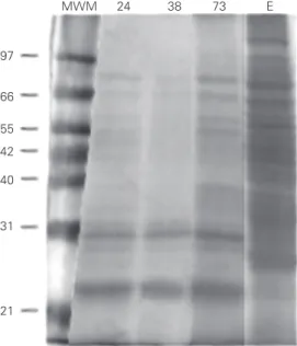
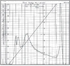
Culture supernatants, harvested at three different culture times (24, 38 and 73 days), and cell extracts were fractionated by FPLC anion-exchange chromatography with a linear gradient of 0-0.42 M NaCl. As indicated in Figure 1, nonfractionated supernatant and ex-tracts were complex mixtures of several indi-vidual proteins. In the culture supernatant frac-tions, proteins of <10, 20, 22, 24, 31, 32, 40, 43, 45, 50, 65 and 75 kDa were observed. In cell extracts, heavily stained bands were seen at 10, 14, 16, 20, 22, 24, 31, 40, 66 and 71 kDa. When culture supernatants and cell ex-tract preparations were separated by anion-exchange chromatography, four (24-day cul-ture supernatant) to three (73-day culcul-ture supernatant and cell extract) major peaks were observed in the gradient zone. A chro-matogram from a 73-day culture supernatant is shown in Figure 2 as an example. Protein profiles for each fraction were determined by SDS-PAGE. Figure 3 shows the fractions from a 38-day culture supernatant.
Fractions having a similar protein pro-file, from each chromatogram peak, were pooled and dialyzed and their concentration was measured and used for LPA using PBMC from four animals experimentally infected with M. bovis. Two uninoculated animals of the same age, sex and source were used as uninfected controls.
Figure 2. A representative an-ion-exchange chromatogram of a 73-day culture supernatant. Ordinate: absorbance at 280 nm. Abscissa: fraction num-bers.
Figure 3. SDS-PAGE analysis of the anion-exchange chromatog-raphy elution profile of 38-day AN5 culture supernatants. The fraction number is indicated above each lane. AN5 = culture supernatants before chroma-tography. Note the relatively large amounts of low MW pro-teins in fractions 30 to 35. Num-bers on the left indicate the molecular mass in kDa. 66
14 21 31 45
AN5 5 10 11 12 13 14 15 16 17 18 19 20 23 24 25 26 27 30 31 32 33 34 35 83 84
T-cell responses to chromatographic fractions
The FPLC profile of the 24-day culture supernatant contained five peaks. Pools from these regions were prepared, representing fractions 11-17, 20-24, 25-28, 32-36, and 81-84 (Table 1). Some of these fractions showed an SI equal to or higher than that of PPD. The mean reactivity of pools was 25-28 > 20-24 = 32-36 > 81-84 > 11-17, except for animal 4 where pool 81-84 was the more reactive. SDS-PAGE analysis of the most stimulatory pool (25-28) showed proteins of <10 kDa and a less intense band of 20 kDa. Fraction 20-24 had intense bands of 20, 25, 32 and 41 kDa. The 32-36 pool had a <10-kDa protein, while the 81-84 pool had a 20-kDa protein and the lowest stimulatory pool (11-17) showed bands of 20, 31 and 32 kDa and additional weaker bands.
sepa-except for animal 1 where 83 was the more reactive pool. SDS-PAGE analysis (Figure 3) of the low stimulatory pool (17-18) showed prominent bands of 16, 20 and 32 kDa, as well as additional bands of 24, 40, 43 and 45 kDa. Pool 25-26 had proteins of 14, 16, 20, 24, 31, and 44 kDa (doublet), and less in-tense low molecular weight proteins. The most stimulatory pool (30-34) showed an intense <10-kDa band (Figure 3) and a 24-kDa protein, with minor bands of 43 and 45 kDa as well as other bands. Fraction 83 had proteins of 14 and 16 kDa (Table 1).
The T-cell-proliferative response was similar for all six pools analyzed (14-16, 17-18, 22-23, 25-28, 29-31, 81) from the 73-day culture supernatant (Table 1). SDS-PAGE analysis showed that all of them, except fraction 81, contained protein bands <10 kDa, with little difference in the protein profile of all these pools. Major bands were seen at <10, 14.5, 20, 22, 24, 25, 32, 40, 44 and 46 kDa.
Using whole cell extract, six peaks were observed. Pools were prepared from frac-tions 6-14, 20-24, 25-29, 30-32, 33-40 and 82-84. Many fractions showed an SI equal to PPD or higher. The reactivity order was roughly 30-32 > 25-29 > 33-40 > 20-24 > 82-84 > 6-14 for animals 1 and 2, and 20-24 > 30-32 > 33-40 > 25-29 > 82-84 = 6-14 for animals 3 and 4. SDS-PAGE analysis of fraction 30-32 showed proteins of <10, 19, 25, 40 and 70 kDa. Fraction 20-24 contained proteins of <10, 12, 14, 19, 25, 30, 40 and 70 kDa. Fraction 6-14 contained proteins of <10, 19, 25, 30, 40, 45, 66 and 70 kDa. Finally, fraction 82-84 showed several heavily stained and smeared bands.
When PBMC from healthy, noninfected animals were stimulated with pools of the fraction, very low SI were observed (Table 1, animals 88 and 99).
To assess the functionality of the meth-odology (PBMC preparation and LPA) throughout the experiment, we analyzed the indices of concanavalin A stimulation for all ration of the 38-day culture supernatant. Pools
were prepared from fractions 17-18, 25-26, 30-34, and 83 (Table 1). The reactivity order of the pools was 30-34 > 83 > 25-26 > 17-18,
Table 1. Stimulation indices obtained in a lymphoproliferation assay.
Antigen Stimulation index
Infected animals Noninfected animals
1 2 3 4 88 99
Culture supernatant - 24 days
PBS 1 (158) 1 (63) 1( 67) 1 (103) 1 (234) 1 (170)
Con A 13 49 97 200 352 277
PPD B 13 23 86 217 1.71 2.02
Pool 11-17 5 5 21 17 1.06 1.70
Pool 20-24 25 57 27 294 1.37 2.00
Pool 25-28 28 68 24 303 1.06 1.96
Pool 32-36 26 56 26 177 1.77 1.52
Pool 81-84 9 27 9 345 1.26 2.29
Culture supernatant - 38 days
PBS 1 (433) 1 (330) 1 (477) 1 (389) 1 (234) 1 (170)
Con A 126 234 148 119 352 277
PPD B 109 55 71 64 1.71 2.02
TP 123 50 49 48 1.70 1.74
Pool 17-18 32 12 12 89 1.10 1.04
Pool 25-26 80 45 21 73 0.70 1.24
Pool 30-34 64 91 14 136 1.07 1.32
Pool 83 118 67 nd 108 2.23 3.29
Culture supernatant - 73 days
PBS 1 (433) 1 (330) 1 (477) 1 (389) 1 (234) 1 (170)
Con A 126 234 148 119 352 277
PPD B 109 55 71 64 1.71 2.02
TP 120 59 40 105 2.70 1.04
Pool 14-16 110 55 28 75 1.42 1.24
Pool 17-18 107 62 31 50 2.08 1.72
Pool 22-23 107 93 30 57 0.98 1.52
Pool 25-28 98 73 31 97 1.09 1.92
Pool 29-31 97 68 32 113 2.10 1.46
Pool 81 96 54 15 103 1.76 2.97
Cell extract
PBS 1 (98) 1 (68) 1 (567) 1 (200) nd 1 (325)
Con A 542 980 540 607 nd 250
PPD B 109 55 71 64 nd 2.30
Pool 6-14 113 25 34 16 nd 3.09
Pool 20-24 225 93 234 408 nd 2.40
Pool 25-29 623 178 76 52 nd 2.47
Pool 30-32 660 186 98 57 nd 3.25
Pool 33-40 606 161 102 46 nd 3.42
Pool 82-84 457 30 43 11 nd 1.26
four animals, which were found to be highly uniform. Variations in LPA from animal to animal and within individual animals throughout the experiments were determined by analyzing LPA results when PPD was used as antigen.
Identification of proteins in antigenic fractions
Six proteins were identified in fractions 25-28 from 24-day culture supernatants and in fractions 30-34 from 38-day culture su-pernatants. They were CFP10 (11 kDa), ESAT6 (9 kDa), TRB-B (36 kDa), 85B (32 kDa), TPX (17 kDa), and MPB70 (20 kDa) (Table 2). N-terminal sequences of 85B, MPB70 and TRB-B are compatible with sig-nal sequences. Specific antisera were used to confirm the identification of MPB70 and 85B by Western blot (data not shown). ESAT6 was identified only by Western blot (data not shown).
Several cell extract fractions from 38-day cultures showed stimulatory properties. We concentrated on one of them (fraction 30-32) that showed high T-cell reactivity. Since the N-terminus seemed to be blocked in most proteins, we used MALDI-TOF for protein identification. Two proteins were identified: Rv3747 (13 kDa) and L7/L12 (13 kDa) (Table 2). Proteins corresponding to three other spots could not be identified.
Discussion
In this study we used a novel approach to detect T-cell-stimulating antigens from M. bovis, i.e., fractionation of antigens using anion-exchange chromotagraphy. Other in-vestigators have used anion-exchange col-umns to purify antigens already identified in M. bovis (11). Compared to protein separa-tion by SDS-PAGE, the advantage of the present methodology is that antigens are maintained in the native state. In addition, we screened antigens from cell extracts as
Table 2. Identification of Mycobacterium bovis in antigenic fractions.
Antigen N-terminal Position Identification Confirmation by
sequence in ORF by MALDI-TOF Western blot
CFP10 AEMKTD 2 -
-85B FSRPGL 41 - +
MPB70 GDLVGP 31 + +
ESAT6 - - + +
TPX AQITLR 2 -
-TRB-B TELTGA 16 -
-L7/L12 - - +
-Rv3747 - - +
-well as culture supernatant proteins. Finally, we harvested culture supernatant proteins at various times instead of using a single sample to test whether the bacteria might secrete different proteins during the growth phases. Since cellular responses are the major immune mechanism in tuberculosis, T-cell reactivity toward well-characterized myco-bacterial antigens has been extensively stud-ied (5,12,13). However, the direct screening of antigens with immune cells is technically much more complicated compared to anti-gen-antibody interactions. Several ap-proaches have been used previously to test soluble antigens prepared from a nitrocellu-lose membrane or “T-cell Western blot” (14-17). Mustafa et al. (18) performed direct screening of an M. tuberculosis expression library with PBMC, and identified a protein reacting with T cells but not with antibodies. Electroelution from SDS-PAGE gels was first used by Gulle et al. (19) to obtain soluble antigens. Using a similar technique, Andersen and Heron (20) identified M. tuberculosis antigens related to memory response. Gulle et al. (21) screened BCG cellular and se-creted proteins for T-cell stimulant fractions using PBMC from cattle immunized with ei-ther viable or gamma-irradiated BCG. In con-trast, in the present study, a virulent strain was used to infect cattle and to prepare proteins.
con-tained several protein bands, some of them shared with other fractions while others were not. This allowed us to deduce which bands confer reactivity to the fraction. In 24- and 38-day culture supernatants, fractions asso-ciated with a high SI contained low molecu-lar weight proteins (<10 kDa) together with other proteins ranging from 20 to 35 kDa. In an attempt to identify individual antigens in antigenic fractions using a combination of methods, we determined that these stimulant fractions consisted of 85B, TRB-B, MPB70, TPX, CFP10 and ESAT6. 85B (22), ESAT6 (6,23) and MPB70 (24-26) are well known T-cell-stimulating antigens, while the anti-genicity of CFP10 has only recently been demonstrated (23,25,27). These results sup-port the view that low molecular weight proteins such as ESAT6 (6) play an impor-tant role in bovine immune responses to M. bovis. TRB-B and TPX are novel candidate antigens. While there are no previous refer-ences concerning TRB-B, TPX has been already identified as a protein by Rosen-krands et al. (28) and Weldingh et al. (29), who named it CFP20. Only two proteins, Rv3747 and L7/L12, could be identified in cell extracts. Rv3747 is a small protein with unknown function and L7/L12 is a riboso-mal protein previously described as a major component of PPD (30). To fulfill one of our objectives of identifying M. bovis immuno-dominant antigens, the proteins described here should be made by recombinant meth-ods and the antigenicity evaluated in in-fected cattle. These studies are underway in our laboratories.
Differences in the immunodominance of certain fractions were less marked in protein fractions derived from culture supernatants obtained at later times. An explanation may be the appearance in the culture supernatant of multiple antigens (proteins, complex lip-ids) released from the cells due to cell lysis. These antigens may mask the antigenicity of
secreted low molecular weight proteins. Another explanation based on a lower con-tent of low molecular weight proteins in late culture supernatants is less probable because we did not observe a decrease of low molec-ular weight proteins in late culture superna-tants in SDS-Tricine-PAGE gels (data not shown). Subsequent close examination of gels indicated that the low molecular weight fraction is composed of several proteins, as demonstrated by SDS-Tricine-PAGE gels (data not shown). Our results differ from those of Diaz et al. (31) who worked with M. bovis AN5 culture supernatant proteins sepa-rated by isoelectrofocusing and screened with PBMC from naturally infected animals. These investigators did not report low molecular weight proteins in stimulating fractions, a fact possibly explained by apparent differ-ences in SDS-PAGE conditions.
References
1. de Kantor IN & Ritacco V (1994). Bovine tuberculosis in Latin Ame-rica and the Caribbean: current status, control and eradication pro-grams. Veterinary Microbiology, 40: 5-14.
2. Pritchard DG (1988). A century of bovine tuberculosis 1888-1988, conquest and controversy. Journal of Comparative Pathology, 99: 357-399.
3. Monaghan ML, Doherty ML, Collins JD, Kazda JF & Quinn PJ (1994). The tuberculin test. Veterinary Microbiology, 40: 111-124. 4. Orme IM & Collins FM (1983). Protection against Mycobacterium
tuberculosis infection by adoptive immunotherapy. Requirement for T-cell deficient recipients. Journal of Experimental Medicine, 158: 74-83.
5. Kaufmann SHE (1990). Immunity to mycobacteria. Research in Mi-crobiology, 141: 765-768.
6. Pollock JM & Andersen P (1997). Predominant recognition of the ESAT-6 protein in the first phase of interferon with Mycobacterium bovis in cattle. Infection and Immunity, 65: 2587-2592.
7. Laemmli UK (1970). Cleavage of structural proteins during the as-sembly of the head of bacteriophage T4. Nature, 227: 680-685. 8. Morrissey JH (1981). Silver stain for proteins in polyacrylamide gels:
a modified procedure with enhanced uniform sensitivity. Analytical Biochemistry, 117: 307-310.
9. O’Farrell PH (1975). High resolution two-dimensional electrophore-sis of proteins. Journal of Biological Chemistry, 250: 4007-4021. 10. Kyhse-Andersen J (1984). Electroblotting of multiple gels: a simple
apparatus without buffer tank for rapid transfer of proteins from polyacrylamide to nitrocellulose. Journal of Biochemical and Bio-physical Methods, 10: 203-209.
11. Fifis T, Rothel JS & Wood PR (1994). Soluble Mycobacterium bovis
protein antigens: studies on their purification and immunological evaluation. Veterinary Microbiology, 40: 65-81.
12. Young DB, Kaufmann SHE, Hermans PWM & Thole JER (1992). Mycobacterial protein antigens: a compilation. Molecular Microbiol-ogy, 6: 133-145.
13. Andersen P (1997). Host responses and antigens involved in protec-tive immunity to Mycobacterium tuberculosis. Scandinavian Jour-nal of Immunology, 45: 115-131.
14. Lamb JR & Young DB (1987). A novel approach to the identification of T-cell epitopes in M. tuberculosis using human T-lymphocyte clones. Immunology, 60: 1-5.
15. Havlir DV, Wallis RS, Boom WH, Daniel TM, Chervenak K & Ellner JJ (1991). Human immune response to Mycobacterium tuberculosis
antigens. Infection and Immunity, 59: 665-670.
16. Carlucci S, Beschin A, Tuosto L, Ameglio F, Gandolfo GM, Cocito C, Fiorucci F, Saltini C & Piccolella E (1993). Mycobacterial antigen complex A60-specific T-cell repertoire during the course of pulmo-nary tuberculosis. Infection and Immunity, 61: 439-447.
17. Torres M, Mendez-Sampeiro P, Jimenez-Zamudio L, Teran L, Camarena A, Quezada R, Ramos E & Sada E (1994). Comparison of the immune response against Mycobacterium tuberculosis antigens between a group of patients with active pulmonary tuberculosis and healthy household contacts. Clinical and Experimental Immunology, 96: 75-78.
18. Mustafa AS, Oftung F, Deggerdal A, Gill HK, Young RA & Godal T (1988). Gene isolation with human T lymphocyte probes. Isolation of a gene that expresses an epitope recognized by T-cells specific for Mycobacterium bovis BCG and pathogenic mycobacteria.
Jour-nal of Immunology, 141: 2729-2733.
19. Gulle H, Schoel B & Kaufmann SHE (1990). Direct blotting with viable cells of protein mixtures separated by two dimensional elec-trophoresis. Journal of Immunological Methods, 133: 253-261. 20. Andersen P & Heron I (1993). Simultaneous electroelution of whole
SDS-polyacrylamide gels for the direct cellular analysis of complex protein mixtures. Journal of Immunological Methods, 161: 29-39. 21. Gulle H, Fray LM, Gormley EP, Murray A & Moriarty KM (1995).
Responses of bovine T cells to fractionated lysate and culture filtrate proteins of Mycobacterium bovis BCG. Veterinary Immunol-ogy and ImmunopatholImmunol-ogy, 48: 183-190.
22. Wiker HG & Harboe M (1992). The antigen 85 complex: a major secretion product of Mycobacterium tuberculosis. Microbiological Reviews, 56: 648-661.
23. Vordermeier HM, Whelan A, Cockle PJ, Farrant L, Palmer N & Hewinson RG (2001). Use of synthetic peptides derived from the antigens ESAT-6 and CFP-10 for differential diagnosis of bovine tuberculosis in cattle. Clinical and Diagnostic Laboratory Immunol-ogy, 8: 571-578.
24. Pollock JM, Girvin RM, Lightbody KA, Clements RA, Neill SD, Buddle BM & Andersen P (2000). Assessment of defined antigens for the diagnosis of bovine tuberculosis in skin test-reactor cattle. Veteri-nary Record, 146: 659-665.
25. Rhodes SG, Gavier-Widen D, Buddle BM, Whelan AO, Singh M, Hewinson RG & Vordermeier HM (2000). Antigen specificity in experimental bovine tuberculosis. Infection and Immunity, 68: 2573-2578.
26. Lightbody KA, Girvin RM, Mackie DP, Neill SD & Pollock JM (1998). T-cell recognition of mycobacterial proteins MPB70 and MPB64 in cattle immunized with antigen and infected with Mycobacterium bovis.Scandinavian Journal of Immunology, 48: 44-51.
27. van Pinxteren LA, Ravn P, Agger EM, Pollock J & Andersen P (2000). Diagnosis of tuberculosis based on the two specific anti-gens ESAT-6 and CFP10. Clinical and Diagnostic Laboratory Immu-nology, 7: 155-160.
28. Rosenkrands I, Weldingh K, Jacobsen S, Hansen CV, Florio W, Gianetri I & Andersen P (2000). Mapping and identification of Myco-bacterium tuberculosis proteins by two-dimensional gel electropho-resis, microsequencing and immunodetection. Electrophoresis, 21: 935-948.
29. Weldingh K, Rosenkrands I, Jacobsen S, Rasmussen PB, Elhay MJ & Andersen P (1998). Two-dimensional electrophoresis for analysis of Mycobacterium tuberculosis culture filtrate and purification and characterization of six novel proteins. Infection and Immunity, 66: 3492-3500.
30. Kitaura H, Kinomoto M & Yamada T (1999). Ribosomal protein L7 included in tuberculin purified protein derivative (PPD) is a major heat-resistant protein inducing strong delayed-type hypersensitiv-ity. Scandinavian Journal of Immunology, 50: 580-587.
31. Diaz F, Masso F, Paez A, Varela E, Suarez-Guemes F & Montano LF (1999). Secretion of IFN-gamma by bovine peripheral blood mono-nuclear cells stimulated with Mycobacterium bovis protein frac-tions obtained by isoelectric-focusing. Veterinary Immunology and Immunopathology, 67: 203-212.