Introduction
Cephalosporins structurally differ from pen-cillins by the heterocyclic ring system. Cephalosporins are pencillinase-resistant antibiotics with significant activity against both gram-positive and gram-negative bacteria. The key intermediate for semisynthetic production of a large number of cephalosporins is 7-aminocephalosporanic acid, which is formed by hydrolysis of cephalosporin C produced by fermentation [1]. A few thousand semi-synthetic cephalosporins have been described in the scientific literature, but only a small number of these has shown clinical importance.
Cefotaxime, ceftriaxone, cefradine and cephalexin are β-lactam antibiotics possessing a broad spectrum of antibacterial properties [2,3]. Several methods have been reported for the quan-titative determination of cephalosporins. These
include fluorimetric [4], polarographic [5] and isotachophoretic methods [6]. Some reported spectrophotometric methods for the determina-tion of these analytes are chloranilic acid [3], paramolybdate anion [7], molybdophosphoric acid [8] formation of a complex with Cu(II) [9], a reaction with potassium iodate in acidic medium [10]. Cefotaxime, ceftriaxone and cefradine were also determined in pharmaceuticals preparations [11-15], Urine [14,16-19] and human serum [20]. Recently a rapid development of chromatograph-ic determination methods of pharmaceutchromatograph-icals has been observed too [21,22].
The hydrolysis of ß-lactum ring, which is the common feature for cephalosporins and pencillins, has been achieved by the sodium hydroxide addi-tion. Major difficulties in the determination of cephalosporin have been encountered at the β -lac-tum ring hydrolysis step [23]. Aβ-lactum enzyme www.scielo.br/eq
www.ecletica.iq.unesp.br
Volume 33, número 2, 2008
A simple method for the spectrophotometric determination of
cephalosporins in pharmaceuticals using variamine blue
C. Pasha and B. Narayana*
Department of Post Graduate Studies and Research in Chemistry, Mangalore University, Mangalagangothri – 574 199, Karnataka, India.
Abstract: A simple spectrophotometric method for the determination of cefotaxime, ceftriaxone, cefadroxil and cephalexin with variamine blue is presented. The determination is based on the hydrol-ysis of β-lactam ring of cephalosporins with sodium hydroxide which subsequently reacts with iodate to liberate iodine in acidic medium. The liberated iodine oxidizes variamine blue to violet colored species of maximum absorption at 556 nm. The absorbance is measured within the pH range of 4.0-4.2. Beer’s law is obeyed in the range of 0.5-5.8 µg mL–1, 0.2-7.0 µg mL–1, 0.2-5.0µg mL–1 and 0.5-8.5 µg mL–1 for cefotaxime, ceftriaxone, cefadroxil and cephalexin respectively. The analytical param-eters were optimized and the method is successfully applied for the determination of cefotaxime, cef-triaxone, cefadroxil and cephalexin in pharmaceuticals.
[24] has been used for the hydrolysed product of the analyte reacts with iodate in acid medium and liber-ates iodine. The liberated iodine oxidizes variamine blue to the violet colored species is the basis for the spectrophotometric determination of the analytes. The reaction mechanism followed the course simi-lar to the one described for pencillins [25]. The pro-posed method has been successfully applied for the determination of cefotaxime, ceftriaxone, cefadrox-il and cephalexin in pharmaceuticals.
Experimental
Apparatus
A Systronics 2201 UV-VIS Double Beam Spectrophotometer with 1 cm quartz cell was used for the absorbance measurements and a WTW pH 330, pH meter was used.
Reagents and solutions
All chemicals used were of analytical grade and doubly distilled water was used for dilution of the reagents and samples. Cefotaxime, ceftriaxone, cefadroxil, and cephalexin stock solutions (1000
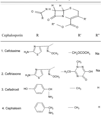
µg mL–1) were prepared by dissolving standard sodium cefotaxime (Alkem Lab. Ltd. Mumbai) or sodium ceftriaxone (Aristo Pharmaceuticals Ltd. Mumbai) or standard cefadroxil (Alkem Lab. Ltd. Mumbai) or standard cephalexin (Ranbaxy, India) in water. These compounds chosen to represent cephalosporins. They were prepared freshly, as required, by dissolving an appropriate amount of each antibiotic in water to provide a 1 µg mL–1 solution. The standard solution must be protected from contact with light. The structures of the cephalosporins studied are listed in table 1. Sodium hydroxide 0.1 mol L–1 aqueous solution, hydrochloric acid (Merck Limited, Mumbai) 1 mol L–1 aqueous solution, potassium iodate (S.D. fine – Chem Limited, Mumbai) 0.1 mol L–1 aqueous solution were used through out.
Taxim (Alkem Lab. Ltd. Mumbai), Monocef (Aristo Pharmaceuticals Ltd. Mumbai), Cefadrox (Aristo Pharmaceuticals Ltd. Mumbai) and Sporidex (Ranbaxy, India) were examined. A 0.05% solution of Variamine Blue (E-Merck Limited, Mumbai) in (75: 25) water-ethanol mix-ture was used and stored in an amber bottle.
Table 1. Structures of the cephalosporins studied.
Procedure
Analysis of injection solution
An appropriate amount of each antibiotic was dissolved in water so as to prepare 1mg mL–1 solution and then the recommended procedure was followed without modification. The presence of other substances caused no significant inter-ference with the determination of antibiotics.
Analysis of formulations
Weighed an amount of the sample equiva-lent to about 250 mg cephalosporin and was dis-solve in a sufficient amount of distilled water. The solution was shaken and filtered off through Whatman No. 1 filter paper and washed with water. The filtrate was diluted up to the mark with distilled water and made upto 100 mL. The general procedure was applied with no modifica-tion and the presence of excipients in the sample such as glucose, fructose, lactose, sucrose, calci-um or starch caused no interference in the deter-mination and process of separation was not required.
Results and discussion
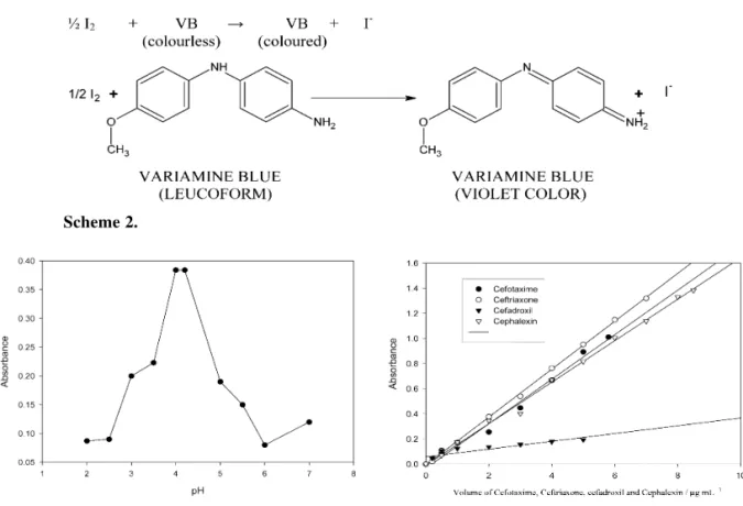
This method is based on the hydrolysis of ß-lactum ring of the analytes on heating with sodium hydroxide and the reaction of the hydrolysed product with potassium iodate in acidic medium to which liberates iodine. The liberated iodine oxidizes variamine blue to vio-let colored species of maximum absorption at 556 nm. The reagent blank had negligible absorbance at this wavelength. Beer’s law is obeyed in the range of 0.5-5.8 µg mL–1, 0.2-7.0 µg mL–1, 0.2-5.0µg mL–1 and 0.5-8.5 µg mL–1 for cefotaxime, ceftriaxone, cefadroxil and cephalexin respectively. Determination of cefo-taxime, ceftriaxone, cefadroxil, and cephalexin are represented in scheme 1. The absorption spectra of the oxidized form of variamine blue are presented in figure 1, the absorption spectra of colored species of variamine blue with cefo-taxime, ceftriaxone, cefadroxil and cephalexin against reagent blank in the range 300 – 800 nm are illustrated in figure 2. The maximum absorption is at 556 nm and reaction systems are presented in scheme 2.
Figure 1. Absorption spectra of the oxidized form of variamine blue with reagent blank.
Effect of sodium hydroxide concentration
The effect of sodium hydroxide concen-tration on the absorbance was studied with 2 µg mL– 1of cephalosporins. Volumes from 0.5 – 2.0 mL of 0.1 mol L–1NaOH solutions were exam-ined. The investigation showed that 1.0 – 1.5 mL of 0.1 mol L–1 NaOH solution gave maximum absorbance and 1.0 mL of 0.1 mol L–1 NaOH solution was chosen for the procedure.
Figure 2. Absorption spectra of colored species of variamine blue with cefotaxime, ceftriaxone, cefadroxil and cephalexin against reagent blank: c (variamine blue) = 0.05%, c (cephalosporins) = 2
Effect of temperature, time and pH
The effect of different variables such as temperature, time and pH on the coloration was studied with 2 µg mL–1of cephalosporins. It was observed that the optimum reaction temperature is 80°C – 90°C, lower or higher temperature gives inaccurate results, and the reaction time for
complete hydrolysis of ß-lactum ring was 10–15 min. Constant and maximum absorbance values were obtained in the pH = 4.0 – 4.2 hence the pH of the reaction system was maintained at pH = 4.0–4.2 throughout the study by adding 2 mL of 1 mol L–1sodium acetate solution. Effect of pH on color stability is presented in figure 3.
Calibration Graph
To the aqueous sample solutions containing 0.5 – 5.8 µg mL–1, 0.2-7.0 µg mL–1, 0.2-5.0µg mL–1 and 0.5-8.5 µg mL–1 of cefotaxime, ceftriaxone, cefadroxil and cephalexin respectively, the reagents were added as described above. Within the studied concentration ranges, the measured absorbance val-ues changed linearly. The correlation coefficients for cefotaxime, ceftriaxone, cefadroxil and cephalexin were found to be 0.9980, 0.9992, 0.9996 and 0.9991 respectively. The following regression coefficients were calculated: for cefotaxime α = 0.2239 b = 0.014, for ceftriaxone ? =0.1809 b = 0.0129, for cefadroxil α= 0.1622 b = 0.0065 and for cephalexin α= 0.1630 b = 0.0080. The follow-ing relative molar absorption coefficients were obtained: 1.07 x 10⁵Lmol-1cm–1, 1.02 x 10⁵Lmol -1cm–1, 2.68 x 10⁴Lmol-1cm–1and 5.90 x 10⁴Lmol
-1cm–1 for cefotaxime, ceftriaxone, cefadroxil and
cephalexin respectively. Calibration graphs for the determination of cefotaxime, ceftriaxone, cefadrox-il and cephalexin are presented in figure 4.
Scheme 2.
Figure 3. Effect of pH on color intensity for
cefo-taxime (2 µ g mL-1).
Effect of foreign substances
The influence of foreign substances was examined by the proposed method. The maximum tolerance (in mg) in the determination of 100 µg mL– 1cephalosporins was 54.0 for glucose, 35.5 for fructose, 56.5 for lactose, 32.4 for sucrose, 8.4 for starch and 22.0 for calcium. The tolerance limits of foreign substances are summarized in table 2.
Figure 4. Calibration graphs of cefotaxime,
ceftriax-one, cefadroxil and cephalexin:
c (cefotaxime) = 0.5 – 5.8 µg mL-1, c ( ceftriaxone) = 0.2 – 7.0 µg mL-1, c (cefadroxil) = 0.2 – 5.0 µg mL-1, c (cephalexin) = 0.5 – 8.5 µg mL-1.
Table 2. Maximum amount tolerance of excipients for the determination of cephalosporins.
Common excipient Tolerance limit (mg)
of high stability of the reaction system (4 hours). The proposed method was applied to the determi-nation of cephalosporins in pharmaceuticals.
Acknowledgements
Authors are thankful to the department of science and technology, New Delhi for financial support through FIST.
Received 10 March 2008 Accepted 20 May 2008
References
[1] W.O. Foye, ‘Principles of Medicinal Chemistry’, Lea and Febiger, Philadelpia, USA, (1975) p.726.
[2] J.K. Podlewski, P.A. Chwalibogowaka, Drugs of the Modern Therapy, Split Trading,(Warsaw) (1999) pp 45-47, 125-132 (in Polish).
[3] C.S.P. Sastry, T.E. Divakar, U.V. Prasad, Chem. Anal., (Warsaw) 32 (1987) 301.
[4] J. Yang, , G. Zhou, N. Jie, R. Han, C. Li, , J. Hu,. Anal. Chim. Acta, 325 (1996) 195.
[5] B. Ogoreve, V. Hudnik, S. Gomiseek, . Fresenius’ Z. Anal. Chem., 330 (1988) 59.
[6] A. Cieslak, E.Gwozdz, W. Holska, Chem. Anal.,(Warsaw) 36 (1991) 363.
[7] P.B. Issopoulos, J. Pharm.Biomed. Anal.,7 (1989) 619. [8] P.B. Issopoulos, Analyst, 113 (1988) 1083.
[9] P.B. Issopoulos, J. Pharm. Biomed. Anal, 6 (1988) 321. [10] S.A. Nabi, S.M. Eyad, A Nameh, I.H.H. Murad, Chem. Anal.,(Warsaw), 42 (1997) 881.
[11] M.M. Ayad, A.A. Shalaby, H.E. Abdellatef, H.M. Elsaid, J. Pharm.Biomed.Anal., 20 (1999) 557.
[12] A. Shalaby, J. Liq. Chromatrogr. Relat. Technol., 21(1998) 3161.
[13] S.S. Zarapkar, S.A. Shivalkar, A.A. Dhanvate, P.M. Deshpande, S.S. Kolte, Indian, Drugs, 32 (1995) 232. [14] M. Hefnawy, Y. El-Shabrawy, F. Belal, J. Pharm. Biomed. Anal., 21 (1999) 703.
[15] Y.M. Issa, A. S. Amin, Mikrochim. Acta, 124 (1996) 203. [16] H. Tan, R. Wang, S. Wang, W. Wei, S. Yao, Anal. Lett., 31(1998) 949.
[17] H. Fabre, M.D Blanchin, D. Lerner, B. Mandrou, Analyst, 110 (1985) 775.
[18] J.A. Murill, J.M. Lemus, L.F. Garci, Anal. Lett., 27 (1994) 1875.
[19] M. Azza, M. Ali, Bioelectrochem; Bioenerg., 33 (1994) 201. [20] P. Izquierdo, M.C Gutierrez, H.A. Gomez, B.D.Perez, Anal. Lett., 23 (1990) 487.
[21] H.T. Pan, P. Kumari, J. Lim, C.C. Lin, J. Pharm. Sci.,81 (1992) 663.
[22] K. Matsubayashi, H. Tachzawa, J.Chromatogr, 515 (1990) 547.
[23] Z.H. Earle, D.T. Hurst, M. Viney, J. Chem. Soc., (1969) C 2093.
[24] B.M. Frantz, J. Pharm. Sci., 65 (1976) 887.
[25] M.I.H. Halaleh, N. Rahman, R.M.A.Q. Jamhour, Chem. Anal., (Warsaw), 42 (1997) 265.
[26] F. Buhl, B.S. Sroka, Chem. Anal., (Warsaw) 48 (2003) 145.
Application
The proposed method has been successful-ly applied to the determination of studied antibi-otics in pharmaceuticals. Cefotaxime was deter-mined in 1 g- vials of taxim, ceftriaxone in 250 mg vials of monocef, cefadroxil in 250 mg tablets of cefodrox and cephalexin in 125 mg tablets of sporidex. Their contents in the investigated drug samples were calculated from the calibration curves mentioned above are found to be in a good agreement with the labelled amounts (table 3). The results, listed in the table 3 compared favorably with those from a reference method [26]. The pre-cision of the proposed method was evaluated by replicate analysis of 3 samples containing cephalosporins at different concentrations.
Conclusions
A simple method for the determination of ß -lactum antibiotics is described. The method is based on the reaction of iodate with the hydrolysed product of ß -lactum antibiotics which liberates iodine, subsequently oxidizes variamine blue into violet colored species, and measured at 556nm. The developed method does not involve any strin-gent reaction conditions and offers the advantages
Table 3. Determination of cefotaxime, ceftriaxone, cefadroxil, cephalexin and cephalexin in pharma-ceuticals preparations.
Pharmaceutical Declared Found in the
Quantity sample a (µg mL–1 )
(µg mL–1 ) ± S.D
TAXIM 1 2.00 1.984 ± 0.03
TAXIM 2 4.00 3.975 ± 0.02
TAXIM 3 5.50 5.454 ± 0.02
MONOCEF 1 1.00 0.986 ± 0.03
MONOCEF 2 3.00 2.992 ± 0.04
MONOCEF 3 5.00 4.954 ± 0.02
MONOCEF 4 7.00 6.966 ± 0.025
CEFADROX 1 2.00 1.994 ± 0.01
CEFADROX 2 4.00 3.986 ± 0.02
SPORIDEX 1 2.00 1.984 ± 0.015
SPORIDEX 2 4.00 3.896 ± 0.01
SPORIDEX 3 6.00 5.982 ± 0.04
SPORIDEX 4 8.00 7.940 ± 0.08