472
1 - Assistant professor 2 - Professor 3 - Assistant Professor 4 - Tutor
Work carried out in the Kasturba Medical College. Mangalore, India Correspondence address:
Dr. Mangala M. Pai, Department of Anatomy, Center for basic sciences, Kasturba medical college, Bejai, Mangalore- 575004, Karnataka, India.
E-mail: [email protected]
Mangala M. PAI1, Latha V. PRABHU2, PRAKASH3, Varsha NAYAK4
Braz J Cardiovasc Surg 2006; 21(4): 472-475 CASE REPORT
RBCCV 44205-857
Mal formação ílio-femoral
Iliofemoral arterial malformation
Abstract
During routine dissection, an Iliofemoral arterial malformation was noticed in a 65 year old male cadaver. The abdominal aorta was considerably laterally displaced and also bifurcated higher up. The common iliac artery divided one vertebral level higher and the femoral artery gave the profunda femoris artery about 1.2 cm below the inguinal ligament, which is considerably proximal to its usual level of origin. A brief review of literature and embryological basis of the anomalies are discussed.
Descriptors: Arteries, abnormalities. Abdominal aorta.
Iliac artery. Femoral artery.
Resumo
Durante uma dissecção de rotina realizada em um cadáver do sexo masculino com 65 anos de idade foi constatada malformação arterial iliofemoral. A aorta abdominal estava consideravelmente deslocada lateralmente e também bifurcava em nível mais alto. A artéria ilíaca comum dividia-se uma vértebra acima do nível normal e a artéria femoral dava origem à artéria femoral profunda aproximadamente l,2 cm abaixo do ligamento inguinal, o que é consideravelmente proximal ao seu nível normal. Aqui nós apresentamos uma breve revisão de literatura e base embriológica dessas anomalias.
Descritores: Artérias, anormalidades. Aorta abdominal.
Artéria ilíaca. Artéria femoral.
473 PAI, MM - Iliofemoral arterial malformation Braz J Cardiovasc Surg 2006; 21(4): 472-475
INTRODUCTION
The abdominal aorta begins at the median, aortic hiatus of the diaphragm descending anterior to the lumbar vertebrae to end at the fourth lumbar, a little left of the midline, dividing into right and left common iliac arteries. Gray’s reports that, in three-fourths of a large number of cases, the aorta bifurcated either upon the fourth lumbar vertebra, or upon the fibrocartilage between it and the fifth; the bifurcation being, in one case out of nine, below, and in one out of eleven, above this point [1]. The common iliac arteries diverge as they descend to divide into external and internal iliac arteries near the level of lumbosacral intervertebral disc, anterior to the sacroiliac joint. The point of division is subject to great variety [2].
The external iliac arteries are larger than the internal arteries and extend from the common iliac bifurcation to the mid-inguinal point, enter the thigh posterior to the inguinal ligament to become the femoral artery. Apart from small muscular branches to psoas, the artery has no branches until the inferior epigastric and deep circumflex iliac arise near to its termination. About 1 cm distal to the inguinal ligament the superficial circumflex iliac, superficial epigastric and superficial external pudendal take origin from the femoral artery. The arteria profunda femoris [PFA] is a large branch arising laterally from the femoral about 3.5 cm distal to the inguinal ligament. Its origin is sometimes medial or rarely posterior on the femoral artery; if the former, it may cross anterior to the femoral vein and then pass backwards around its medial side [1].
In 430 thighs, Quain found that the distance from the inguinal ligament of the origin of the profunda was between 2.5 and 5.1 cm in 68%; of these it was between 2.5 and 3.8 cm in 42.6%. This distance was less than 2.5 cm in 24.6% of the thighs and more than 5.1 cm in only 7.4% [3]. Here we describe a rare case of high origin of common iliac, external and internal iliac and PFA associated with other arterial variations.
CASE REPORT
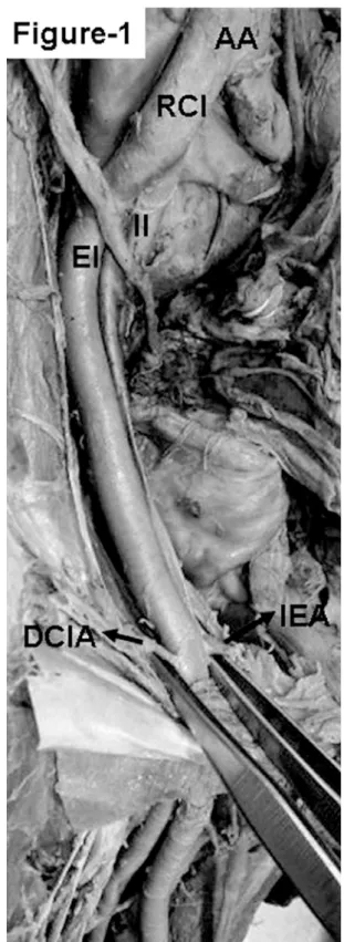
During routine dissection, an abnormal arterial pattern was noticed in a 65 year old male cadaver. The abdominal aorta was considerably laterally displaced and also it bifurcated higher up, at the level of L3 vertebrae into common iliac arteries. The right common iliac artery followed a similar pattern and terminated into external and internal iliac arteries at the level of L4 vertebrae. The right external iliac gave its usual branches and continued as the femoral artery beyond inguinal ligament. The PFA originated as the first branch of the femoral artery about 1.2 cm below the inguinal ligament. The lateral and medial circumflex femoral arteries branched from the PFA as usual but were very tortuous.
474
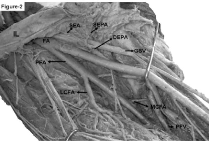
A thin twig originated from the femoral artery distal to the PFA from its anteromedial aspect which divided into three branches namely, the superficial epigastric, and the superficial and deep external pudendal arteries. The superficial circumflex was conspicuously absent. The other branches of femoral artery and PFA were normal. The femoral and the profunda femoral vein were as per the standard description.
DISCUSSION
Interesting anomalies in the origin and course of the principal arteries of the lower limbs have long received the attention of anatomists and surgeons [4]. They usually result from embryologic abnormalities of the arterial network of the lower limb. The embryologic development of the vascular plexus of the lower limb is based on an unusual selection of channels, some of which enlarge while the others contract and disappear, thereby establishing the final pattern. Before pelvic and femoral arteries appear as independent blood vessels from the rete pelvicum and rete femorale respectively, the blood flow destined for this territory makes an unexpected choice of source channels [5, 6].
In about 80 per cent of the cases the aorta bifurcated within 1.25 cm. above or below the level of the crest of the ilium; more frequently below than above [2]. In the present described case the aorta bifurcated at the level of L3 vertebrae which lies above the supracristal plane. In two-thirds of a large number of cases the division of common iliac arteries was between the last lumbar vertebra and the upper border of the sacrum; being above that point in one case out of eight, and below it in one case out of six [2]. The level of division of common iliac artery was at L4, one vertebral level higher up than its usual level of termination. The origin of PFA was also higher up, 1.2 cm below the inguinal ligament. Quian’s data state that the origin of the PFA 0-1.3 cm below the inguinal ligament, 13; 1.3-2.5 cm below, 86 cases [3].
This shows that only in 3% of the cases the PFA has a very high origin. The knowledge of the site of origin of the profunda helps in avoiding iatrogenic femoral arterio-venous fistula while performing femoral artery puncture [7]. Further the preferred site of femoral artery ligation is proximal to the origin of profunda femoris to ensure vascularity of lower extremity via the anastomoses of profunda femoris complex with the branches of popliteal artery.
Arteriography still remains the main line investigation in peripheral occlusive arterial diseases. Peripheral arteriograms are used for imaging of vascular malignancies, demonstration of the vascularity of malignancies and to identify diseases inherent to the arterial system [7]. The
REFERENCES
1. Annister LH, Berry MM, Collins P. Gray’s anatomy. In: Cardiovascular system. 38th ed. London: Churchill Livingstone;1995. p.1558-68.
Fig. 2 - The photograph of the right lower extremity. The femoral artery (FA) giving the profunda femoris artery (PFA) 1.2 cm below the inguinal ligament (IL). The other branches of FA; namely the superficial epigastric (SEA), superficial external pudendal (SEPA) and deep external pudendal artery (DEPA) are marked. The DEPA is going deep to the great saphenous vein (GSV). Lateral and medial circumflex femoral arteries are tortuous (LCFA and MCFA). The profunda femoris vein (PFV) is drain into the femoral vein at the usual level
femoral and PFA are used for various imaging techniques. In view of the expanding scope of interventional radiology this case report with multiple arterial variation gains importance.
The unexpected presence of the variant vessels can become a matter of great concern to orthopaedicians, urologists, gynaecologists and general surgeons, who may perform surgical procedures in this area.
475
2. Gray H. Anatomy of the human body. The common iliac arteries. Accessed on August 25, 2006. Disponível em: http:// www.bartleby.com/107/155.html
3. Bergman RA, Thompson SA, Afifi AK, Saadeh FA. Compendium of human anatomic variation: catalog, atlas and world literature. Baltimore:Urban & Schwarzenberg;1988.
4. Sarikcioglu L, Sindel M. Multiple vessel variation in the retropubic region. Folia Morphol. 2002;61(1):43-5.
5. Bilgic S, Sahin B. Rare arterial variation: a common trunk from the external iliac artery for the obturator, inferior epigastric and profunda femoris arteries. Surg Radiol Anat. 1997;19(1):45-7.
6. Sanudo JR, Roig M, Rodriguez A, Ferreira B, Domenech JM.
Rare origin of the obturator, inferior epigastric and medial circumflex femoral arteries from a common trunk. J Anat. 1993;183(Pt 1):161-3.
7. Dixit DP, Mehta LA, Kothari ML. Variations in the origin and course of profunda femoris. J Anat Soc India. 2001;50(1):6-7.
NOTA DO EDITOR
O presente artigo é anatômico e trata de uma anomalia rara. Estamos publicando na RBCCV em virtude do acesso femoro-ilíaco ser importante em casos de cirurgia cardíaca e na introdução de próteses endovasculares. Conhecer as variações anatômicas é importante para os cirurgiões cardiovasculares.