1984-1462/© 2015 Sociedade de Pediatria de São Paulo. Published by Elsevier Editora Ltda. All rights reserved.
REVISTA PAULISTA
DE PEDIATRIA
Rev Paul Pediatr. 2015;33(2):230–240www.rpped.com.br
REVIEW ARTICLE
Near-infrared spectroscopy as an auxiliary tool
in the study of child development
Suelen Rosa de Oliveira*, Ana Carolina Cabral de Paula Machado,
Débora Marques de Miranda, Flávio dos Santos Campos,
Cristina Oliveira Ribeiro, Lívia de Castro Magalhães,
Maria Cândida Ferrarez Bouzada
Universidade Federal de Minas Gerais (UFMG), Belo Horizonte, MG, Brazil
Received 28 April 2014; accepted 7 August 2014
KEYWORDS Child development; Spectroscopy near-infrared;
Hemodynamics
Abstract
Objective: To investigate the applicability of Near-Infrared Spectroscopy (NIRS) for corti-cal hemodynamic assessment tool as an aid in the study of child development.
Data source: Search was conducted in the PubMed and Lilacs databases using the fo-llowing keywords: ‘‘psychomotor performance/child development/growth and develo-pment/neurodevelopment/spectroscopy/near-infrared’’ and their equivalents in Por-tuguese and Spanish. The review was performed according to criteria established by Cochrane and search was limited to 2003 to 2013. English, Portuguese and Spanish were included in the search.
Data synthesis: Of the 484 articles, 19 were selected: 17 cross-sectional and two longitu-dinal studies, published in non-Brazilian journals. The analyzed articles were grouped in functional and non-functional studies of child development. Functional studies addressed the object processing, social skills development, language and cognitive development. Non-functional studies discussed the relationship between cerebral oxygen saturation and neurological outcomes, and the comparison between the cortical hemodynamic res-ponse of preterm and term newborns.
Conclusions: NIRS has become an increasingly feasible alternative and a potentially use-ful technique for studying functional activity of the infant brain.
© 2015 Sociedade de Pediatria de São Paulo. Published by Elsevier Editora Ltda. All rights reserved.
DOI of original article: http://dx.doi.org/10.1016/j.rpped.2015.03.003 *Corresponding author.
Espectroscopia de luz próxima ao infravermelho como ferramenta auxiliar no estudo do desenvolvimento infantil
Resumo
Objetivo: Investigar a aplicabilidade da espectroscopia de luz próxima ao infravermelho (NIRS) para avaliação da hemodinâmica cortical como ferramenta auxiliar no estudo do desenvolvimento infantil.
Fontes de dados: Revisão integrativa de literatura feita nas bases de dados PubMed e Li-lacs, a partir da combinação das palavras-chave: ‘‘psychomotor performance/child develo-pment/growth and development/neurodevelopment/NIRS/spectroscopy/near-infrared’’ e seus correspondentes em português e espanhol. A pesquisa seguiu protocolo adaptado dos critérios estabelecidos pela Cochrane e teve como limite temporal de 2003 a 2013. Foram incluídas publicações nos idiomas inglês, português e espanhol.
Síntese dos dados: Foram localizados 484 artigos, dos quais 19 foram selecionados, 17 transversais e dois longitudinais, todos publicados em periódicos estrangeiros. A análise dos artigos permitiu agrupá-los, quanto à sua abordagem, em estudos funcionais e es-tudos não funcionais do desenvolvimento infantil. Os eses-tudos funcionais abordaram o processamento de objetos eo desenvolvimento de habilidades sociais, da linguagem e cognitivo. Os estudos não funcionais discutiram a relação entre a saturação de oxigênio cerebral e o desfecho neurológico e a comparação entre a resposta hemodinâmica corti-cal de recém-nascidos prematuros e a termo.
Conclusões: A NIRS se torna, cada vez mais, uma opção viável e uma técnica potencial-mente útil para estudos de atividade funcional do cérebro infantil.
© 2015 Sociedade de Pediatria de São Paulo. Publicado por Elsevier Editora Ltda. Todos os direitos reservados.
PALAVRAS-CHAVE Desenvolvimento infantil;
Espectroscopia de luz próxima ao infravermelho; Processos hemodinâmicos
Introduction
Near Infrared Spectroscopy (NIRS) represents a break-through in techniques used for brain function assessment. This tool has been considered promising for the evaluation of children’s cerebral cortex function, contributing to the increase in knowledge related to neurodevelopment and cognition in children.1-4
The action mechanism of spectroscopy is based on the fact that neural activity is accompanied by changes in blood oxygenation, cerebral blood flow and volume. Thus, different wavelengths within the near infrared spectrum (780-2,500 nm) are used, capturing different characteris-tics of light absorption and dispersion in biological tissue. The light originates from a source, migrates through the tissue and is captured by a detector. Considering that tis-sue dispersion is a constant, the attenuation of the amount of light captured by the detector can be calculated, pro-viding an indirect measure of activity in this tissue. That is, variations in the concentration of oxyhemoglobin
(HbO2), deoxyhemoglobin (HHb) and total
hemoglo-bin (HBT) are calculated, which allows a quantitative and qualitative assessment of hemodynamics and neuronal activation.5,6
Compared with other neuroimaging techniques, NIRS has the advantage of being a noninvasive, portable, quiet, rel-atively low-cost and safer method, less sensitive to motion artifacts, as it does not require a tracer or carrier sub-stance to be injected into the blood stream and does not
require irradiation.1 Additionally, it allows children to move
on their caregiver’s lap, where they remain more comfort-able and, therefore, more likely to complete the examina-tion (Fig. 1). Another advantage is that as newborns and infants tend to have fine hair and their skulls are thin and small, the ratio of loss of signal due to dispersion is less than that for participating adults.6
Although the assessment of cerebral hemodynamics seems to be advantageous, it is important to identify how the methodology has been used and in what kind of research in child-related areas. The aim of this study was to carry out an integrative review of the literature published in indexed journals in the period of 2003-2013, on the use of NIRS to assess cerebral hemodynamics as an auxiliary tool in the study of normal childhood development.
Method
An integrative review was carried out following an adapta-tion of the Cochrane criteria, which included: definiadapta-tion of the study databases, definition of target audience, time limit, definition of keywords, inclusion criteria for the selection of studies, study quality assessment, synthesis and interpretation of results.
232 Oliveira SR et al.
Near-Infrared” and their equivalents in Portuguese and Spanish.
The inclusion criteria for articles were: type of study (cohort, case-control, cross-sectional, randomized trials), target audience (children 0-7 years), language of publication (English, Portuguese and Spanish), available as full-text in digital media and temporal limits (June 2003 to June 2013).
The titles were selected by reading the abstracts in order to determine whether they addressed the subject of this research and if they met the inclusion criteria. The next step consisted in recovering the articles and read them in full. This step was performed in two stages: first, two researchers read and selected the articles inde-pendently and, second, the information was cross-checked and the articles were selected in agreement.
The next step was to identify the central ideas of each study, which were then grouped according to recurring themes, into thematic categories. These categories were analyzed, allowing the articulation among the assessed topics and the development of the knowledge synthesis.
Results
Based on the combination of the previously mentioned descriptors and databases, 484 articles were located. After applying the inclusion criteria, 19 articles were
selected, of which 17 were cross-sectional and two longi-tudinal studies.
The difference between the number of located publica-tions and the number of selected publicapublica-tions is due to the fact that most of the identified studies consisted of review articles, with samples at age ranges above the one that was established for this study, studies carried out in animals, studies that were limited to exploring methodological aspects of the spectroscopy technique, studies published prior to the period established in the inclusion criteria, articles in other languages and not available in digital form.
All selected articles were published in 12 international journals, a heterogeneous distribution, highlighting the predominance of publications in the NeuroImage journal (26.3%). As for the distribution of articles by year of publi-cation, there were four (21%) in 2012 and three (15.8%) each year in 2011, 2010, 2009 and 2007. The others were published in 2006 (5.3 %) and 2008 (10.5%).
Study analysis allowed us to group them concerning the use or not of stimulation paradigms to investigate cortical activation. Studies evaluating the neural activation during performance of stimulation paradigms were called func-tional studies. Funcfunc-tional studies addressed four topics related to child development, namely: object processing (Table 1), development of social skills and cognitive devel-opment (Table 2) and language develdevel-opment (Table 3). Non-functional studies were those that did not use specific stimulation paradigms, assessing only the spontaneous fluc-tuations of cortical hemodynamics (Table 4).
Regarding the methodology, all studies used multi-chan-nel, continuous-wave NIR equipment with two wave-lengths and most followed the standardization of the international 10-20 system of electroencephalography to locate optodes, with reported sample loss rate between 3% and 80%. The justifications for the losses, as specified by the authors, were: motion artifacts, obstruction by the hair, failure in the experimental protocol, crying and agi-tation, difficulty obtaining optical signal and intolerance to the equipment.
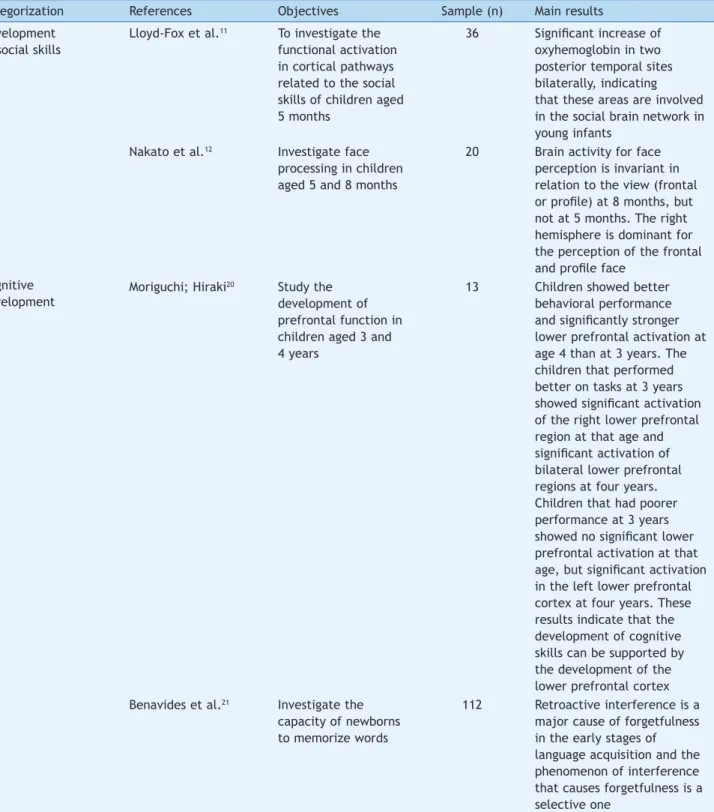
The studies on object processing were developed with children aged two to 12 months old and aimed to investi-gate cortical functional organization related to the visual perception of objects (color, shape and movement). The main results showed that, between two and three months old, it is already possible to identify functionally differen-tiated cortical regions for visual perception.7-10
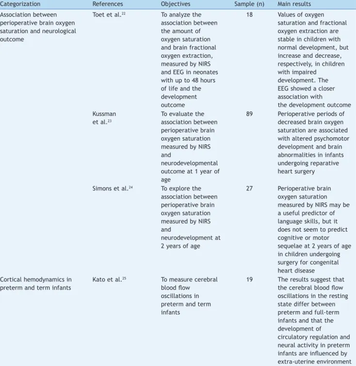
About the development of social skills, the studies assessed functional activation in cortical pathways related to social skills of children between five and eight months of age, using images of the human face in different plans and different facial expressions. The results indicated that, at five months of age, there is already a specialized area of
the temporal cortex activated by social stimuli.11,12
Furthermore, right hemisphere dominance was verified in the frontal plane perception and facial profile,12 whereas
cortical activation occurs bilaterally for the perception of dynamic social stimuli.11
Studies on language development were carried out with children aged two months to four years, with the aim to analyze brain functional processing of language, mainly related to phonetic processing,13-15 prosody,16,17 the
lateral-Figure 1 Nine-month-old child undergoing near-infrared spectroscopy (NIRS) on the mother’s lap.
ization of speech13,14,16,18 and the influence of the speaker’s
familiarity in speech perception.19 The results were
consis-tent with those obtained through other neurophy siological methods, emphasizing that: responses to specific language phonemic contrasts are present at six months of age; how-ever, these become consistent and lateralized only after 12 months.13 Differences in proso dic patterns were
discrimi-nated by infants between one and nine days of life17 and
functional specialization of the right hemisphere for proso-dy processing is present, with a response pattern similar to that of the adult, at four years of age.16
The approach of cognitive development involved the study of cognitive flexibility in children aged three and four years, and memory in newborns. The main findings indicat-ed that the development of cognitive flexibility skill is related to the development of the inferior prefrontal cor-tex and suggest that children develop prefrontal
activa-tions between the ages of three and four years.20 About
neonatal memory, a study investigated the capacity of new-borns to memorize words, focusing on the causes of
forget-fulness in early childhood, testing the capacity of neonates to recognize words after a period of familiarization. It was observed that newborns are already capable of memorizing words hours after birth.21
The non-functional studies discussed the association between cerebral oxygen saturation and neurological
out-come;22-24 and the comparison between the cortical
hemo-dynamic response of preterm and full-term infants.25 All
used continuous wave equipment, ranging from two to 24 channels and only one used the international 10-20 system for positioning of the optodes with sample loss ranging from 14% to 56%. The reported causes for these losses were: neonatal death, diagnosed syndrome, follow-up losses and failure in the experimental protocol.
Children younger than 12 months were evaluated and the main results suggest that brain oxygen saturation values and oxygen extraction fraction may be related to neurolog-ical outcomes22-24 and that the oscillations in cerebral blood
flow in the resting state differ between preterm and full-term infants.25
Table 1 Characteristics of functional studies of cortical hemodynamics using NIRS to assess processing of objects.
Categorization References Objectives Sample (n) Main results
Objects processing Watanabe et al.7 To investigate the
functional cortical development through hemodynamic responses measured by NIRS during the presentation of
speciic visual stimuli
in children aged 2 months
40 Cortical functional regions appear at 2-3 months for visual perception
Wilcox et al.8 To investigate the
applicability of NIRS in assessment of brain objects processing in children aged 6.5 months
35 NIRS is sensitive enough to assess the neural basis of object processing in infants
Wilcox et al.9 To evaluate the
functional
organization of visual areas of objects processing in children aged 3-5 months and 11-12 months
111 The child’s cortex is functionally specialized for processing objects at the
beginning of the irst year, but
the activation patterns change between 3 and 12 months,
which may relect a functional
reorganization of the immature cortex or be related to differences in age-related cognitive processes Watanabe et al.10 To study the visual
perception and to understand the functional cortical organization in children aged 3 months
234 Oliveira SR et al.
Table 2 Characteristics of functional studies of cortical hemodynamics using NIRS to assess the development of social skills and cognitive development.
Categorization References Objectives Sample (n) Main results
Development of social skills
Lloyd-Fox et al.11 To investigate the
functional activation in cortical pathways related to the social skills of children aged 5 months
36 Signiicant increase of
oxyhemoglobin in two posterior temporal sites bilaterally, indicating that these areas are involved in the social brain network in young infants
Nakato et al.12 Investigate face
processing in children aged 5 and 8 months
20 Brain activity for face perception is invariant in relation to the view (frontal
or proile) at 8 months, but
not at 5 months. The right hemisphere is dominant for the perception of the frontal
and proile face
Cognitive development
Moriguchi; Hiraki20 Study the
development of prefrontal function in children aged 3 and 4 years
13 Children showed better behavioral performance
and signiicantly stronger
lower prefrontal activation at age 4 than at 3 years. The children that performed better on tasks at 3 years
showed signiicant activation
of the right lower prefrontal region at that age and
signiicant activation of
bilateral lower prefrontal regions at four years. Children that had poorer performance at 3 years
showed no signiicant lower
prefrontal activation at that
age, but signiicant activation
in the left lower prefrontal cortex at four years. These results indicate that the development of cognitive skills can be supported by the development of the lower prefrontal cortex Benavides et al.21 Investigate the
capacity of newborns to memorize words
112 Retroactive interference is a major cause of forgetfulness in the early stages of language acquisition and the phenomenon of interference that causes forgetfulness is a selective one
Discussion
Before the advent of neuroimaging techniques, the associ-ation between brain regions and neurodevelopment was obtained primarily by clinical neuropsychological investiga-tions of patients with brain injury and post-mortem exam-inations. With the advancement of this technology, it
became possible to investigate not only the brain areas involved in a particular skill, but also the neural circuits involved in a particular function.26
fluctua-235
Table 3 Characteristics of functional studies of cortical hemodynamics using NIRS to assess language development.
Categorization References Objectives Sample (n) Main results Development of language Minagawa-Kawai et al.13 To investigate the development
of the neural adjustment process to phonemic contrasts,
speciic of language in children
aged 3-28 months
57 Phonemic-speciic brain response was found
from 6-7 months of age, but there was no
lateralization. A consistent, phonemic-speciic
and lateralized response was only observed after 12 months
Minagawa-Kawai et al.14 To investigate the nature of the
functional brain language processing at 4 months of age
12 At 4 months, the human brain is more responsive to the native language and speech processing is based on an interaction between generic audio systems and learning
mechanisms Wartenburger et al.16 Determine brain activations
correlated with the perception and processing of prosody at 4 years
51 The results showed that the speciic language
processes in children depend on inter-hemispheric specialization, with left hemispheric dominance for the segmental information processing (phonological) and right hemispheric dominance for
suprasegmental processing (prosodic). At the age of 4 years, the functional specialization of the right hemisphere for prosody
processing is present, with a response pattern similar to the adult
Bortfeld et al.18 Evaluate the usefulness of NIRS
as a technique to study infant speech processing in children aged 6 and 9 months
21 Results showed signiicant hemodynamic
changes in the left temporal cortex in response to audiovisual stimulus when compared to visual stimulus alone, as well as relative changes in all stimulus conditions in homologous regions of the right cortex in the same children. The results are consistent with those obtained through other
236
Oliveira SR et al.
Table 3 (Continued).
Categorization References Objectives Sample (n) Main results
Petitto et al.15 To explore, with NIRS, early
phonetic processing in monolingual and bilingual children aged 2 to 16 months
61 Monolingual and bilingual children showed activation in the same language areas classically observed in adults, including the left superior temporal gyrus and the left inferior frontal cortex, with an intriguing temporal difference of development: activation of the left superior temporal gyrus was observed earlier and remained with stable activity over time, while the left inferior frontal cortex showed greater increase in neural activation in older babies, corresponding to the precise age at which
babies reach the irst universal milestone of
language acquisition. Bilingual babies maintained linguistic sensitivity open for a longer time
Saito et al.17 To assess, using NIRS, how the
frontal lobe of newborns responds to changes in prosody
20 Newborns between 1 and 9 days can already discriminate differences in prosodic patterns
Naoi et al.19 To analyze the hemodynamic
brain responses to speech directed to the child, using NIRS in children aged between 4 and 13 months
Table 4 Characteristics of non-functional studies of cortical hemodynamics using NIRS.
Categorization References Objectives Sample (n) Main results
Association between perioperative brain oxygen saturation and neurological outcome
Toet et al.22 To analyze the
association between the amount of oxygen saturation and brain fractional oxygen extraction, measured by NIRS and EEG in neonates with up to 48 hours of life and the development outcome
18 Values of oxygen
saturation and fractional oxygen extraction are stable in children with normal development, but increase and decrease, respectively, in children with impaired
development. The EEG showed a closer association with
the development outcome Kussman
et al.23
To evaluate the association between perioperative brain oxygen saturation measured by NIRS and
neurodevelopmental outcome at 1 year of age
89 Perioperative periods of decreased brain oxygen saturation are associated with altered psychomotor development and brain abnormalities in infants undergoing reparative heart surgery
Simons et al.24 To explore the
association between perioperative brain oxygen saturation measured by NIRS and
neurodevelopment at 2 years of age
27 Perioperative brain oxygen saturation measured by NIRS may be a useful predictor of language skills, but it does not seem to predict cognitive or motor sequelae at 2 years of age in children undergoing surgery for congenital heart disease
Cortical hemodynamics in preterm and term infants
Kato et al.25 To measure cerebral
blood low
oscillations in preterm and term infants
19 The results suggest that
the cerebral blood low
oscillations in the resting state differ between preterm and full-term infants and that the development of
circulatory regulation and neural activity in preterm
infants are inluenced by
extra-uterine environment
tions (electroencephalogram, EEG and amplitude-integrat-ed electroencephalography, aEEG) that occur in neural activity; while others, such as functional magnetic reso-nance imaging (fMRI) and functional near-infrared spectros-copy (fNIR spectrosspectros-copy or fNIRS), measure local changes in cerebral hemodynamic activity, which can be used to make inferences about the underlying neural activity.1
Many of these techniques, which are well-established for use in adults, have restrictions for use in children. Among all brain imaging techniques, the fMRI is considered the
“gold standard” for noninvasive functional mapping of the
human brain.26 This technique stands out from the others
238 Oliveira SR et al.
limited to the study of children while sleeping, sedated or very young ones.
For many years, the first choice for neuroimaging studies in children while awake was EEG, a technique with high temporal resolution, but relatively low spatial resolution.6
In this field, continuous brain monitoring through aEEG has been used in neonates to assess real-time brain function and for long periods, allowing a better classification of the encephalopathy severity, early detection of subclinical
sei-zures and the monitoring of treatment response.27
Abnormalities found in the aEEG early in life have strong predictive values of abnormal results at one year of age.28
Compared with the aforementioned techniques, NIRS offers a new direction for the study of child development, as it has the following advantages over these methods: bet-ter temporal resolution, betbet-ter safety level, it is silent and less sensitive to motion artifacts, requiring less rigid stabi-lization of the head and body without the need for a labeled or carrier substance to be injected into the bloodstream.2
The most commonly used and simpler NIRS method involves measuring the intensity of diffusely reflected light with sources that emit light continuously. Instruments that acquire such measurements are referred to as continu-ous-wave systems.29 All studies discussed here resorted to
this method, which, although it does not provide quantita-tive measurements of absolute concentrations of different types of hemoglobin, provides estimates of changes in their levels from a baseline value, thus reflecting variations in tissue oxygen use.5
The use of multiple channels with different combinations of sources and detectors has been described in the litera-ture in recent years. Until the early 1990s, almost all NIRS systems employed one or two measurement channels, but over time, the number of channels in the available systems
has increased, improving spatial resolution.29 Of all the
reviewed studies, most of them used multichannel acquisi-tion systems, allowing greater coverage of the region of interest.
The advantages of increasing the number of channels are clear. However, this results in the inevitable increase in weight and size of the device that maintains the optodes positioning in the scalp. This may explain the greater pro-portion of optical data loss due to excessive movement arti-facts. The losses reported in the literature vary from 12.5 to 70%,6 similar to the losses found in this review: 3-80% in
functional studies and 14-56% in non-functional studies. A possible explanation for the greater loss in functional stud-ies would be the use of more complex data collection pro-tocols, employing a greater numbers of channels.
Although NIRS has been used for more than 35 years, it was not applied to children up until the mid-1990s. Investigations in this field have expanded rapidly, providing evidence that NIRS can be used to collect information about hemodynamics correlated to the neural activity in children from an early age, using tasks that assess cognitive skills, language acquisition, visual perception, social cogni-tion and other funccogni-tional aspects of the brain during
child-hood.30 Moreover, non-functional NIRS studies have
demon-strated its potential as a prognostic tool, based on the non-invasive monitoring of cerebral hemodynamics and oxygenation.22,31-33
Based on the publications analyzed, it was possible to recognize advances in the use of NIR spectroscopy in the study of child development; however, some methodological obstacles inherent in the use of this technology must be considered. Consistent with the results of other studies reviewing NIRS, it was observed that there is great variabil-ity in the methods used for data acquisition and analysis. The use of different wavelength combinations and different separations between sources and detectors could affect the captured response. In the 19 studies included in the review, we identified seven different wavelength combina-tions used. The separation between optrodes was more uni-form, being two or three centimeters, which is adequate for the pediatric population.
Another question that should be addressed concerns the large differences in sample sizes of the studies (between 12 and 112 subjects) and significant losses due to the qual-ity of the generated signal. While there is a good signal-to-noise ratio for optical imaging, the variation that is inher-ent in infant behavior requires that there be at least ten babies in each age group.34 The reviewed studies analyzed
samples with 12 or more children. However, one study that proposed to evaluate the association between the amount of oxygen saturation, the fractional cerebral tissue oxygen extraction and the result of development,22 with an initial
sample of 18 children, lost nine of them due to death, ending up with a final sample of nine children and found developmental change in only one. Despite the importance of studies like this to extend the current body of knowl-edge that emphasize the validity of NIRS to study infant brain development, the interpretation of results obtained with small sample groups should be very cautious and their projection to other contexts or populations becomes impaired.
Moreover, the difficulty of determining the locations for positioning of optodes using external markers, especially in children, should be considered. A current trend in NIRS studies, and which was identified in 13 of the 19 articles included in this review, is the use of the international 10-20 system for electroencephalography for the positioning of optodes.
It is also necessary to define the number of experiments required to obtain a significant response. Finding the bal-ance between the number of repetitions required to cap-ture a reliable response without making the test long and stressful, seems to be a delicate aspect of research using NIRS in children. This is due to the fact that inadequate signals and motion artifacts often make it necessary to repeat the tests. The studies reviewed here showed not only a large variability in the number of experiments, as well as in the duration of tests, reinforcing the premise that there is yet no consensus in the literature about this aspect. Some authors emphasize that the use of long exper-iments could lead to a diminished response over time, as the body adapts to the stimulus that is repeated many times. Additionally, the study designs in blocks, with long periods of stimulation and rest, has potential risk for false positive/negative changes in the signal due to any low fre-quency fluctuation at baseline or motion artifacts.35 On the
sponta-neous changes in cerebral blood volume are common, for instance, the so-called Mayer waves or slow vasomotion at 0.1 Hz. Such changes are about the same size as the func-tional activations and, therefore, may be misidentified. Repetition allows making averages of time series, reducing the influence of spontaneous changes, as they are not syn-chronized with the stimuli.36
It is worth noting that this review aimed to emphasize NIRS as an auxiliary tool in the study of normal childhood development. For this reason, no studies that addressed developmental disorders were included, due to high speci-ficity of each of them. However, it is necessary to stress that the current literature on NIRS has also emphasized the adequacy of this technology for the study of child develop-ment disorders and there is a growing number of publica-tions in this area. In this context, studies on Autism
Spectrum Disorder,37,38 Attention Deficit Hyperactivity
Disorder39 Cerebral Palsy40-42 and Down Syndrome43 have
been highlighted.
In conclusion, NIRS is increasingly becoming a practical alternative and potentially useful technique for studies of functional activity of the infant brain. The development of equipment more adequate for use in children has increased, so that the results obtained when using NIRS technology are more reliable. It is worth mentioning that the spatial loca-tion of signals will never achieve the accuracy of fMRI, but in conjunction with other techniques, such as EEG, NIRS is emerging as an important non-invasive tool for the study of the developing brain.
Funding
This article is part of the research “Near Infrared Spectroscopy in the Prediction of Neurodevelopment of Preterm Infants at 4 and 8 months Corrected Age,” which received financial support from Fundação de Amparo à Pesquisa de Minas Gerais – FAPEMIG. Process number: APQ-01182-13.
Conlicts of interest
The authors declare no conflicts of interest.
References
1. Gervain J, Mehler J, Werker JF, Nelson CA, Csibra G, Lloyd-Fox S, et al. Near-infrared spectroscopy: a report from the McDonnell infant methodology consortium. Dev Cogn Neurosci. 2011;1:22-46.
2. Nagamitsu S, Yamashita Y, Tanaka H, Matsuishi T. Functional near-infrared spectroscopy studies in children. Biopsychosoc Med. 2012;6:1-7.
3. Kawakubo Y, Kono T, Takizawa R, Kuwabara H, Ishii-Takahashi A, Kasai K. Developmental changes of prefrontal activation in humans: a near-infrared spectroscopy study of preschool children and adults. PLoS One. 2011;6:e25944.
4. Franceschini MA, Thaker S, Themelis G, Krishnamoorthy KK, Bortfeld H, Diamond SG, et al. Assessment of infant brain development with frequency-domain near-infrared spectroscopy. Pediatr Res. 2007;61:546-51.
5. Lima A, Bakker J. Near-infrared spectroscopy for monitoring peripheral tissue perfusion in critically ill patients. Rev Bras Ter Intensiva. 2011;23:341-51.
6. Lloyd-Fox S, Blasi A, Elwell CE. Illuminating the developing brain: the past, present and future of functional near infrared spectroscopy. Neurosci Biobehav Rev. 2010;34:269-84.
7. Watanabe H, Homae F, Taga G. General to speciic development
of functional activation in the cerebral cortexes of 2- to 3-month-old infants. Neuroimage. 2010;50:1536-44.
8. Wilcox T, Bortfeld H, Woods R, Wruck E, Boas DA. Hemodynamic response to featural changes in the occipital and inferior temporal cortex in infants: a preliminary methodological exploration. Dev Sci. 2008;11:361-70.
9. Wilcox T, Stubbs S, Hirshkowitz A, Boas DA. Functional activation of the infant cortex during object processing. Neuroimage. 2012;62:1833-40.
10. Watanabe H, Homae F, Nakano T, Taga G. Functional activation in diverse regions of the developing brain of human infants. Neuroimage. 2008;43:346-57.
11. Lloyd-Fox S, Blasi A, Volein A, Everdell N, Elwell CE, Johnson MH. Social perception in infancy: a near infrared spectroscopy study. Child Development. 2009;80:986-99.
12. Nakato E, Otsuka Y, Kanazawa S, Yamaguchi MK, Watanabe S,
Kakigi R. When do infants differentiate proile face from frontal
face? A near-infrared spectroscopic study. Hum Brain Mapp. 2009;30:462-72.
13. Minagawa-Kawai Y, Mori K, Naoi N, Kojima S. Neural attunement processes in infants during the acquisition of a
language-speciic phonemic contrast. J Neurosci. 2007;27:315-21.
14. Minagawa-Kawai Y, Van der Lely H, Ramus F, Sato Y, Mazuka R, Dupoux E. Optical brain imaging reveals general auditory and
language-speciic processing in early infant development.
Cereb Cortex. 2011;21:254-61.
15. Petitto LA, Berens MS, Kovelman I, Dubins MH, Jasinska K, The Shalinsky M. Perceptual Wedge hypothesis as the basis for bilingual babies’ phonetic processing advantage: new insights from fNIRS brain imaging. Brain Lang. 2012;121:130-43. 16. Wartenburger I, Steinbrink J, Telkemeyer S, Friedrich M,
Friederici AD, Obrig H. The processing of prosody: evidence of interhemispheric specialization at the age of four. Neuroimage. 2007;34:416-25.
17. Saito Y, Kondo T, Aoyama S, Fukumoto R, Konishi N, Nakamura K, et al. The function of the frontal lobe in neonates for response to a prosodic voice. Early Hum Dev. 2007;83:225-30. 18. Bortfeld H, Fava E, Boas DA. Identifying cortical lateralization
of speech processing in infants using near-infrared spectroscopy. Dev Neuropsychol. 2009;34:52-65.
19. Naoi N, Minagawa-Kawai Y, Kobayashi A, Takeuchi K, Nakamura K, Yamamoto J, et al. Cerebral responses to infant-directed speech and the effect of talker familiarity. Neuroimage. 2012;59:1735-44.
20. Moriguchi Y, Hirakic K. Longitudinal development of pre-frontal function during early childhood. Dev Cogn Neurosci. 2011;1:153-62.
21. Benavides-Varela S, Gómez DM, Macagno F, Bion RA, Peretz I, et al. Memory in the Neonate brain. PLoS ONE. 2011;6:e27497. 22. Toet MC, Lemmers PM, Van Schelvenb LJ, Van Bel F. Cerebral
oxygenation and electrical activity after birth asphyxia: the irrelation to outcome. Pediatrics. 2006;117:333-9.
23. Kussman BD, Wypij D, Laussen PC, Soul JS, Bellinger DC, DiNardo JA, et al. Relationship of intraoperative cerebral oxygen saturation to neurodevelopmental outcome and brain MRI at one year of age in infants undergoing biventricular repair. Circulation. 2010;122:245-54.
240 Oliveira SR et al.
25. Kato I, Kusaka T, Nishida T, Koyano K, Nakamura S, Nakamura M,
et al. Extrauterine environment inluences spontaneous
low-frequency oscillations in the preterm brain. Brain Dev. 2013;35:17-25.
26. Cutini S, Moro SB, Bisconti S. Functional near infrared optical imaging in cognitive neuroscience: an introductory review. J Near Infrared Spectrosc. 2012;20:75-92.
27. Toso PA, González AJ, Pérez ME, Kattan J, Fabres JG, Tapia JL, et al. Clinical utility of early amplitude integrated EEG in monitoring term newborns at risk of neurological injury. J Pediatr (Rio J). 2014;90:143-8.
28. Laptook A. Amplitude integrated electroencephalogram (aEEG): has it found its niche in neonatal intensive care unit? J Pediatr (Rio J). 2014;90:102-4.
29. Minagawa-Kawai Y, Mori K, Hebden JC, Dupoux E. Optical imaging of infants’ neurocognitive development: recent advances and perspectives. Dev Neurobiol. 2008;68:712-28. 30. Aslin RN, Mehler J. Near-infrared spectroscopy for functional
studies of brain activity in human infants: promise, prospects, and challenges. J Biomed Opt. 2005;10:11009.
31. Nicklin SE, Hassan IA, Wickramasinghe YA, Spencer SA. The light still shines, but not that brightly? The current status of perinatal near infrared spectroscopy. Arch Dis Child Fetal Neonatal Ed. 2003;88:F263-8.
32. Meek JH, Elwell CE, McCormick DC, Edwards A, Townsend J, Stewart A, et al. Abnormal cerebral haemodynamics in perinatally asphyxiated neonates related to outcome. Arch Dis Child Fetal Neonatal Ed. 1999;81:F110-5.
33. Van Bel FV, Dorrepaal CA, Benders MJ, Zeeuwe PE, Van de Bor MV, Berger HM. Changes in cerebral hemodynamics and
oxy-genation in the irst 24 hours after birth asphyxia. Pediatrics.
1993;92:365-72.
34. Hespos SJ, Ferry AL, Cannistraci CJ, Gore J, Park S. Using optical imaging to investigate functional cortical activity inhuman infants. In: Roe AW, editor. Imaging the brain with optical methods. New York: Springer; 2010. p. 159-76.
35. Taga G, Asakawa K, Maki A, Konishi Y, Koizumi H. Brain imaging in awake infants by near-infrared optical topography. PNAS. 2003;100:10722-7.
36. Wolf M, Greisen G. Advances in near-infrared spectroscopy to study the brain of the preterm and term neonate. Clin Perinatol. 2009;36:807-34.
37. Iwanaga R, Tanaka G, Nakane H, Honda S, Imamura A, Ozawa H. Usefulness of near-infrared spectroscopy to detect brain dysfunction in children with autism spectrum disorder when inferring the mental state of others. Psychiatry Clin Neurosci. 2013;67:203-9.
38. Kikuchi M, Yoshimura Y, Shitamichi K, Ueno S, Hiraishi H, Munesue T, et al. Anterior prefrontal hemodynamic connectivity in conscious 3- to 7-year-old children with typical development and autism spectrum disorder. PLoS One. 2013;8:e56087.
39. Tsujimoto S, Yasumura A, Yamashita Y, Torii M, Kaga M, Inagaki M. Increased prefrontal oxygenation related to
distractor-resistant working memory in children with attention-deicit/
hyperactivity disorder (ADHD). Child Psychiatry Hum Dev. 2013;44:678-88.
40. Chaudhary U, Hall M, Gonzalez J, Elbaum L, Bloyer M, Godavarty A. Motor response investigation in individuals with cerebral palsy using near infrared spectroscopy: pilot study. Appl Opt. 2014;53:503-10.
41. Kurz MJ, Wilson TW, Arpin DJ. An fNIRS exploratory investiga-tion of the cortical activity during gait in children with spastic diplegic cerebral palsy. Brain Dev. 2014. Epub 2014 Feb 5. 42. Tian F, Delgado MR, Dhamne SC, Khan B, Alexandrakis G,
Romero MI, et al. Quantiication of functional near infrared
spectroscopy to assess cortical reorganization in children with cerebral palsy. Opt Express. 2010;18:25973-86.