PERCEPTION OF THE BED PARTNER AND THE INDIVIDUAL
SUFFERING FROM SNORING/OSAS BEFORE AND AFTER
SPEECH THERAPY
A percepção do acompanhante e do indivíduo
com RONCO/SAOS antes e após fonoterapia
Erika Matsumura (1), Gislene A. Barros Rodrigues Tonisi (2), Ana Lúcia Cruz Vecina (3),
Lia Bartieri Inocêncio (4), Kátia C. Carmello Guimarães (5), Nair Kátia Nemr (6)
(1) University of São Paulo, SP, Brazil.
(2) Pontiical Catholic University of São Paulo, SP, Brazil. (3) Instituto Sedes Sapientiae, São Paulo, SP, Brazil. (4) CEV, São Paulo, SP, Brazil.
(5) University of São Paulo, SP, Brazil.
(6) Faculty of Medicine of the University of São Paulo, SP,
Brazil.
Conlict of interest: non existent
(AASM) as the presence of recurring episodes of partial or total obstruction of the upper airway during sleep and is manifested as partial reduc
-tions (hypopneas) or complete cessa-tions (apneas) of the airlow. OSAS is considered a chronic and progressive disease, associated with excessive daytime sleepiness, cardiovascular disease, and snoring 1,2. The presence of obstructive sleep apnea (OSA) is often associated with snoring, which is characterized as a fricative noise in the soft tissues of the upper airways 3. This symptom alone can be described in full polysomnography as isolated or primary snoring, in which snoring occurs without
INTRODUCTION
Obstructive sleep apnea syndrome (OSAS) is deined by the American Academy of Sleep Medicine
ABSTRACT
Purposes: the purpose of this study was to evaluate the perception of bed partners,
auto-evaluate individuals with snoring/obstructive sleep apnea syndrome, collect cervical and abdominal circumferences before and after speech therapy, and conduct a myofunctional evaluation to associate the results with the severity of sleep disorder. Methods: eleven patients between ages 25 and 75
years, of both genders presenting a recent polysomnographic diagnosis of obstructive sleep apnea syndrome of mild to severe and/or primary snoring were selected as subjects. All patients were subjected to speech therapy, myofunctional clinical assessment, and Berlin (adapted) and Epworth questionnaires before and after therapy. Results: the data obtained were statistically analyzed with
the Wilcoxon test (α = 0.05). Two individuals did not adhere to the treatment. No signiicant difference was observed between the initial and the inal cervical and abdominal circumferences (cervical p = 0.069 / abdominal p = 0.789). All the patients improved their suprahyoid muscles tonus, lowering of the back of tongue, soft palate, bilateral chewing, speech, and nasal breathing. The results of the Berlin questionnaire showed a reduction in the perception of the bed partner in snoring intensity (p = 0.005) more so than frequency (p = 0.05). Signiicant reductions of the excessive diurnal somnolence were observed in all the patients (p = 0.000). Conclusions: considering the limitations of this study, it could
be concluded that after speech therapy the perceptions of the patients with obstructive sleep apnea syndrome/snoring and their bed partners was that their sleep and life quality was improved, there was a reduction of snoring intensity, and an improved in their daily activities due to the excessive diurnal somnolence reduction.
aspects or behaviors that predict the presence of a
respiratory sleep disorder19.
There are several treatments suggested for OSAS including surgery, functional oral appliances, and electromyographic stimulation. Increasing the size of the upper airway is recommended in all treatments. However, the indication and success of these treatments depends on the degree of OSAS and the adaptation of the patient. Recurrences may occur. Continuous positive airway pressure (CPAP) is considered the gold standard to treat OSAS as it mechanically opens upper airway structures and prevents their collapse. However, this treatment is often discontinued due to the occurrence of symptoms such as rhinorrhea, nasal congestion, and nasal and oral dryness, in addition to the need for behavioral changes 20.
Speech therapy that is derived of speech therapy is being considered as a new treatment option for OSAS. It involves exercises that recruit oropharyngeal muscles and provides myofunctional changes. The results in the improvements of the upper airway structures signiicantly improves the initial symptoms and improves quality of life, after a short period of time. Patients receiving speech therapy once a week showed a 62.5% improvement in symptoms and presented reduced in interrup
-tions breathing, as well as a reduction in some of the symptoms like snoring and daytime sleepiness after three months of therapy 21. Guimarães et
al. 22 obtained results indicating an improvement in patients with moderate OSAS including a reduction in snoring and daytime sleepiness, and better sleep quality as a result of performing oropharyngeal exercises.
The objectives of this study were to assess the perceptions of the bed partner and the self-evaluation of the individual snoring or suffering from obstructive sleep apnea, on two occasions, before and after speech therapy, to collect abdominal (WC) and cervical (CC) circumference data at the beginning and end of the speech therapy process and to perform myofunctional assessments in order to correlate the results with the severity of the sleep
disorder observed.
METHODS
This study was approved by the Research Ethics Committee of CEFAC, under the protocol number 016/10.
This longitudinal study was developed in clinics located in the cities, Itajubá on the Minas Gerais State, Guaratinguetá and Piracicaba on the São Paulo State. Eleven patients participated in this study; the objectives and procedures required for evidence of OSA, hypoventilation, awakenings,
or insomnia 2. Secondary snoring is accompanied by disruptions of the airlow such as apneas and hypopneas, that is, apnea syndrome and mild to severe obstructive sleep apnea-hypopnea syndrome (OSAHS).
The upper airway structures have several functions including being the muscles 50% respon
-sible for the resistance required for adequate breathing 4. During sleep, the pharyngeal muscles responsible for the maintenance of upper airway structures relax. In individuals suffering from OSAS and snoring, this reduction in the tone of dilator muscles results in a decrease in size and volume, thus generating a pressure imbalance and increasing negative pressure in the oropharynx during inspiration. This leads to resistance in upper airway structures, noises (snoring), reduction (hypopnea), or complete cessation of the airlow (apnea), and frequent sleep awakenings 5-8. Other
factors that cause obstruction of the upper airway structures include anatomical changes, poor craniofacial formations, poor dental occlusion 9, and functional changes such as obesity 7. Obesity is the main risk factor for OSAS as about 2/3 of patients with this condition are obese 2,10,11. It is estimated that obstructive sleep apnea syndrome affects 4% of male and 2% of female adults over age 40 2,6.
People who snore or suffer from OSAS may affect the quality of life of their partners 2. Sharief et al. (2008) 12 observed lower sleep quality, and conse -quently, a poorer quality of life in their spouses 13. Therefore, a quick improvement of these factors is observed when spouses sleep in a different room than their partner who snores or has OSAS 14.
To make a subjective assessment of the param
-eters of snoring/OSAS, questionnaires/scales were given to patients and/or their partners. A variety of questionnaires were used including the Berlin Questionnaire, the Pittsburgh Sleep Quality Index (PSQI), and the Epworth Sleepiness Scale (ESS). The Berlin Questionnaire and Epworth Sleepiness Scale deserve more attention as they are simple methods used to diagnose OSAS in the general
population 15,16. The Epworth Sleepiness Scale (ESS) provides estimates of the level of daytime sleepiness of the individual. It includes 8 questions that assess the likelihood of falling asleep in situa
-tions commonly encountered in daily life 17. This risk of falling sleep is deined as excessive daytime sleepiness (EDS), which is an increased tendency to sleep and a subjective compulsion to sleep, affecting from 0.5% to 14% of the population 18. The Berlin
4 - Speech therapy
The speech therapy was based on the method developed by Guimarães (1999) 23, which was derived from a specialty in orofacial myology to treat OSAS in 12 individual sessions lasting 40 minutes each. The aim of the sessions is to work on mobility and to change the tonus of upper airway muscles, with emphasis on the oropharyngeal region through the tongue, soft palate, facial muscle exercises, breathing, exercising stomatognathic functions (like swallowing, chewing, sucking), and relaxing the cervical muscles.
The exercises focus on the following areas.
Soft palate. This requires pronouncing an oral
vowel intermittently (isotonic exercise) and continu
-ously (isometric exercise). The palatopharyngeus, palatoglossus, uvula, tensor veli palatini, and levator veli palatini muscles are recruited in this exercise. The isotonic exercises also recruits the lateral walls of the pharynx. These exercises are repeated for 3 minutes daily and supervised once a week.
Tongue. These exercises include: (1) brushing
the lateral and superior surfaces of the tongue while it is positioned on the loor of the mouth (ive times each movement, three times a day); (2) positioning the tip of the tongue against the front of the palate and sliding it backward (three minutes per day); (3) force the tongue sucking upward against the palate, pressing the entire tongue against the palate (three minutes per day); (4) force the back of the tongue against the loor of the mouth while keeping the tip of the tongue in contact with the inferior incisive teeth (three minutes per day).
Facial. Facial muscle exercises used in facial
mimics work the orbicularis oris muscle, bucci
-nator, major zygomaticu and minor zygomaticus muscles, levator labii superioris muscles, levator anguli oris muscles, and the lateral and medial pterygoid muscles. These exercises include the following. (1) Orbicularis oris muscle pressure with the mouth closed (isometric exercise), close with pressure for 30 seconds and then perform the next exercise; (2) Suction movements by contracting only the buccinator muscle. These exercises were performed in repetitions (isotonic) while maintaining position (isometric). (3) Exercising the buccinator muscle against the inger inserted into the oral cavity by pressing the buccinator muscle out. (4) Alternating elevation of the mouth angle muscle (isometric exercise) and then in repetitions (isotonic exercise). The patient should complete three sets of ten elevations cycles intermittently. (5) Lateral jaw movements with alternating elevation of the mouth angle muscle (isometric exercise).
Stomatognathic functions:
the study were verbally explained to each of them and all patients gave written informed consent.
1 - Selection criteria
The inclusion criteria used were: 1) ages between 25 to 75 years, 2) male or female, 3) diagnosed with mild to severe OSAS and/or primary snoring through a recent full polysomnography with a maximum interval of three months between the date of the exam and the date in which the speech evaluation and myofunctional assessment began, and 4) having been referred by a doctor. The presence of any one of the following aspects were considered exclusion criteria: 1) body mass index (BMI) equal to or higher than 40 kg/m2, 2) regular use of psychotropic medication, 3) use of CPAP, 3) hypothyroidism, 4) previous stroke, cardiac or neuro
-muscular disorders, 5) severe nasal obstruction, 6) craniofacial malformation, 7) temporomandibular dysfunction, 8) severe systemic disease, or 9) previous surgical procedures to correct OSAS, due to the potential interference of anatomical and tissue changes in the performance of the exercises proposed in this study.
2 - Speech assessment and myofunctional evaluation
For the speech assessment, we followed the protocol described in a previous study 23, which consisted of assessing the structures, mobility and motility of the oropharyngeal region, temporoman
-dibular joint function, oral motor sensory system or stomatognathic system, and a respiratory evalu
-ation. Anthropometric measurements of BMI were obtained, derived from weight and height, as well as abdominal and cervical circumference measure
-ments for comparative analysis of the indings before and after speech therapy.
3 - Administration of the questionnaires
To assess individuals with OSAS and the percep
-tions of their bed partners, an adapted version of the Berlin Questionnaire 23 was used; it had only two questions on intensity and frequency. Scores for snoring intensity ranged from 1 to 3, where: 1 = as
loud as breathing, 2 = as loud as talking, and 3 =
very loud and can be heard from another room.
Snoring frequency was determined on a scale of 0 to 4, where: 0 = never or almost never, 1 = once to
twice a month, 2 = once to twice a week, 3 = three
to four times a week, and 4 = almost every day. To
RESULTS
1 - Description of the study population
Eleven patients with a polysomnographic diagnosis of OSAS and/or snoring were selected for speech therapy, but two were excluded for non-adherence to treatment.
Among the patients studied, 55.55% (5) were female and 44.44% (4) were male, aged between 39–72 years (mean age: 55.1 years). BMIs remained stable throughout the study, with the lowest BMI being 22.65 and the highest being 31.3 (mean BMI: 27.27). Changes found from the polysomnography revealed 4 patients diagnosed with severe OSAS (44.44%), 2 patients with moderate OSAS (22.22%), 1 patient with mild OSAS (11.11%), and 2 patients with primary snoring (22.22%).
2 - Comparison of cervical and abdominal circumference measurements
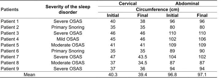
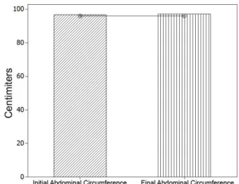
Cervical (CC) and abdominal circumference (AC) measurements were conducted at the beginning and end of the speech therapy process and the results were related to the severity of the sleep disorder observed, as shown in Table 1. According to Figures 1 and 2, no statistical differences were observed between initial CC and AC measurements and inal CC and AC measurements had a signiicance level of 5% (CC p = 0.069/CA p = 0.789).
1. Breathing and speech: (1) Forced nasal inspiration and oral expiration in conjunction with phonation of an open vowel, while sitting; (2) Balloon inlation with nasal inspiration and forced oral expiration, repeated ive times without taking the balloon away from the mouth.
2. Swallowing and chewing: 1) Alternating bilateral chewing and swallowing using the tongue on the palate, with teeth clenched, without perioral contraction, whenever feeding. 2) The supervised exercise consists of alternate bread mastication.
These exercises aimed for correct positioning of the tongue while eating in order to target the adaptation of tongue and jaw movements. Patients were instructed to use mastication pattern when eating.
5 - Analysis of the data obtained
The effect of speech therapy on the cervical and abdominal circumferences obtained, the results of the Berlin Questionnaires before and after therapy, were statistically analyzed using the Wilcoxon test (α = 0.05) since the data were dependent and nonpara
-metric. For the Epworth questionnaire, the paired Student’s t-test was used since the points obtained in this questionnaire were normally distributed. Descriptive analyses of the myofunctional aspects were also carried out, correlating those with the data obtained from the polysomnographic.
Table 1 – Comparison of cervical and abdominal circumference measurements observed with severity of sleep disorder
Patients Severity of the sleep
disorder
Cervical Abdominal
Circumference (cm)
Initial Final Initial Final
Patient 1 Severe OSAS 40 38 96 96
Patient 2 Primary Snoring 35 35 80 80
Patient 3 Severe OSAS 46 46 110 110
Patient 4 Mild OSAS 45 46 102 106
Patient 5 Moderate OSAS 41 41 109 109
Patient 6 Primary Snoring 35 35 89 90
Patient 7 Severe OSAS 47 43.5 104 102
Patient 8 Moderate OSAS 37 34.5 87 87
Patient 9 Severe OSAS 37 36 94 94
Mean 40.3 39.4 96.8 97.1
lowering of the back of tongue, high soft palate, bilateral chewing, speech, and nasal breathing. There were no changes related to the tip of the tongue on the loor of the mouth at either of the times evaluations were performed.
3 - Myofunctional Evaluation
Table 2 shows the results of the initial and inal myofunctional evaluation. In the inal evaluation, all patients presented an improvement of the following aspects: tonus adequacy of the suprahyoid muscles, Wilcoxon test: p = 0.069 (not signiicant)
Figure 1 – Comparison of initial and inal cervical
circumference
Wilcoxon test: p = 0.789 (not signiicant)
Figure 2 – Comparison of initial and inal
abdominal circumference
Table 2 – Analysis of myofunctional adequacy before and after speech therapy.
Function/Position Evaluated
Before speech therapy Severe
OSAS (n = 4)
Moderate OSAS (n = 2)
Mild OSAS (n = 1)
Primary Snoring (n = 2)
Lowering of the back of the tongue 0% 0% 0% 0%
Tip of the tongue on the loor 75% 50% 0% 50%
Soft palate elevation 0% 0% 0% 0%
Nasal breathing 25% 0% 100% 50%
Bilateral chewing 50% 100% 100% 100%
Adequacy of the tonus of suprahyoid muscles 0% 0% 100% 0%
Speech adequacy 75% 100% 100% 100%
Function/Position Evaluated
After speech therapy Severe
OSAS (n = 4)
Moderate OSAS (n = 2)
Mild OSAS (n = 1)
Primary Snoring (n = 2)
Lowering of the back of the tongue 100% 100% 100% 100%
Tip of the tongue on the loor 75% 50% 0% 50%
Soft palate elevation 100% 100% 100% 100%
Nasal breathing 100% 100% 100% 100%
Bilateral chewing 100% 100% 100% 100%
Adequacy of the tonus of suprahyoid muscles 100% 100% 100% 100%
Speech adequacy 100% 100% 100% 100%
Legend: OSAS - Obstructive Sleep Apnea Syndrome
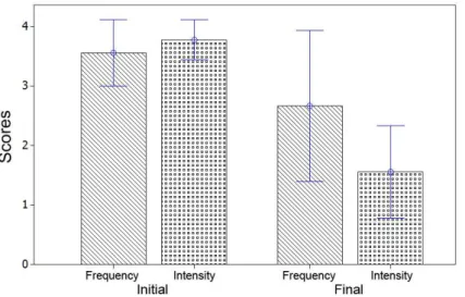
Intensity – Wilcoxon Test: p=0,005 (signiicant) Frequency – Wilcoxon Test: p=0,05 (signiicant)
Figure 3 – Comparison of the scoreson the adapted Berlin Questionnaire before and after speech therapy
4 - Berlin Questionnaire and Epworth Sleepiness Scale
Based on the perception of the bed partner, the results of the Berlin Questionnaire showed a higher reduction in intensity (p = 0.005) than in frequency
of snoring (p = 0.05), represented by Figure 3,
regardless of the severity of the sleep disorder presented by the patient.
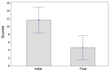
The results of the ESS found that 66% of the
patientsevaluated at the beginning of the study had
scores higher than 10, which indicates the presence of EDS with an increased tendency to sleep during the day and a subjective compulsion to sleep. After completing the myofunctional exercises, there was a signiicant reduction (p = 0.000) of EDS in all
patients and values close to zero in those who did not present EDS at the time of the irst evaluation, as shown in Table 3 and Figure 4.
Table 3 – Epworth Sleepiness Scale results
Patients Severity of the sleep disorder Initial Final
Patient 1 Severe OSAS 5 0
Patient 2 Primary Snoring 8 1
Patient 3 Severe OSAS 11 7
Patient 4 Mild OSAS 15 7
Patient 5 Moderate OSAS 16 10
Patient 6 Primary Snoring 14 10
Patient 7 Severe OSAS 15 3
Patient 8 Moderate OSAS 15 4
Patient 9 Severe OSAS 6 0
Mean 11,67 4,67
Legenda : OSAS – Obstructive Sleep Apnea Syndrome
DISCUSSION
The propaedeutic proposed by Guimarães (2008) 23 and used in this study aim to work on stomatognathic functions such as sucking, swallowing, chewing, breathing, and speech, which are closely related to the muscles present in upper airway structures.
At the initial myofunctional evaluation, most of the patients in the study presented changes in positioning in the back of the tongue, soft palate, and tonus of the suprahyoid muscles, as reported in the literature 8,23. These changes modify the oropha
-ryngeal space and encourage the occurrence of tissue collapse during sleep 23,25-27.
By the end of the sessions, all patients presented improvements in the aspects described above including chewing and breathing patterns, which suggest an increase in the muscle tonus of the stomatognathic system, as well as of the orofacial, oropharyngeal, and cervical regions 6,23, as noted in the inal evaluation using the protocol described in the study of Guimarães’s (2008) 23.
The only aspect with no changes observed between before and after evaluations using the protocol was the tip of the tongue on the loor of the mouth. This inding, however, does not suggest it impairs the eficiency of the procedure since the back of the tongue appeared to be adequate.
In this study, no relationship was found between the severity of OSAS and/or snoring and the myofunctional changes shown, which may be explained by the absence of signiicant anatomical differences in computed tomography (CT) images of
Paired Student’s t-test: p = 0.000 (signiicant)
Figure 4 – Comparison of the Epworth questionnaire score
patients with OSAS and those who only snore 27. However, studies with a larger number of partici
-pants should be performed to verify this fact. The inal results of ESS showed an absence of EDS in all study participants. These after speech therapy results indicate better sleep eficiency, and consequently, an improvement in the quality of life of the participants, characterized by reducing the impact of the disease on the ability to carry out daily activities 28. However, it should be noted that ESS
is a subjective test that may be inluenced by other factors when applied.
Some patients did not show any possible relationship between the type of sleep disorder and the score obtained, unlike indings presented in other studies 15,24. Two patients with severe OSAS showed no indices indicative of EDS. This data may be linked to a behavior, already described in the literature, of patients with OSAS underestimating their tendency to sleep during of clinical presen
-tation, being accustomed to a state of sleepiness slowly progressive over the years 17.
The quality of life of the bed partners of OSAS/ snoring patients also changedwith the occurrence of nocturnal sleep interruptions due to snoring12,14. Studies on quality of life suggest that the self-reported information relects the extent of the damage related to the disease on the general state of health; the information obtained from the partner of the patient is valuable for the clinical evalu -ation13. The evaluation of bed partners enables the avoidance of variations in the self-evaluations of patients, who may being accustomed to this state or
The use of the Berlin Questionnaire as a tool for bed partners to assess the sleep disorders of their partners proved to be eficient in identifying improve
-ments in the sleep disorders of the patients, which was conirmed by the results of ESS.
In this study, no statistically signiicant differences were observed between initial CC and AC and inal CC and AC measurements. This allows us to infer that CC and AC measurements are not related to a reduction in the intensity and frequency of snoring assessed by the Berlin Questionnaire. Therefore, there were no reports found in the literature that supported a possible relationship between CC, intensity, and frequency of snoring. Further studies with larger samples would be required to assess this relationship.
Understanding the pathophysiology of OSAS and snoring has been discussed in several scientiic studies without any conclusions on the relationship between the type of sleep disorder in different individuals, genders, and age ranges 27,29. There have also been no conclusions on the improvement of early detection measurements of OSAS and its treatment 24,30. Juliano et al. 29 describe the existence of a cephalometric pattern found in adults with sleep apnea that resembles a cephalometric pattern of oral breathing found in children, reinforcing the need for the identiication and early treatment of
respiratory disorders.
Speech therapy brings a new form of treatment for sleep disorders to the scientiic community, since it has as one of its focuses of actuation, the orofacial myology area.
CONCLUSIONS
The perceptions of the bed partners of patients with OSAS and/or snoring in this study have shown an improvement in the quality of life, a reduction in the intensity of snoring, better sleep eficiency, and a reduction in the impact on their daily activities since there was a reduction of EDS in all study participants. All patients presented an improvement of myofunctional aspects after speech therapy, thus making it a therapeutic instrument and assessment tool for patients with OSAS also in speech-language clinics.
ACKNOWLEDGMENTS
The authors are grateful to FAPESP grant agency (2013/10281-7) for their support during this work. We would like to thank the Barros clinic on behalf of Dr. Carmen Freire de Barros for providing the space that rendered this study possible. We would also like to thank Ms. Lucas Hian da Silva for his help with statistical analysis and reviewing the manuscript.
RESUMO
Objetivo: veriicar a percepção do acompanhante e a auto-avaliação do indivíduo com ronco/sín
-drome da apnéia obstrutiva do sono; coletar medidas das circunferências abdominal e cervical antes e após fonoterapia, além de realizar avaliação miofuncional para relacionar os resultados com a gra -vidade do distúrbio do sono observado. Métodos: participaram deste estudo onze indivíduos entre
25 e 75 anos de ambos os sexos com diagnóstico polissonográico recente de síndrome da apnéia obstrutiva do sono leve a severo e/ou ronco primário. Os sujeitos receberam fonoterapia, exame clínico fonoaudiológico, aplicação de questionários de Berlim (adaptado) e Epworth nas fases pré e pós-fonoterapia. Os dados obtidos foram analisados estatisticamente por meio do teste de Wilcoxon (α = 0,05). Resultados: dois indivíduos não aderiram ao tratamento. Não foi veriicada diferença
signiicante entre circunferências cervical e abdominal inicial e inal (cervical p=0,069 / abdominal p=0,789). Todos os pacientes apresentaram melhora no tônus da musculatura supra-hióidea, rebaixa
-mento de dorso de língua, elevação do palato mole, mastigação bilateral, fala e respiração nasal. Os resultados do questionário de Berlim mostraram redução na percepção do acompanhante na intensi
-dade (p=0,005) do ronco maior do que na frequência (p=0,05). Houve redução signiicante (p=0,000) da sonolência diurna excessiva em todos os pacientes. Conclusão: Considerando-se as limitações
deste estudo, conclui-se que após a fonoterapia as percepções dos acompanhantes e dos pacientes com síndrome da apnéia obstrutiva do sono/ronco ilustraram melhora efetiva do sono, da qualidade de vida, redução de intensidade do ronco e do comprometimento nas atividades diárias decorrentes da redução da sonolência diurna excessiva.
REFERENCES
1. Balbani APS, Formigoni GGS. Ronco e síndrome da apnéia obstrutiva do sono. Rev Ass Med Brasil. 1999;45(3):273-8.
2. Noal RB, Menezes AMB, Canani SF, Siqueira FV. Ronco habitual e apnéia obstrutiva observada em adultos: estudo de base populacional. Rev Saúde Pública. 2008;42(2):224-33.
3. Kushida CA, Littner MR, Morgenthaler T, Alessi CA, Bailey D, Coleman J, Jr., et al. Practice parameters for the indications for polysomnography and related procedures: an update for 2005. Sleep. 2005;28(4):499-521.
4. Series F. Upper airway muscles awake and
asleep. Sleep Med Rev. 2002;6(3):229-42.
5. Ito FA, Ito RT, Morae NM, Sakima T, Bezerra MLS, Meirelles RC. Condutas terapêuticas para tratamento da Síndrome da Apnéia e Hipopnéia Obstrutiva do Sono (SAHOS) e da Síndrome da Resistência das Vias Aéreas Superiores (SRVAS) com enfoque no Aparelho Anti-Ronco (AAR-ITO). Rev Dent Press Ortodon Ortop Facial.
2005;10(4):143-56.
6. Silva LMP, Aureliano FTS, Motta AR. Atuação fonoaudiológica na síndrome da apnéia e hipopnéia obstrutiva do sono: relato de caso. Rev CEFAC.
2007;9(4):490-6.
7. Ishiguro K, Kobayashi T, Kitamura N, Saito C. Relationship between severity of sleep-disordered breathing and craniofacial morphology in Japanese male patients. Oral Surg Oral Med Oral Pathol Oral
Radiol Endod. 2009;107(3):343-9.
8. Rosa EPS, Oliveira SMA, Alves VAM, Barbosa PG. Fonoaudiologia e apnéia do sono: uma revisão. Rev CEFAC. 2010;12(5):850-8.
9. Ueda H, Almeida FR, Chen H, Lowe AA. Effect of 2 jaw exercises on occlusal function in patients with obstructive sleep apnea during oral appliance therapy: a randomized controlled trial. Am J Orthod Dentofacial Orthop. 2009;135(4):430 e1-7; discussion -1.
10. Silva GA, Giacon LAT. Síndrome das Apnéias/ Hipopnéias Obstrutivas do Sono (SAHOS). Rev Medicina. 2006;39(2):185-94.
11. Al-Shawwa BA, Badi AN, Goldberg AN, Woodson BT. Deining common outcome metrics used in obstructive sleep apnea. Sleep Med Rev. 2008;12(6):449-61.
12. Sharief I, Silva GE, Goodwin JL, Quan SF. Effect of sleep disordered breathing on the sleep of bed partners in the Sleep Heart Health Study. Sleep. 2008;31(10):1449-56.
13. Breugelmans JG, Ford DE, Smith PL, Punjabi NM. Differences in patient and bed partner-assessed
quality of life in sleep-disordered breathing. Am J
Respir Crit Care Med. 2004;170(5):547-52.
14. Beninati W, Harris CD, Herold DL, Shepard JW, Jr. The effect of snoring and obstructive sleep apnea on the sleep quality of bed partners. Mayo Clin Proc. 1999;74(10):955-8.
15. Bertolazzi NA. Tradução e adaptação cultural e validação de 2 instrumentos de avaliação do sono escala de sonolência de Epworth e Índice de qualidade do sono de Pittsburg [Dissertação]. Porto Alegre (RS): Universidade Federal do Rio Grande do Sul; 2008.
16. Jesus EVS, Dias FEB, Mota BM, Souza L, Santos CM, Rocha JBGea. Suspeita de Apneia Obstrutiva do Sono deinida pelo Questionário de Berlim prediz eventos em pacientes com Síndrome Coronariana Aguda. Arq Bras Cardiol. 2010;95(3):313-20. 17. López PG, Gil FC, Gallego MEQ, Pradera MAF, Bernal CC, Armegol AS. Valoración mediante escal de Epworth de la somnolencia diurna en pacientes con sospecha de síndrome de apneas obstructivas durante el sueño. Diferencias entre los pacientes y sus parejas. Arch Bronconeumol. 2000;36(11):608-11.
18. Canani SF, Barreto SSM. Sonolência e acidentes automobilísticos. J Pneumol. 2001;27(2):94-6. 19. Netzer NC, Stoohs RA, Netzer CM, Clark K, Strohl KP. Using the Berlin Questionnaire to identify patients at risk for the Sleep Apnea Syndrome. Ann Intern Med. 1999;131:485-91.
20. Massie CAS, Hart RW, Perales K, Richards GN. Effects of humidiication on nasal symptoms and compliance in sleep apnea patients using continuous positive airway pressure. Chest. 1999;116(2):403-8. 21. Guimarães KC. Alterações no tecido mole de orofaringe em portadores de apnéia do sono obstrutiva [monograia]. São Paulo (SP): CEFAC - Saúde e Educação; 1999.
22. Guimaraes KC, Drager LF, Genta PR, Marcondes BF, Lorenzi-Filho G. Effects of oropharyngeal exercises on patients with moderate obstructive sleep apnea syndrome. Am J Respir Crit Care Med.
2009;179(10):962-6.
23. Guimarães KC. Efeitos dos exercícios orofaríngeos nos pacientes com apnéia obstrutiva do sono moderada: um estudo controlado e randomizado [Tese]. São Paulo (SP): Universidade de São Paulo; 2008.
24. Johns MW. A new Method for measuring daytime sleepiness: the Epworth sleepness scale. Sleep.
1991;14(6):540-4.
25. Soares EB, Pires JB, Menezes MA, Santana
SKS, Fraga J. Fonoaudiologia X ronco/apnéia do
(WHOQOL-100): características e perspectivas. Ciênc saúde coletiva. 2000;5(1):33-8.
29. Juliano ML, Machado MAC, Carvalho LBC, Brado LBF. Mouth breathing children have cephalometric patterns similar to those of adults patients with obstructive sleep apnea syndrome. Arq Neuropsiquiatr. 2009;67(3-B):860-5.
30. Abreu GA, Oliveira LCL, Nogueira AR, Bloch KV. Quadro Clínico: reconhecimento do paciente com apnéia obstrutiva do sono. Rev Bras Hipertens. 2009;16(3):164-8.
26. Ernest AOE. Can Singing exercises reduce snoring? A Pilot study. Complement Ther Med. 2000;8(3):151-6.
27. Koren A, Groselj LD, Fajdiga I. CT comparison of primary snoring and obstructive sleep apnea syndrome: role of pharyngeal narrowing ratio and soft palate-tongue contact in awake patient. Eur Arch Otorhinolaryngol. 2009;266(5):727-34.
28. Fleck MPA. O instrumento de avaliação de qualidade de vida da Organização Mundial da Saúde
Received on: October 30, 2012 Accepted on: March 04, 2013
Mailing address: Erika Matsumura
Rua São Carlos, 105
São José dos Campos – SP – Brasil
CEP: 12240-230