www.reumatologia.com.br
REVISTA BRASILEIRA DE
REUMATOLOGIA
* Corresponding author.
E-mail: [email protected] (A.F. Zimmermann).
0482-5004/$ - see front matter. © 2013 Elsevier Editora Ltda. All rights reserved. http://dx.doi.org/10.1016/j.rbre.2013.01.001
Review article
Update on the etiopathogenesis of systemic sclerosis
Adriana Fontes Zimmermann
a,*, Marcia Margaret Menezes Pizzichini
ba Division of Rheumatology, Department of Internal Medicine, Hospital Universitário da Universidade Federal de Santa Catarina, Santa
Catarina, SC, Brazil
b Division of Pneumology, Department of Internal Medicine, Hospital Universitário da Universidade Federal de Santa Catarina, Santa
Cata-rina, SC, Brazil
a r t i c l e i n f o
Article history: Received 24 June 2012 Accepted 28 February 2013
Keywords: Systemic sclerosis Etiopathogenesis Immune system Angiopathy Extracellular matrix
a b s t r a c t
Systemic Sclerosis (SSc) is an autoimmune disease of multifactorial etiology, triggered by a combination of genetic and environmental factors. Its varied clinical expression re-sults from the complex physiopathogenic interaction of three main elements: prolifera-tive vasculopathy, immune dysregulation and abnormal deposition and remodeling of the extracellular matrix (ECM), of which the characteristic disease i brosis is the result. Early physiopathogenic events appear to be endothelial injury and imbalance in vascular repair with the activation of endothelial cells, the immune system and platelets, with the release of multiple mediators such as TH2 proinl ammatory cytokines and growth factors, triggering a sequence of simultaneous or cascading events that involve several intracel-lular signaling pathways.
The most important result of these events is the hyperactivation of i broblasts, the main effector cells of i brosis, which will then produce large amounts of ECM constituents and secrete multiple growth factors and cytokines that perpetuate the process. In this article we review the main factors potentially involved in the etiology of SSc and reexamine the current knowledge about the most important mechanisms involved in the development of lesions that are characteristic of the disease. A better understanding of these physiopatho-genic mechanisms will help identify potential therapeutic targets, which may result in advances in the management of this complex and debilitating disease.
© 2013 Elsevier Editora Ltda. All rights reserved.
Atualização na etiopatogênese da esclerose sistêmica
Palavras-chave: Esclerose sistêmica Etiopatogênese Sistema imune Angiopatia Matriz extracelular
r e s u m o
cresci-mento, desencadeando uma sequência de eventos simultâneos ou em cascata que envolve diversas vias de sinalização intracelular. O resultado mais importante desses eventos é a hiperativação dos i broblastos, as principais células efetoras da i brose, as quais passam a produzir grandes quantidades de constituintes da MEC e a secretar múltiplos fatores de crescimento e citocinas que perpetuam o processo. Neste artigo apresentamos uma revisão dos principais fatores potencialmente implicados na etiologia da ES e revisitamos os conhecimentos atuais sobre os mais importantes mecanismos envolvidos no desenvol-vimento das lesões características da doença. O melhor entendimento desses mecanismos i siopatogênicos possibilita identii car potenciais alvos terapêuticos, o que pode resultar em avanços no manejo dessa complexa e debilitante doença.
© 2013 Elsevier Editora Ltda. Todos os direitos reservados.
Introduction
Systemic sclerosis (SSc) is a connective tissue disease of au-toimmune origin, characterized by vascular alterations and progressive skin and visceral i brosis affecting mainly the lungs, gastrointestinal tract, heart, and kidneys.1 The disease
is extremely heterogeneous in its phenotypic expression, and its prognosis is determined by the predominant clinical manifestations, especially regarding visceral involvement.2-4
Despite advances in knowledge about the mechanisms responsible for the disease onset, there is still a lack of un-derstanding regarding the pathogenic process is initiated and undergoes intensii cation in each subgroup of patients, leading to different clinical expressions. As in other autoim-mune diseases, especially those that are not organ specii c, it is known that the process results from the interaction of multiple factors, both individual and those related to the surrounding environment.5
However, the mechanisms that trigger the disease in genetically susceptible individuals have not yet been eluci-dated. In this article, the main factors potentially implicated in the etiopathogenesis of SSc are reviewed, as well as the current knowledge on the most important mechanisms in-volved in the development of SSc characteristic lesions, and the potential treatments directed at molecular and cell tar-gets that have emerged from recent research are listed.
SSc etiology: environmental factors
In the great majority of cases, SSc is considered to be idio-pathic. However, in some situations, environmental factors likely play an important role in its development. These associ-ations have been recognized for many decades, and the most important are those related to exposure to silica and organic solvents. The combined relative risk estimate (CRRE) for silica, calculated in a meta-analysis, was 3.20 (95% CI: 1.89-5.43) for men. Regarding exposure to organic solvents, the CRRE found in a meta-analysis was 2.91 (95% CI: 1.60-5.30),6 and,
accord-ing to another more recent meta-analysis,7 the latter is a
fac-tor associated with increased risk of SSc in men.
The possible association between silicone breast implants and connective tissue disorders, especially scleroderma, has been the subject of great controversy, generated by case re-ports that suggested this association.8 However, a
meta-anal-ysis9 and a recent and comprehensive systematic review10 of
the available epidemiological evidence did not coni rm such association.
Other environmental factors that have been implicated in the etiology of SSc are exposures to infectious agents, espe-cially viruses. Among them, parvovirus B19 is an example of a very prevalent infection in patients with SSc.11 Regarding
the Epstein-Barr virus, it has been demonstrated that spe-cii c adaptive immunity against the virus (the activation of suppressor T lymphocytes) was dei cient in scleroderma pa-tients.12 Cytomegalovirus (CMV) infects endothelial cells and
monocytes, leading to the production of proi brotic cytokines, causing vascular lesions and activating i broblasts, and may therefore trigger pathogenic processes associated with SSc development.13
An association between positivity to anti-CMV antibodies and prevalence of specii c autoantibodies in patients with SSc has been reported.14 These data suggest an association
be-tween infectious and environmental factors and the disease, but more studies are still necessary to determine the possible causal role of external agents in triggering SSc.
SSc etiology: hormonal and genetic factors
Considering the marked predominance of SSc in women,(7:1) a hypothesis was raised that hormonal factors are relevant to the disease development. However, this has not been proven. For instance, an increased incidence of the disease in women who use oral contraceptives has not been observed.15
Among the host factors involved in the etiopathogenesis of autoimmune diseases, genetic susceptibility is perhaps the most relevant, and therefore has been the target of extensive research. In SSc, as in almost all other systemic rheumatic diseases, polymorphisms of the main histocompatibility sys-tem (human leukocyte antigen [HLA]) have been associated to the disease development, with the main associations being with class II HLA antigens.
A recent study involving a fairly signii cant sample showed a positive association of haplotypes HLA-DRB1_1104, DQA1_0501, DQB1_0301 and, in contrast, the protective fac-tor of haplotypes HLA-DRB1_0701, DQA1_0201, DQB1_0202, and DRB1_1501 in whites and Hispanics with SSc; however, in blacks, the association was with HLA-DRB1_0804, DQA1_0501, and DQB1_0301 alleles.16 Moreover, the association of the
been studied. The same abovementioned study demon-strated the association of anti-Scl70 antibodies with HLAD-PB1_1301 antigens, of anti-centromere antibodies (ACA) with HLADQB1_0501 and DQB1_26, and of anti-RNA polymerase antibodies with HLA-DRB1_0404, DRB1_11, and DQB1_03 al-leles(16), which illustrates the multiple inl uences that this gene system can exert on SSc and its clinical and immuno-logical proi le.
Many other genes have been implicated in SSc suscep-tibility, mostly those encoding proteins responsible for the regulation and transduction of signals that comprise the au-toimmunity and inl ammation mechanisms involved in the pathogenesis of SSc. One of the main associations found in different populations was with the gene encoding for the signal transducer and activator of transcription 4 (STAT4),17
which promotes the differentiation of type-1 T helper lym-phocytes and negatively regulates the Th2 type.
The association of B-cell scaffold protein with ankyrin re-peats (BANK1), which binds the B-cell receptor to intracellular signaling proteins such as kinases,18 and with the interferon
regulatory factor i ve (IRF-5), regulator of transcription of type I interferon genes, were also coni rmed.19 Moreover, positive
associations have also been reported of SSc with candidate interleukin 23 (IL-23R) receptor genes involved in expression of the IL-23, a cytokine which expands the population of TH17 lymphocytes, but SSc has only been associated with positivity to anti-scleroderma 70 (anti-Scl70) autoantibody, and it acts as a protective factor in relation to pulmonary hypertension.20
Another gene related to SSc susceptibility is the connective tissue growth factor (CTGF), which induces cell proliferation, increasing production of ECM and chemotaxis of mesenchy-mal cells.21
Recently, it has been observed that interactions between genes are also important, such as the apparent additive ef-fect in SSc exerted by the simultaneous presence of STAT4, IRF-5, and BANK1 on the susceptibility and the development of the diffuse clinical form and pulmonary i brosis.17,22
Anoth-er intAnoth-eresting i nding was the sharing of genetic risk factors between different autoimmune diseases, exemplii ed by the protein tyrosine phosphatase 22 (PTPN22) non-receptor gene, involved in SSc susceptibility23 as well as in diabetes mellitus
type I,24 systemic lupus erythematous (SLE),25 and rheumatoid
arthritis (RA).(25) Other shared genes, such as BANK1, IRF5, and STAT4 have also been reported as involved in SLE and RA susceptibility.26-28
Another genetic factor of SSc susceptibility was recently demonstrated in white European populations.29 It is the
mac-rophage migration inhibitory factor (MIF)-173, a cytokine with immunoregulatory functions and a mediator of innate and adaptive immunity, which has been implicated in the patho-genesis of vasculopathy in SSc.(30) In addition to the asso-ciation with susceptibility, the MIF-173C allele was associated with the diffuse form of the disease.29
Physiopathogenic mechanisms of systemic
sclerosis
The current accumulated knowledge regarding the phys-iopathogenesis of SSc derives from extensive research in
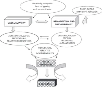
different areas, and indicates that the pathological process arises from the complex interrelation between three main components: vascular dysfunction, innate and adaptive im-munity dysregulation, and excess activation of i broblasts and related cells, which culminates in the development of i brosis (Fig. 1).31
Proliferative vasculopathy
The occurrence of Raynaud’s phenomenon in almost all pa-tients with SSc, with high severity in many of them, dem-onstrates the importance of vasculopathy in the pathogenic context of the disease. It is believed that the vasculopathy may originate after injury and activation of endothelial cells by unknown factors.32 The endothelial activation has been
demonstrated in several studies that detected high plasma levels and increased expression of von Willebrand factor, in-tercellular adhesion molecules, endothelin-1, and thrombo-modulin, which also represent evidence of excessive apopto-sis of endothelial cells.33-35
Chronic endothelial injury results in platelet adhesion and activation of the i brinolytic system, as well as in increased vascular permeability and leukocyte adhesion to the vascular wall.34 Increased levels of endothelin-1, a potent
vasoconstric-tor, have been detected in plasma and bronchoalveolar lavage l uid of patients with SSc.36 This protein is responsible for
in-creasing leukocyte adhesion to the endothelium, promoting migration and proliferation of smooth muscle cells into the intima layer of vessels and activating i broblasts.
This process promotes the synthesis and deposition of ECM molecules, leading to i brosis with loss of elasticity and progressive reduction of the vascular lumen, causing progres-sive tissue necrosis and hypoxia.37
Tissue hypoxia promotes angiogenesis (formation of new vessels from remaining functional vessels) and
vasculogen-'ĞŶĞƟĐĂůůLJƐƵƐĐĞƉƟďůĞ ŚŽƐƚнƚƌŝŐŐĞƌŝŶŐ
ĞŶǀŝƌŽŶŵĞŶƚĂůĨĂĐƚŽƌ d>zDW,Kzdͬ >zDW,Kzdd/sd/KE
VASCULOPATHY
,^/KEDK>h>^͕ EKd,>/EͲϭ͕ Zd/sKyz'E^W/^
zdK</E^͕'ZKtd, &dKZ^͕ ,DK</E^͕ hdKEd/K/^
&/ZK>^d^͕ WZ/zd^͕ DzK&/ZK>^d^
INFLAMMATION AND AUTO-IMMUNITY
TISSUE REMODELING
FIBROSIS
esis (formation of new vessels from endothelial progenitor cells),38 in order to restore cell oxygen supply. Due to
multi-ple causes not yet understood, these processes are dei cient in SSc, and there is an imbalance between angiogenic and angiostatic factors and ineffective vascular repair.39-40
The result is the formation of morphologically aberrant and dysfunctional capillaries, which can be viewed in the nailfold capillaroscopy examination. It is now known that tissue hypoxia present in SSc plays an important patho-genic role in the disease through different factors, such as the production of hypoxia-induced factor (HIF-1), whose production and dei cient regulation is ineffective in restor-ing normal blood pressure levels of oxygen. This dei ciency creates a vicious circle, with increased activation of immune cells, increased activation of i broblasts, increased levels of transforming growth factor β (TGFβ), and ECM deposition, which in turn aggravate the hypoxic environment. Further-more, the increase in oxidative stress caused by hypoxia has a proinl ammatory and proi brotic effect, exerted by reactive oxygen species (ROS).41
An additional factor that contributes to microvasculopa-thy in SSc is the pericytes, cells very close to the endothe-lium that normally inhibit cell migration and vascular pro-liferation. These cells are hyperplastic and hyperactivated in SSc, inhibiting the process of angiogenesis and transdiffer-entiating into myoi broblasts, producing excess ECM, which accumulates in the perivascular area and aggravates the proliferative vasculopathy of the disease.38 Table 1
summa-rizes the most relevant physiopathological events in relation to SSc vasculopathy.
Autoimmunity and inl ammation
Another important factor implicated in the pathogenesis of SSc is the involvement of both humoral and cellular im-mune system, whose mediators are the connection between vascular disease and tissue i brosis, as they are involved in both processes.32 The involvement of the humoral immune
system is demonstrated by the presence of B lymphocyte ini ltrates with expression of chronic activation markers
(CD19, CD85), which have been detected in patients with SSc skin using DNA microarray techniques.42 The
homeo-stasis of circulating B lymphocytes is altered, expanding the population of non-activated cells (naïve) and reduc-ing the number, but increasreduc-ing the activation of memory B cells.43 This knowledge has led to the use of therapies
di-rected against B cells (monoclonal antibodies belimumab and rituximab) in the treatment of patients with SSc, as well as to the blocking of the costimulation between the latter and T lymphocytes.44
The specii c autoantibodies, which represent one of the attributes of the disease, characterize its clinical forms and have important prognostic associations; they are the most obvious expression of the humoral immune system involve-ment in the etiopathogenesis of SSc. The main autoanti-bodies whose pathogenic potential has been established, in addition Scl70 and ACA, are those directed against endo-thelial cells and against the platelet-derived growth factor (PDGF) receptor.32
ACA are characteristic of limited SSc, which is gener-ally more benign than the diffuse form, but is associated with later development of pulmonary hypertension, one of the most severe complications of the disease, as well as digital ulcers and important gastrointestinal involvement. Patients with limited SSc may have anti-Th/To antibod-ies (more frequent in men, with interstitial pulmonary in-volvement and earlier mortality),45 anti-PM/Scl antibodies
(associated with myositis, calcinosis, acro-osteolysis and interstitial lung disease),46 and the anti-U1RNP antibodies
(that characterize overlapping forms between SSc, SLE, and myositis).47
Conversely, the presence of anti-Scl-70 is associated with the diffuse form of SSc, with interstitial pulmonary involve-ment and increased risk of renal crisis, especially in the early years of the disease. Musculoskeletal complications, such as l exion contractures of the i ngers, are common and very disabling.47 More rarely, the anti-RNA polymerase III
(POL3) antibodies occur in the diffuse form, whose patients have a high frequency of renal crisis, rapidly progressive skin i brosis, and little pulmonary and gastrointestinal in-volvement.48 The diffuse form can also show the presence
of anti-U3RNP (i brillarin), which is more common in black individuals and has worse prognosis, with severe pulmo-nary i brosis, arterial pulmopulmo-nary hypertension, pigmenta-tion alterapigmenta-tions, and joint contractures.49
The pathogenic role of cell immunity, represented main-ly by T main-lymphocytes, is evidenced in SSc by the increase in endothelial transmigration of T CD4 + cells originating from perivascular inl ammatory ini ltrates and oligoclonal acti-vation of these cells, resulting in proi brotic cytokine pro-duction.50 The oligoclonal activation of T-lymphocytes was
demonstrated by the increased levels of soluble IL-2 recep-tor in the serum of SSc patients, which showed a strong association with skin involvement (Rodnan skin score) in the disease.51
The predominant production of these cytokines (IL-4, IL-5, IL-6, IL-10, IL-13, and monocyte chemoattractant tein-1 [MCP-1]) results from an imbalance between the pro-i les of T helper type 1 (TH1) and type 2 (TH2) cells, derpro-ived from TH2- cells, which is the predominant proi le in SSc
Table 1 – Physiopathogenesis of vasculopathy in systemic sclerosis.
Effector cells Physiopathogenic processes
Molecules involved
Endothelial cells Smooth muscle
cells of vessels Pericytes
• Lesion and apoptosis of endothelial cells • Oxidative stress • Increased vascular
permeability • Platelet activation and
thrombosis
• Structural vasculopathy (progressive reduction of vascular caliber due to hypertrophy and i brosis) Impaired
neovascularization
• Von Willebrand factor • HIF-1 • Endothelin- 1 • VCAM-1 • ELAM-1 • VEGF
and promotes worsening of tissue i brosis.52 Current
knowl-edge allows for the inference that the processes triggered by proi brotic cytokines derived from the interaction be-tween T lymphocytes and i broblasts play a key role in the development of i brosis (Table 2).
Development of i brosis: i broblast activation
The third key pathogenic mechanism involved in SSc, and the most representative of the disease, is i brosis, which lends its name to the disease and affects the skin, lungs, heart, digestive tract, kidneys, and musculoskeletal system. The extent of skin i brosis is associated with mortality by SSc, as recently demonstrated both in the European and Brazilian populations.3-4
Fibrosis results from complex interactions between mul-tiple concurrent or cascading processes, in which dozens of cell mediators, cytokines and their receptors, chemokines, growth factors, and intracellular signaling molecules are present (Table 3). The result of this interaction is the ex-cessive production and accumulation of insoluble material in the tissues that constitutes the ECM. Fibrillar collagens types I and II, type VII collagen, and elastin and i brillin i -brils are mainly found in ECM, as well as enzymes that pro-mote the production of collagen crosslinks.53
Regarding i brosis cell mediators, the i broblast is the cell primarily responsible for the production and remodel-ing of the ECM, and its main physiological importance lies in the healing process of connective tissue injuries, having a self-limited action, depending on the injury extent.53 In
SSc, as in other i broproliferative diseases, there is a state in which the i broblast is permanently hyperactivated, with altered expression of genes that determine ECM overpro-duction.
In SSc, one of the mechanisms responsible for this hy-peractivation is the mechanotransduction abnormality, in which i broblasts and myoi broblasts would be able to perceive tensional forces and change their intracellular signaling in response, leading to altered homeostasis and remodeling of the ECM.54 In addition to the i broblasts,
myo-i broblasts also partmyo-icmyo-ipate myo-in the process of myo-i brosmyo-is forma-tion in SSc. These contractile and ECM-producing cells orig-inate from the transdifferentiation of i broblasts, epithelial
cells, and pericytes.55 Unlike the normal healing process, in
which myoi broblasts occur only transiently in granulation tissue, in SSc these cells become permanent, producing ar-eas of i brosis with contracture of the ECM.56
The bone marrow also contributes to the increase of the amount of proi brotic cells, with the release of pluripotent mesenchymal cells and i broblast progenitors (i brocytes). The latter physiologically replenish the population of i bro-blasts maintaining tissue homeostasis, but they also exert a pathogenic role in SSc. Fibrocyte precursors migrate and accumulate in tissues by gradient of chemokine receptors (CRs), such as CCR3, CCR5, and CXCR4.32 These receptors
and their ligands have been found at high levels in the skin of patients with SSc.57 Furthermore, tissueini ltrating i
-brocytes have also been implicated in the pathogenesis of pulmonary i brosis disease.58
The main effector proteins involved in the i brosis mech-anisms in SSc are the cytokines, chemokines, and the fam-ily of ECM growth factors, which induce i brogenic cell re-sponse by increasing the expression of genes encoding ECM constituents.59 Proi brotic cytokines play a pathogenic role
in SSc. The best-known and most often studied cytokine re-garding its mechanisms in i brosis is IL-4, which stimulates the synthesis of TGFβ and CTGF; it also promotes the migra-tion, proliferamigra-tion, and synthesis of collagen by i broblasts. High levels of IL-4 were found in serum and tissues of pa-tients with SSc, and a larger number of T cells that produce this cytokine were detected in these patients.60-61 It is also
known that IL-13 induces the expression of type I collagen gene and that its levels are elevated in patients with SSc, but its possible role in the induction of i brosis is not well established.60 Very recently, a study found increased levels
of IL-6 in serum and tissues of patients with diffuse SSc, which was associated with more severe skin involvement and increased mortality after three years,62 suggesting that
IL-6 antagonism would be a promising therapeutic target in this situation.
Perhaps the most important of all effector proteins of i -brosis is TGFβ, considered to be the orchestrator of the
phys-Table 2 – Autoimmunity and inl ammation in the pathogenesis of systemic sclerosis.
Effector cells
Physiopathogenic processes
Molecules involved
T Lymphocytes B Lymphocytes
• General and local activation of T and B lymphocytes, oligoclonal expansion • Transendothelial migration
of CD4+ activated T lymphocytes
• Production and release of pro-i brotic autoantibodies and cytokines
• Autoantibodies (PDGF, anti-endothelial cells, Scl70, anti-centromere) • Proi brotic
cytokines: IL-4, IL-6, IL-10, IL-13 • IL-2 receptor
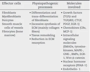
Table 3 – Physiopathogenesis of i brosis in systemic sclerosis.
Effector cells Physiopathogenic processes
Molecules involved
Fibroblasts Myoi broblasts Pericytes Smooth muscle
cells of vessels Fibrocytes (bone
marrow)
• Differentiation and trans-differentiation of i broblasts • Excessive synthesis of
ECM (mainly collagen i bers)
• Tissue remodeling • Reduction in ECM
resorption
• Growth factors (TGFβ and TGFβRII, CTGF, PDGF, EGR-1) • Chemokines (CRs,
MCP-1) • Intracellular
signaling molecules (SMADs, tyrosine-kinases, MAPK-, GNK-, BMPs, EGR-1, Wnt-β-catenin • Nuclear hormone
iological process of tissue repair and known to be involved in the development of pathological i brosis, as in SSc.59 TGFβ
is produced by i broblasts, T cells, monocytes, and platelets, and it is released as a latent complex that is activated by tis-sue injury to its biologically active form within the ECM, ex-erting its function by binding to a specii c receptor (TGFβRII) on the surface of different cell types.37
After this binding, an intracellular signaling cascade starts, which results in the expression of target genes such as type I collagen, CTGF, and plasminogen activator inhibitor-1, among others.63 The main control of TGFβ intracellular
sig-naling system is exercised by the SMAD system (SMAD2 and SMAD3 activators, as well as endogenous inhibitor SMAD7 and its cofactors), whose dysregulation results in increased i brogenesis, a phenomenon already demonstrated in SSc.64
Recently, other regulatory mechanisms of TGFβ were studied and their importance was demonstrated in the pathogenesis of SSc, such as c-Abl kinase (c-Abelson, a tyrosine kinase im-plicated in the pathogenesis of chronic myeloid leukemia), a potent regulator of the TGFβ-induced proi brotic response in i broblasts.65
Imatinib, an inhibitor of c-Abl, reduces i brotic responses in animal models and have shown to be promising in the treatment of patients with SSc.44,66 Also relevant as
me-diators of proi brotic cell response to TGFβ are genes from the early growth response (EGR) family, particularly EGR-1, whose expression is induced by chemical or mechanical tissue injury and whose levels are elevated in skin lesions of SSc, as demonstrated in a murine model of bleomycin-induced scleroderma.67
Another relevant mechanism in the process of i brosis that has recently been demonstrated, but whose role still re-quires elucidation, is the Wnt-β-catenin, an important intra-cellular signaling pathway in the embryonic period during organogenesis whose posterior and aberrant reactivation is associated with disease (cancer, for instance) by increasing i broblast activity and promoting transdifferentiation. In bi-opsies of scleroderma patients, this pathway was activated and found responsible for signii cant production of i brosis and lipoatrophy.68
In addition to TGFβ, other growth factors are important in the pathogenesis of SSc, such as CTGF, whose effects are similar to those of TGFβ and whose levels are increased in lesions of patients with SSc. Its expression is stimulated by TGFβ itself, by hypoxia, and by ET-1.37 PDGF is produced by
i broblasts, platelets, macrophages, and endothelial cells, with important mitogen and chemoattractant activity for i -broblasts, inducing their production of collagen, i bronectin, and proteoglycans, and the release of proi brotic mediators IL-6 and MCP-1. This is another growth factor implicated in the interaction with TGFβ and CTGF, which was detected at high levels in the lungs of patients with SSc.69
In addition to all the aforementioned mechanisms, which help to promote excessive i brogenesis, in SSc there are also abnormalities in the physiological counterregulation sys-tems that normally prevent the occurrence of i brosis. This was demonstrated in the case of SMAD7, signaling mole-cules that exert an antii brotic role through negative regula-tion of TGFβ action.64
Searching for antii brotic mechanisms that could be ex-plored for therapeutic purposes in SSc, the peroxisome pro-liferator activated receptor-gamma (PPAR-ϒ) was identii ed. This molecule is a key hormonal nuclear receptor in lipid and glucose metabolism, which has been increasingly asso-ciated with ECM remodeling and i brosis, capable of modu-lating TGFβ signaling and mesenchymal cell plasticity. It was found that this receptor is involved in an important anti-i brotanti-ic mechananti-ism, and anti-its expressanti-ion and actanti-ivanti-ity were found to be reduced in patients with SSc.70 Natural or
syn-thetic PPAR-ϒ agonists, such as the antidiabetic drugs piogli-tazone or rosiglipiogli-tazone, or tissue stimulation of its expres-sion could, therefore, play a role in the treatment of i brosis. Table 3 summarizes the elements involved in the physio-pathological process of establishment i brosis in SSc.
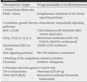
In spite of the fact that SSc is considered to be incurable, in recent years intensive research has led to the identii ca-tion of antii brotic cellular and molecular targets and the development of therapeutic options directed against them. Table 4 lists some treatments aimed at targets (mostly dis-cussed above) that have emerged from in vitro studies, ani-mal models, and early trials in humans.44 A better
under-standing of the physiopathological mechanisms of SSc has shed light on the factors that most likely initiate the process and lead to its perpetuation.
Considering that the vascular lesion and its defective re-pair are known early events, it is possible that the clinical expression of the disease depends more on the magnitude of vascular changes and its chronicity than on the trigger-ing event itself.41 Therefore, efforts are needed in order to
diagnose the early establishment of vasculopathy and to institute treatment in order to halt the progression of the process. In addition, the elucidation of the molecular mech-anisms involved in SSc may contribute to the identii cation of other potential targets to be assessed in the search for more specii c and effective treatments for this severe and debilitating disease.
Table 4 – Therapies directed against targets in systemic sclerosis.
Therapeutic target Drugs (available or in development)
• Intracellular Molecules
PPARϒ, Notch Rosiglitazone, inhibitors of the Notch signaling pathway
• Cytokines, growth factors, chemokines, intracellular signaling pathways
MCP-1/CCR2 CCR2 inhibitors (PF-04136309, BMS-741672, MLN1202)
TGFβ, CTGF, IL-13, IL-6 Monoclonal antibodies (CAT-192, FG3019, QAX576, tocilizumab) Chemokines (CXCL12,
CCL4)
CXCR4, CCR2 inhibitors
Wnt signaling pathway PRI-724 inhibitors, resveratrol
• Blocking of the coagulation system activation Thrombin Inhibitor: dabigatran
• Therapies directed against T and B cells T/B co-stimulation Abatacept (CTLA4-Ig)
Conl icts of interest
The authors declare no conl icts of interest.
R E F E R E N C E S
1. LeRoy EC, Black C, Fleischmajer R, Jablonska S, Krieg T, Medsger TA, Jr., et al. Scleroderma (systemic sclerosis): classification, subsets and pathogenesis. J Rheumatol. 1988 Feb;15(2):202-5.
2. Steen VD, Medsger TA. Changes in causes of death in systemic sclerosis, 1972-2002. Ann Rheum Dis. 2007 Jul;66(7):940-4.
3. Sampaio-Barros PD, Bortoluzzo AB, Marangoni RG, Rocha LF, Del Rio AP, Samara AM, et al. Survival, causes of death, and prognostic factors in systemic sclerosis: analysis of 947 Brazilian patients. J Rheumatol. 2012 Oct;39(10):1971-8. 4. Tyndall AJ, Bannert B, Vonk M, Airo P, Cozzi F, Carreira
PE, et al. Causes and risk factors for death in systemic sclerosis: a study from the EULAR Scleroderma Trials and Research (EUSTAR) database. Ann Rheum Dis. 2010 Oct;69(10):1809-15.
5. Tan FK. Systemic sclerosis: the susceptible host (genetics and environment). Rheum Dis Clin North Am. 2003 May;29(2):211-37.
6. Aryal BK, Khuder SA, Schaub EA. Meta-analysis of
systemic sclerosis and exposure to solvents. Am J Ind Med. 2001 Sep;40(3):271-4.
7. Kettaneh A, Al Moufti O, Tiev KP, Chayet C, Toledano C, Fabre B, et al. Occupational exposure to solvents and gender-related risk of systemic sclerosis: a metaanalysis of case-control studies. J Rheumatol. 2007 Jan;34(1):97-103. 8. Levy Y, Rotman-Pikielny P, Ehrenfeld M, Shoenfeld Y.
Silicone breast implantation-induced scleroderma: description of four patients and a critical review of the literature. Lupus. 2009 Nov;18(13):1226-32.
9. Janowsky EC, Kupper LL, Hulka BS. Meta-analyses of the relation between silicone breast implants and the risk of connective-tissue diseases. N Engl J Med. 2000 Mar 16;342(11):781-90.
10. Lipworth L, Holmich LR, McLaughlin JK. Silicone breast implants and connective tissue disease: no association. Semin Immunopathol. 2011 May;33(3):287-94.
11. Zakrzewska K, Corcioli F, Carlsen KM, Giuggioli D, Fanci R, Rinieri A, et al. Human parvovirus B19 (B19V) infection in systemic sclerosis patients. Intervirology. 2009;52(5):279-82.
12. Kahan A, Menkes CJ, Amor B. Defective Epstein-Barr virus specific suppressor T cell function in progressive systemic sclerosis. Ann Rheum Dis. 1986 Jul;45(7):553-60.
13. Radic M, Martinovic Kaliterna D, Radic J. Infectious disease as aetiological factor in the pathogenesis of systemic sclerosis. Neth J Med. 2010 Nov;68(11):348-53.
14. Neidhart M, Kuchen S, Distler O, Bruhlmann P, Michel BA, Gay RE, et al. Increased serum levels of antibodies against human cytomegalovirus and prevalence of autoantibodies in systemic sclerosis. Arthritis Rheum. 1999 Feb;42(2):389-92.
15. Mayes MD. Epidemiologic studies of environmental agents and systemic autoimmune diseases. Environ Health Perspect. 1999 Oct;107 Suppl 5:743-8.
16. Arnett FC, Gourh P, Shete S, Ahn CW, Honey RE, Agarwal SK, et al. Major histocompatibility complex (MHC) class II alleles, haplotypes and epitopes which confer susceptibility or protection in systemic sclerosis: analyses
in 1300 Caucasian, African-American and Hispanic cases and 1000 controls. Ann Rheum Dis. 2010 May;69(5):822-7. 17. Dieude P, Guedj M, Wipff J, Ruiz B, Hachulla E, Diot E, et
al. STAT4 is a genetic risk factor for systemic sclerosis having additive effects with IRF5 on disease susceptibility and related pulmonary fibrosis. Arthritis Rheum. 2009 Aug;60(8):2472-9.
18. Rueda B, Gourh P, Broen J, Agarwal SK, Simeon C, Ortego-Centeno N, et al. BANK1 functional variants are associated with susceptibility to diffuse systemic sclerosis in Caucasians. Ann Rheum Dis. 2010 Apr;69(4):700-5. 19. Dieude P, Guedj M, Wipff J, Avouac J, Fajardy I, Diot
E, et al. Association between the IRF5 rs2004640 functional polymorphism and systemic sclerosis: a new perspective for pulmonary fibrosis. Arthritis Rheum. 2009 Jan;60(1):225-33.
20. Agarwal SK, Gourh P, Shete S, Paz G, Divecha D, Reveille JD, et al. Association of interleukin 23 receptor polymorphisms with anti-topoisomerase-I positivity and pulmonary hypertension in systemic sclerosis. J Rheumatol. 2009 Dec;36(12):2715-23.
21. Fonseca C, Lindahl GE, Ponticos M, Sestini P, Renzoni EA, Holmes AM, et al. A polymorphism in the CTGF promoter region associated with systemic sclerosis. N Engl J Med. 2007 Sep 20;357(12):1210-20.
22. Dieude P, Wipff J, Guedj M, Ruiz B, Melchers I, Hachulla E, et al. BANK1 is a genetic risk factor for diffuse cutaneous systemic sclerosis and has additive effects with IRF5 and STAT4. Arthritis Rheum. 2009 Nov;60(11):3447-54. 23. Dieude P, Guedj M, Wipff J, Avouac J, Hachulla E, Diot E,
et al. The PTPN22 620W allele confers susceptibility to systemic sclerosis: findings of a large case-control study of European Caucasians and a meta-analysis. Arthritis Rheum. 2008 Jul;58(7):2183-8.
24. Onengut-Gumuscu S, Ewens KG, Spielman RS, Concannon P. A functional polymorphism (1858C/T) in the PTPN22 gene is linked and associated with type I diabetes in multiplex families. Genes Immun. 2004 Dec;5(8):678-80. 25. Orozco G, Sanchez E, Gonzalez-Gay MA, Lopez-Nevot MA,
Torres B, Caliz R, et al. Association of a functional single-nucleotide polymorphism of PTPN22, encoding lymphoid protein phosphatase, with rheumatoid arthritis and systemic lupus erythematosus. Arthritis Rheum. 2005 Jan;52(1):219-24.
26. Remmers EF, Plenge RM, Lee AT, Graham RR, Hom G, Behrens TW, et al. STAT4 and the risk of rheumatoid arthritis and systemic lupus erythematosus. N Engl J Med. 2007 Sep 6;357(10):977-86.
27. Graham RR, Kyogoku C, Sigurdsson S, Vlasova IA, Davies LR, Baechler EC, et al. Three functional variants of IFN regulatory factor 5 (IRF5) define risk and protective haplotypes for human lupus. Proc Natl Acad Sci U S A. 2007 Apr 17;104(16):6758-63.
28. Orozco G, Abelson AK, Gonzalez-Gay MA, Balsa A, Pascual-Salcedo D, Garcia A, et al. Study of functional variants of the BANK1 gene in rheumatoid arthritis. Arthritis Rheum. 2009 Feb;60(2):372-9.
29. Bossini-Castillo L, Simeon CP, Beretta L, Vonk MC, Callejas-Rubio JL, Espinosa G, et al. Confirmation of association of the macrophage migration inhibitory factor gene with systemic sclerosis in a large European population. Rheumatology (Oxford). 2011 Nov;50(11):1976-81. 30. Selvi E, Tripodi SA, Catenaccio M, Lorenzini S, Chindamo
D, Manganelli S, et al. Expression of macrophage migration inhibitory factor in diffuse systemic sclerosis. Ann Rheum Dis. 2003 May;62(5):460-4.
32. Kahaleh MB. Raynaud phenomenon and the vascular disease in scleroderma. Curr Opin Rheumatol. 2004 Nov;16(6):718-22.
33. Scheja A, Akesson A, Geborek P, Wildt M, Wollheim CB, Wollheim FA, et al. Von Willebrand factor propeptide as a marker of disease activity in systemic sclerosis (scleroderma). Arthritis Res. 2001;3(3):178-82.
34. Cerinic MM, Valentini G, Sorano GG, D’Angelo S, Cuomo G, Fenu L, et al. Blood coagulation, fibrinolysis, and markers of endothelial dysfunction in systemic sclerosis. Semin Arthritis Rheum. 2003 Apr;32(5):285-95.
35. McHugh NJ, Distler O, Giacomelli R, Riemekasten G. Non organ based laboratory markers in systemic sclerosis. Clin Exp Rheumatol. 2003;21(3 Suppl 29):S32-8.
36. Cambrey AD, Harrison NK, Dawes KE, Southcott AM, Black CM, du Bois RM, et al. Increased levels of endothelin-1 in bronchoalveolar lavage fluid from patients with systemic sclerosis contribute to fibroblast mitogenic activity in vitro. Am J Respir Cell Mol Biol. 1994 Oct;11(4):439-45. 37. Wei J, Bhattacharyya S, Tourtellotte WG, Varga J. Fibrosis in
systemic sclerosis: Emerging concepts and implications for targeted therapy. Autoimmunity Reviews. [doi: DOI: 10.1016/j.autrev.2010.09.015]. 2011;10(5):267-75.
38. Muller-Ladner U, Distler O, Ibba-Manneschi L, Neumann E, Gay S. Mechanisms of vascular damage in systemic sclerosis. Autoimmunity. 2009 Nov;42(7):587-95. 39. Koch AE, Distler O. Vasculopathy and disordered
angiogenesis in selected rheumatic diseases: rheumatoid arthritis and systemic sclerosis. Arthritis Res Ther. 2007;9 Suppl 2:S3.
40. van Hal TW, van Bon L, Radstake TR. A system out of breath: how hypoxia possibly contributes to the pathogenesis of systemic sclerosis. Int J Rheumatol. 2011;2011:824972.
41. Geyer M, Muller-Ladner U. The pathogenesis of systemic sclerosis revisited. Clin Rev Allergy Immunol. 2011 Apr;40(2):92-103.
42. Whitfield ML, Finlay DR, Murray JI, Troyanskaya OG, Chi JT, Pergamenschikov A, et al. Systemic and cell type-specific gene expression patterns in scleroderma skin. Proc Natl Acad Sci U S A. 2003 Oct 14;100(21):12319-24.
43. Sato S, Fujimoto M, Hasegawa M, Takehara K. Altered blood B lymphocyte homeostasis in systemic sclerosis: expanded naive B cells and diminished but activated memory B cells. Arthritis Rheum. 2004 Jun;50(6):1918-27. 44. Maurer B, Distler O. Emerging targeted therapies in
scleroderma lung and skin fibrosis. Best Pract Res Clin Rheumatol. 2011 Dec;25(6):843-58.
45. Mitri GM, Lucas M, Fertig N, Steen VD, Medsger TA, Jr. A comparison between anti-Th/To- and anticentromere antibody-positive systemic sclerosis patients with limited cutaneous involvement. Arthritis Rheum. 2003 Jan;48(1):203-9.
46. Oddis CV, Okano Y, Rudert WA, Trucco M, Duquesnoy RJ, Medsger TA, Jr. Serum autoantibody to the nucleolar antigen PM-Scl. Clinical and immunogenetic associations. Arthritis Rheum. 1992 Oct;35(10):1211-7.
47. Steen VD, Powell DL, Medsger TA, Jr. Clinical correlations and prognosis based on serum autoantibodies in patients with systemic sclerosis. Arthritis Rheum. 1988 Feb;31(2):196-203.
48. Santiago M, Baron M, Hudson M, Burlingame RW, Fritzler MJ. Antibodies to RNA polymerase III in systemic sclerosis detected by ELISA. J Rheumatol. 2007 Jul;34(7):1528-34. 49. Okano Y, Steen VD, Medsger TA, Jr. Autoantibody to U3
nucleolar ribonucleoprotein (fibrillarin) in patients with systemic sclerosis. Arthritis Rheum. 1992 Jan;35(1):95-100. 50. Sakkas LI, Xu B, Artlett CM, Lu S, Jimenez SA, Platsoucas
CD. Oligoclonal T cell expansion in the skin of patients
with systemic sclerosis. J Immunol. 2002 Apr 1;168(7):3649-59.
51. Steen VD, Engel EE, Charley MR, Medsger TA, Jr. Soluble serum interleukin 2 receptors in patients with systemic sclerosis. J Rheumatol. 1996 Apr;23(4):646-9.
52. Wynn TA. Fibrotic disease and the T(H)1/T(H)2 paradigm. Nat Rev Immunol. 2004 Aug;4(8):583-94.
53. Cox TR, Erler JT. Remodeling and homeostasis of the extracellular matrix: implications for fibrotic diseases and cancer. Dis Model Mech. 2011 Mar;4(2):165-78.
54. Huang C, Ogawa R. Fibroproliferative disorders and their mechanobiology. Connect Tissue Res. 2012;53(3):187-96. 55. Abraham DJ, Eckes B, Rajkumar V, Krieg T. New
developments in fibroblast and myofibroblast biology: implications for fibrosis and scleroderma. Curr Rheumatol Rep. 2007 May;9(2):136-43.
56. Desmouliere A, Chaponnier C, Gabbiani G. Tissue repair, contraction, and the myofibroblast. Wound Repair Regen. 2005 Jan-Feb;13(1):7-12.
57. Cipriani P, Franca Milia A, Liakouli V, Pacini A, Manetti M, Marrelli A, et al. Differential expression of stromal cell-derived factor 1 and its receptor CXCR4 in the skin and endothelial cells of systemic sclerosis patients: Pathogenetic implications. Arthritis Rheum. 2006 Sep;54(9):3022-33.
58. Phillips RJ, Burdick MD, Hong K, Lutz MA, Murray LA, Xue YY, et al. Circulating fibrocytes traffic to the lungs in response to CXCL12 and mediate fibrosis. J Clin Invest. 2004 Aug;114(3):438-46.
59. Wynn TA. Cellular and molecular mechanisms of fibrosis. J Pathol. 2008 Jan;214(2):199-210.
60. Hasegawa M, Fujimoto M, Kikuchi K, Takehara K. Elevated serum levels of interleukin 4 (IL-4), IL-10, and IL-13 in patients with systemic sclerosis. J Rheumatol. 1997 Feb;24(2):328-32.
61. Tsuji-Yamada J, Nakazawa M, Minami M, Sasaki T. Increased frequency of interleukin 4 producing CD4+ and CD8+ cells in peripheral blood from patients with systemic sclerosis. J Rheumatol. 2001 Jun;28(6):1252-8.
62. Khan K, Xu S, Nihtyanova S, Derrett-Smith E, Abraham D, Denton CP, et al. Clinical and pathological significance of interleukin 6 overexpression in systemic sclerosis. Ann Rheum Dis. 2012 Jul;71(7):1235-42.
63. Chizzolini C, Brembilla NC, Montanari E, Truchetet M-E. Fibrosis and immune dysregulation in systemic sclerosis. Autoimmunity Reviews. [doi: DOI: 10.1016/j. autrev.2010.09.016]. 2011;10(5):276-81.
64. Varga J. Scleroderma and Smads: dysfunctional Smad family dynamics culminating in fibrosis. Arthritis Rheum. 2002 Jul;46(7):1703-13.
65. Bhattacharyya S, Ishida W, Wu M, Wilkes M, Mori Y, Hinchcliff M, et al. A non-Smad mechanism of fibroblast activation by transforming growth factor-beta via c-Abl and Egr-1: selective modulation by imatinib mesylate. Oncogene. 2009 Mar 12;28(10):1285-97.
66. Gordon JK, Spiera RF. Targeting tyrosine kinases: a novel therapeutic strategy for systemic sclerosis. Curr Opin Rheumatol. 2010 Nov;22(6):690-5.
67. Wu M, Melichian DS, de la Garza M, Gruner K, Bhattacharyya S, Barr L, et al. Essential roles for early growth response transcription factor Egr-1 in tissue fibrosis and wound healing. Am J Pathol. 2009 Sep;175(3):1041-55.
69. Ludwicka A, Ohba T, Trojanowska M, Yamakage A, Strange C, Smith EA, et al. Elevated levels of platelet derived growth factor and transforming growth factor-beta 1 in bronchoalveolar lavage fluid from patients with scleroderma. J Rheumatol. 1995 Oct;22(10):1876-83.