www.reumatologia.com.br
REVISTA BRASILEIRA DE
REUMATOLOGIA
* Corresponding author.
E-mail: [email protected] (V. F. Azevedo).
0482-5004/$ - see front matter. © 2013 Elsevier Editora Ltda. All rights reserved. http://dx.doi.org/10.1016/j.rbre.2013.06.002
Article original
Evaluation of sub-clinical atherosclerosis and plasma levels
of minimally modii ed LDL in patients with ankylosing
spondylitis and its correlation with disease activity
Fernanda Teles Ceccon
a, Valderílio Feijó Azevedo
b,*, Carlos A. Engelhorn
a,
Dulcinéia S. P. Abdalla
c, Tanize E. S. Faulin
c, Luis Cesar Guarita-Souza
a,
Roberto Pecoits-Filho
a, José Rocha Faria-Neto
a a Pontifícia Universidade Católica do Paraná, Curitiba, PR, Brazilb Hospital de Clínicas, Universidade Federal do Paraná, Curitiba, PR, Brazil
c Faculty of Pharmaceutical Sciences, Universidade de São Paulo, São Paulo, SP, Brazil
a r t i c l e i n f o
Article history:
Received on 21 March 2012 Accept on 24 June 2013
Keywords: Atherosclerosis Inl ammation Ankylosing spondylitis Minimally modii ed LDL Carotid Intimal-media thickness
a b s t r a c t
Introduction: Accelerated atherosclerosis has been shown in some autoimmune diseases, mainly in Systemic Lupus Erythematosus and Rheumatoid Arthritis. Although high preva-lence of corticosteroids use may be a confounding factor due to their detrimental effects on several risk factors, systemic inl ammation per se is supposed to play an important role in atherogenesis in these patients.
Methods: We have evaluated sub-clinical atherosclerosis and plasma levels of circulating electronegative LDL, which represents the fraction of LDL that is minimally modii ed, in patients with ankylosing spondylitis (AS). Fourteen patients who fuli lled the modii ed New York criteria for AS were compared with 13 paired controls. Carotid intimal-media thick-ness (IMT) was assessed by ultrasonography bilaterally in common carotid artery, internal carotid artery and in the bifurcation. Groups were homogeneous regarding cardiovascular risk factors. Only a single patient in AS group was in use of corticosteroid.
Results: The presence of active inl ammation was demonstrated by elevated BASDAI and higher CRP levels and in patients versus controls (12.36 vs. 3.45 mg/dl, P = 0.002). No dif-ference was found in carotid IMT between both groups, in any site of artery. Averaged IMT (6 measurements, at 3 pre-specii ed sites bilaterally) was 0.72 ± 0.28 in AS group and 0.70 ± 0.45 mm in controls (P = 0.91). Minimally modii ed LDL did not differ signii cantly either between patients and controls (14.03 ± 17.40 vs. 13.21 ± 10.21; P = 0.88).
Conclusions: Patients with AS did not show increased carotid IMT in comparison to con-trols. In the same way, circulating plasma levels of LDL (-), did not differ signii cantly in both groups.
Avaliação da aterosclerose subclínica e de níveis plasmáticos de LDL minimamente modii cada em pacientes com espondilite anquilosante e sua correlação com a atividade da doença
Palavras-chave: Aterosclerose Inl amação
Espondilite anquilosante LDL minimamente modii cada Espessura da íntima-média da
carótida
r e s u m o
Introdução: A aterosclerose acelerada foi demonstrada em algumas doenças autoimunes, principalmente lúpus eritematoso sistêmico e artrite reumatóide. Embora a alta prevalên-cia do uso de corticosteróides possa ser um fator complicador, por causa de seus efeitos prejudiciais em diversos fatores de risco, acredita-se que, nesses pacientes, a inl amação sistêmica per se desempenhe papel importante na aterogênese.
Métodos: Avaliamos a aterosclerose subclínica e os níveis plasmáticos de LDL eletronegativa circulante em pacientes com espondilite anquilosante (EA). Catorze pacientes que aten-diam aos critérios de Nova York modii cados para EA foram comparados com 13 controles equiparados. Avaliamos a espessura da íntima-média (EIM) na carótida por ultrassonogra-i a bultrassonogra-ilateral da artérultrassonogra-ia carótultrassonogra-ida comum, artérultrassonogra-ia carótultrassonogra-ida ultrassonogra-interna e na bultrassonogra-ifurcação. Os grupos foram homogêneos, no que tange a fatores de risco cardiovasculares. Apenas um paciente no grupo de EA estava sendo medicado com corticosteróide.
Resultados: A presença de inl amação ativa foi demonstrada por BASDAI elevado e níveis mais elevados de PCR em pacientes versus controles (12,36 vs. 3,45 mg/dl, P=0,002). Não ob-servamos diferença na EIM da carótida entre os dois grupos, em qualquer local da artéria. A média de EIM (6 mensurações em 3 locais pré-especii cados, bilateralmente) foi 0,72 ± 0,28 no grupo de EA e 0,70 ± 0,45 mm nos controles (P=0,91). Também não observamos diferença signii cativa na LDL minimamente modii cada entre pacientes e controles (14,03 ± 17,40 vs. 13,21 ± 10,21; P=0,88).
Conclusões: Pacientes com EA não demonstraram aumento na EIM da carótida, em com-paração com controles. Do mesmo modo, os níveis plasmáticos circulantes de LDL(-) não diferiram signii cativamente nos dois grupos.
© 2013 Elsevier Editora Ltda. Todos os direitos reservados.
Introduction
Atherosclerosis is a progressive disease of the large and medi-um size arteries involving inl ammation, lipid accmedi-umulation, cell death, and thrombosis in vessel wall.1 The “response to
injury hypothesis” postulates that long-term endothelial cell injury alters endothelial permeability to low density lipopro-teins (LDL) and induces leukocyte adhesion and migration to sub-endothelial space.2 Regardless of the risk factor inducing
endothelial disfunction, the following inl ammation process will lead to plaque formation. The uptake of oxidized LDL (Ox-LDL), but not native LDL, by macrophages in vessel wall will lead to the formation of foam cells that are not only a reser-voir of modii ed lipids, but also a source of proinl ammatory mediators contributing to plaque progression.3
Therefore, proinl ammatory OxLDL may be a unifying link between lipid accumulation and inl ammation.4 Although
most of the oxidation of LDL occurs in the vessel wall, lipopro-teins can be minimally modii ed in plasma becoming more prone to oxidation on a subsequent entry into the intima.5
A more electronegative subfraction of LDL, called LDL(-) has been subfractionated by high resolution ion exchange chro-matography (IE-HPLC) and seems to represent circulating minimally modii ed LDL in plasma.6
The recognition that inl ammation is the main feature of atherosclerotic disease has led to a series of studies report-ing high prevalence of atherosclerosis in chronic inl amma-tory diseases, such as rheumatoid arthritis (RA) and systemic
lupus erythematosus (SLE).7,8 There is a twofold increased
risk for myocardial infarction and stroke in patients with RA, with risk increasing to nearly threefold in those who have the disease for 10 years or more.9 These increased morbidity and
mortality due to atherosclerosis seem to depend not only on traditional RF, that may be negatively affected by corticoste-roid use. Inl ammatory mechanisms seem to be associated with worse cardiovascular outcomes in this patients.10-12
Al-though some controversial results have been published, an-other plausible mechanism for accelerated atherosclerosis in these patients may be an increased level of OxLDL.13
Ankylosing spondylitis (AS) is a chronic rheumatic disease that compromises mainly the spine and sacroiliac joints. De-spite its inl ammatory origin like AR and SLE, it is not totally clear if atherosclerosis accounts for higher mortality in these patients.14,15 As steroids are not commonly part of clinical
treatment of these patients, AS may be a better model to eval-uate the role of inl ammation in atherosclerosis. In this study we evaluated subclinical atherosclerosis (carotid intimal-me-dia thickness – IMT) and plasma levels of minimally modii ed LDL (LDL(-)) in patients with AS in comparison to controls.
Methods
Study population
Written informed consent was obtained from all subjects and the research protocol was approved by the Ethical Committee of the Catholic University of Paraná.
The essentials of diagnosis of AS were inl ammatory back pain (IBP) in young adults, generally worst in the morning; progressive limitation of back motion and chest expansion; peripheral arthritis; anterior uveitis; diagnostic radiographic changes in sacroiliac joints and elevated erythrocyte sedi-mentation rate (ESR) or C-reactive protein (CRP). Patients were recruited in the ADORE Association, a Brazilian as-sociation of rheumatic patients. All enrolled patients had axial disease without peripheral arthritis or extra-articular manifestations. Controls were enrolled in a 1:1 ratio matched for age, sex and CVD risk factors status. Subjects were also questioned about lifestyle and clinical information. Age, gen-der, BASDAI and BMI were recorded. Blood samples were col-lected for the biochemical measurements of complete lipid proi le, fasting blood glucose, hematology evaluation, eryth-rocyte sedimentation rate, CRP and minimally modii ed LDL. Samples were collected in the morning, after a fasting period of 10-12h; serum and plasma were separated and frozen at -22°C. Cholesterol and triglyceride levels were determined by fully enzymatic techniques. LDL cholesterol was calculated as described by Friedwald formula. High sensitivity CRP was as-sessed by turbidimetry methodology.
Minimally modifi ed LDL (LDL(-))
The plasma LDL fraction is comprised of a heterogeneous population of particles that varies with respect to charge,17
density, size, antioxidant content, presence of apoproteins other than apoB and degree of oxidative modii cation.18
Con-centrations of LDL(-) in blood plasma were determined by ELISA using two minimally human LDL monoclonal anti-bodies (MAb 1A3 and MAb 2C7).
Microplates (EIA/RIA, Costar, Cambridge, Mass., USA) were coated with 50μl MAb 1A3 (1μg/well) in carbonate-bicarbon-ate buffer (pH 9.4, 0.1M) and incubcarbonate-bicarbon-ated overnight at 4°C. Then, each microplate was washed three times with phosphate-buffered saline (PBS; Tris-HCl 50mM and NaCl 150mM, pH 7.4) containing tween 20 (0.5%) and blocked with 5% nonfat dry milk for 2 hours at 37°C. The microplates were washed again and incubated with 50μL plasma for 2 hours at 37°C. The plates were washed and incubated with anti-LDL MAb 2C7 bi-otinylated for 2 hours at 37°C. After washing, the microplates were incubated with streptavidin-HRP (horseradish peroxi-dase) conjugate (Rockland Immunochemicals for Research, Gilbertville, Pa., USA) for 1 hour at 37°C. Then, the OPD sub-strate solution was added to each well. The absorbance was determined immediately using a microplate reader (Spectral-Count Microplate Photometer, Packard Instruments Company, Downers Grove, IL, USA). The calibration curve was made with LDL obtained from human plasma. All samples and standards were run in triplicate. The intra-assay and interassay varia-tions for this ELISA test were 8% and 15%, respectively.
Carotid intima-media thickness assessment
Carotid IMT is associated with the risk of coronary artery disease, stroke and myocardial infarction and predicts the
progression of CVD19. Measurement of IMT was taken at the common distal carotid (1-2cm proximal to carotid bifurca-tion), bilaterally in the bifurcation and in the internal carotid, as well as at the origin of the right subclavian artery.
During the analysis, the greatest right and left carotid IMT values were considered, as well as the value measured at the origin of the right subclavian artery. The right subcla-vian artery was easily evaluated since it is more superi cial than the contra-lateral subclavian artery; however, this does not denote advantages or technical limitations relative to the carotid arteries. The measurement of the intima-media com-plex was performed with Siemens Sonoline Elegra® vascular ultrasonography equipment. A 7.5 mHz linear transducer was used, with a frequency range of 7-9 mHz, longitudinal section and B-mode images. Thickness measurement was performed at the anterior or posterior artery wall, as the distance be-tween two echogenic lines corresponding to the lumen-inti-ma and media-adventitia interfaces of the artery wall.
Statistical analysis
All analyses were performed with GraphPad Prism (Version 3.02, GraphPad Software Incorporated). Continuous variables are presented as means ± standard deviation, and categori-cal variables as number and percentages. The two groups were compared using the Student t-test. Chi-square statistics were used to assess differences between categorical variables. Pearson’s correlation analysis was used to test univariate re-lations. Prediction of independent variables was obtained by stepwise, forward, multiple regression model including po-tential confounders. A P value of < 0.05 was considered sig-nii cant.
Results
Initially 33 subjects were screened for the study. Twenty seven of those were found eligible and willing to participate. The baseline characteristics of patients and controls are present-ed in Table 1.
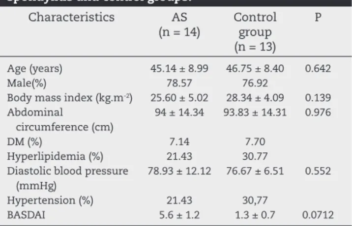
Subjects were predominantly male, and mean age of the patients was 45.14 years ± 8.99 vs 46.75 years ± 8.40 of the controls (P = 0.642). Systolic and diastolic blood pressure was similar in both groups. Body mass index (kg/m2), was 25.60
± 5.02 for AS group and 28.34 ± 4.09 for controls (P = 0.139). The abdominal circumference in patients and controls was, respectively, 94.0 ± 14.3 cm vs. 93.8 ± 14.3 (P = 0.976). As ex-pected, BASDAI was higher in AS patients than in controls (5.6 ± 1.2 vs 1.3 ± 0.7; P = 0.047). Prevalences of hypertension and diabetes were similar in both groups. Mean time of diagnosis was 12 years for the patients with AS. Only a single patient was in use of corticosteroids. No patient was using anti-TNFs.
AS (334,000 ± 106,000 vs. 252,000 ± 66,000; P = 0.028), as have been previously demonstrated in other rheumatic diseases.20
Although subjects were matched for diabetes, glucose level was higher in control than in AS patients (95.43 ± 10.53 vs. 109.1 ± 13.33; P = 0.009). LDL(-) did not differ signii cantly be-tween patients with AS and controls (14.03 ± 17.40 vs. 13.21 ± 10.21; P = 0.88).
Data regarding carotid IMT are summarized in Table 3. Regardless of the location (internal, bifurcation or common carotid) and side (left or right) of the artery where IMT was as-sessed, no difference was found between the two groups. As previously described, right subclavian was also evaluated and no difference was found either.
Discussion
Atherosclerosis is an inl ammatory condition and inl amma-tion experienced in chronic immune diseases may contrib-ute to accelerate atherosclerosis in this situations.21 In this
study, despite of active inl ammation as demonstrated by high CRP levels and BASDAI, patients with AS did not show higher prevalence of sub-clinical atherosclerosis, as
demon-strated by carotid IMT. In the same way, circulating plasma levels of LDL(-), which represents the fraction of LDL that is minimally modii ed, did not differ signii cantly in both groups.
Our study is in accordance with a previous study that also demonstrated lack of correlation between AS and increased IMT.22 In the study by Sari et al., young age of patients (mean
age, 37 years old) could have explained, at least in part, the lack of correlation if inl ammation was really assumed as an important factor for atherosclerosis development. In the same study, a compromised endothelial function, as dem-onstrated by impaired l ow mediated dilation of brachial ar-tery, was also shown. This could be interpreted as evidence that maybe patients were evaluated in a very initial phase of atherosclerosis development, since endothelial dysfunction precedes the morphological changes in arterial wall.23
Whether age (mean, 45 years old) was a limiting factor in our study is not known. Considering patients with other rheumatologic conditions, not only IMT is already increased in the fourth decade,24 but also the length of disease (the
mean time of diagnosis in our population was 12 years) is a determinant of thickening.25
In a publication where IMT was also assessed in patients with AS, only a trend towards increased thickness was seen.26 Similarly to our study, patients in control group had a
slight worse metabolic proi le than patients in AS, specially glucose levels.26 An interesting i nding in this study was that
arterial stiffness, another marker of subclinical atheroscle-rosis, was not modii ed by TNF-α inhibitors, suggesting that
modulation of inl ammatory response has no effect on ar-terial wall injury.26 None of our patients were being treated
with TNF-α inhibitors.
This was the i rst study to evaluate plasma levels of LDL(-) in patients with AS. Although elevated levels of oxidized LDL have been previously reported in patients with other autoimmune diseases,27 little has been published
regard-ing minimally modii ed LDL. These moieties are more prone to be oxidized when into the sub-endothelial space, where most of oxidation process occurs.
Although not as pro-atherogenic as oxidized LDL, mini-mally modii ed forms induce monocyte adhesion to endo-thelial cells and MCP-1 production, critical steps in early atherogenesis.28,29 The source of these minimally modii ed
forms remains unclear; however, there are possible sourc-es:30 oxidation of LDL in the arterial wall followed by egress
Table 1 – Clinical characteristics of the ankylosing spondylitis and control groups.
Characteristics AS (n = 14)
Control group (n = 13)
P
Age (years) 45.14 ± 8.99 46.75 ± 8.40 0.642
Male(%) 78.57 76.92
Body mass index (kg.m-2) 25.60 ± 5.02 28.34 ± 4.09 0.139 Abdominal
circumference (cm)
94 ± 14.34 93.83 ± 14.31 0.976
DM (%) 7.14 7.70
Hyperlipidemia (%) 21.43 30.77 Diastolic blood pressure
(mmHg)
78.93 ± 12.12 76.67 ± 6.51 0.552
Hypertension (%) 21.43 30,77
BASDAI 5.6 ± 1.2 1.3 ± 0.7 0.0712
AS, ankylosing spondylitis; DM, diabetes mellitus; BASDAI, Bath Ankylosing Spondylitis Disease Activity Index.
Table 2 – Biochemical parameters of the ankylosing spondylitis and control groups.
Biochemical measurement
AS (n = 14)
Control group (n = 13)
P
Cholesterol (mg/dL) 185.40 ± 34.88 221.60 ± 59.46 0.080 HDL (mg/dL) 46.79 ± 10.64 44.25 ± 11.11 0.559 Triglycerides (mg/dL) 131.20 ± 66.53 196.40 ± 105.20 0.080 LDL (mg/dL) 112.10 ± 29.10 140.70 ± 47.80 0.099 LDL(-) (U/L) 14.03 ± 17.40 13.21 ± 10.21 0.880 Glucose (mg/dL) 95.43 ± 10.53 109.10 ± 13.33 0.009 Leukocytes 9550 ± 2256 6915 ± 1721 0.003 Platelets (x103) 334 ± 106 252 ± 66 0.028 ESR (mm) 21.64 ± 15.01 15.64 ± 12.42 0.285 CRP (mg/L) 12.36 ± 7.99 3.45 ± 4.81 0.002
AS, ankylosing spondylitis; DM, diabetes mellitus; BASDAI, Bath Ankylosing Spondylitis Disease Activity Index.
Table 3 – Doppler ultrasonography assessment of intima-media thickness (IMT) of left (L) and right (R) carotid and right subclavian artery.
Biochemical measurement
AS (n = 14)
Control group (n = 13)
P
Left common carotid (mm) 0.58 ± 0.13 0.69 ± 0.53 0.461 Left bifurcation (mm) 0.86 ± 0.42 0.85 ± 0.35 0.982 Left internal carotid (mm) 0.74 ± 0.54 0.67 ± 0.15 0.623 Right common carotid (mm) 0.55 ± 0.16 0.55 ± 0.12 0.944 Right bifurcation (mm) 0.82 ± 0.37 0.80 ± 0.42 0.889 Right internal carotid (mm) 0.79 ± 0.53 0.70 ± 0.45 0.652 Right subclavian (mm) 1.16 ± 0.62 0.95 ± 0.30 0.287
into the circulation, ingestion of oxidized fats and/or genera-tion from postprandial lipoprotein remnants or direct oxida-tion in blood plasma. Regardless of the source, our i ndings do not support any role of inl ammation in this initial modi-i catmodi-ion of LDL partmodi-icle. Not only plasma levels were smodi-immodi-ilar in AS and control group, but also no correlation was found between minimally modii ed LDL and CRP levels (data not shown). Whether the same concept can be applied to oxi-dized LDL levels in patients with AS remains to be exploited. In patients with SLE and RA, not only oxidized LDL, but also their autoantibodies, are associated to atherosclerosis.31,32
It is not clear why most of the data from patients with AS are in the opposite direction of studies with SLE and RA regarding atherosclerosis. It’s worth mentioning that corti-costeroids treatment is always a confounding factor in these studies. While commonly prescribed in patients with RA and SLE, this is not a i rst line treatment in AS. In our study, a single patient was in use of prednisone. Mainly when used in high doses, corticosteroids have an important detrimen-tal effect on several cardiovascular risk factors. These effects can be in part responsible for the accelerated atherosclerosis seen in patients with SLE and RA.33,34 Further studies
includ-ing higher number of patients with AS takinclud-ing steroids could clarify this issue.
This study has potential limitations. First, a small num-ber of patients were included, and results might be seen as preliminary. The aim of this study was mainly to evaluate differences in minimally modii ed LDL plasma levels be-tween patients and controls. As this biochemical parameter is only used for research purposes, with no “normal” values even in healthy individuals, a sample calculation was not performed by the absence of reference values or even of pre-vious studies in patients with AS.
Another limitation was the fact that patients with more physical impairment, although invited during the screen-ing phase, did not participate. Refusal was always justii ed by difi culties in transportation. This is a frequent bias, re-ported also in studies with RA and SLE. Once patients with worst clinical conditions were not included, we may have lost those with more complications, including atherosclero-sis due to their inl ammatory condition.
In conclusion, despite active inl ammation and higher disease activity, there were no differences regarding plasma levels of minimally modii ed LDL and carotid IMT assessed by ultrasonography between patients with AS and controls. These i ndings are in accordance with previous studies in AS, but further and larger studies are necessary to explore the role of inl ammation per se as an accelerating factor of atherosclerosis in patients with spondyloarthritis.
Confl icts of interest
The authors declare no conl icts of interest.
R E F E R E N C E S
1. Berliner JA, Watson AD. A role for oxidized phospholipids in atherosclerosis. N Engl J Med. 2005;353:9-11.
2. Ross R. Atherosclerosis – An Inl ammatory Disease. N Engl J Med. 1999;340:115-26.
3. Virella G, Thorpe SR, Alderson NL, Stephan EM, Atchley D, Wagner F, et al. Autoimmune response to advanced glycosylation end-products of human LDL. J Lipid Res. 2003;44:487-93.
4. Tsimikas S, Brilakis ES, Miller ER, McConnell JP, Lennon RJ, Kornman KS et al. Oxidized phospholipids, Lp(a) lipoprotein, and coronary artery disease. N Engl J Med. 2005;353:46-57. 5. Witztum JL. The oxidation hypothesis of atherosclerosis.
Lancet1994;344:793-5
6. Cazzolato G, Avogaro P, Bittolo-Bon G. Characterization of a more electronegatively charged LDL subfraction by ion exchange HPLC. Free RadicBiol Med. 1991;11:247-53. 7. Paramo JA, Rodriguez JA, Orbe J. [Atherosclerosis in
inl ammatory diseases]. Med Clin (Barc). 2007;128:749-56. 8. Manzi S, Meilahn EN, Rairie JE, Conte CG, Medsger TA Jr,
Jansen-McWilliams L, et al. Age-specii c Incidence Rates of Myocardial Infarction and Angina in Women with Systemic Lupus Erythematosus: Comparison with the Framingham Study. Am J Epidemiol. 1997;145:408-15.
9. Dhawan SS, Quyyumi AA. Rheumatoid arthritis and cardiovascular disease.CurrAtheroscler Rep. 2008;10:128-33. 10. Troelsen LN, Jacobsen S. [Chronic inl ammation increases the
risk of cardiovascular disease in patients with rheumatoid arthritis]. UgeskrLaeger. 2006;168:3304-8.
11. Sattar N, McInnes IB. Vascular comorbidity in rheumatoid arthritis: potential mechanisms and solutions.
CurrOpinRheumatol. 2005;17:286-92.
12. Turesson C, Jacobsson LT, Matteson EL. Cardiovascular co-morbidity in rheumatic diseases. Vasc Health Risk Manag. 2008;4:605-14.
13. Kim SH, Lee CK, Lee EY, Park SY, Cho YS, Yoo B, et al. Serum oxidized low-density lipoproteins in rheumatoid arthritis. Rheumatol Int. 2004;24:230-3.
14. Azevedo VF, Pecoits-Filho R. Atherosclerosis and endothelial dysfunction in patients with ankylosing spondylitis. Rheumatol Int. 2010;30(11):1411-6.
15. Divecha H, Sattar N, Rumley A, Cherry L, Lowe GDO, Sturrock R. Cardiovascular risk parameters in men with ankylosing spondylitis in comparison with non-inl ammatory control subjects: Relevance of systemic inl ammation. Clinical Science. 2005;109:171-6.
16. Van Der Linden S, Valkenburg HA, Cats A. Evaluation of diagnostic criteria for ankylosing spondylitis.A proposal for modii cation of the New York criteria.Arthritis Rheum. 1984;27:361-8.
17. Demuth K, Myara I, Chappey B, Vedie B, Pech-Amsellem MA, Haberland ME, et al. A cytotoxic electronegative LDL subfraction is present in human plasma. Arteriosclerosis, Thrombosis, and Vascular Biology. 1996;16:773-83.
18. Avogaro P, Bon GB, Cazzolato G. Presence of a modii ed low density lipoprotein in humans. Arteriosclerosis. 1988;8:79-87. 19. Lorenz MW, Markus HS, Bots ML, Rosvall M, Sitzer M.
Prediction of clinical cardiovascular events with carotid intima-media thickness: a systematic review and meta-analysis. Circulation. 2007;115:459-67.
20. Milovanovic M, Nilsson E, Jaremo P. Relationships between platelets and inl ammatory markers in rheumatoid arthritis. ClinChimActa. 2004;343:237-40.
21. Avouac J, Allanore Y. Cardiovascular risk in rheumatoid arthritis: effects of anti-TNF drugs. Expert OpinPharmacother. 2008;9:1121-8.
22. Sari I, Uslu N, Gorgulu S, Nurkalem Z, Eren M. Inferior myocardial infarction and extensive atherosclerosis in a patient with double right coronary artery. Int J Cardiol. 2006;111:321-3.
of atherosclerosis precedes appearance of intimal lesions assessable with intravascular ultrasound. Am Heart J. 1996;131:231-8.
24. Manzi S, Selzer F, Sutton-Tyrrell K, Fitzgerald SG, Rairie JE, Tracy RP, et al. Prevalence and risk factors of carotid plaque in women with systemic lupus erythematosus. Arthritis Rheum. 1999;42:51-60.
25. Del Rincon I, O’Leary DH, Freeman GL, Escalante A. Acceleration of atherosclerosis during the course of rheumatoid arthritis. Atherosclerosis. 2007;195:354-60. 26. Mathieu S, Joly H, Baron G, Tournadre A, Dubost JJ, Ristori JM,
et al. Trend towards increased arterial stiffness or intima-media thickness in ankylosing spondylitis patients without clinically evident cardiovascular disease. Rheumatology (Oxford). 2008;47:1203-7.
27. Frostegard J. Autoimmunity, oxidized LDL and cardiovascular disease. Autoimmun Rev. 2002;1:233-7.
28. Cushing SD, Berliner JA, Valente AJ, Territo MC, Navab M, Parhami F,et al. Minimally modii ed low density lipoprotein induces monocyte chemotactic protein 1 in human endothelial cells and smooth muscle cells. ProcNatlAcadSci USA. 1990;87:5134-8.
29. Shih PT, Elices MJ, Fang ZT, Ugarova TP, Strahl D, Territo MC, et al. Minimally modii ed low-density lipoprotein induces
monocyte adhesion to endothelial connecting segment-1 by activating beta1 integrin. J Clin Invest.1999;103:613-25. 30. Damasceno NRT, Sevanian A, Apolinário E, Oliveira JMA,
Fernandes I, Abdalla DSP. Detection of electronegative low density lipoprotein (LDL-) in plasma and atherosclerotic lesions by monoclonal antibody-based immunoassays. Clinical Biochemistry. 2006;39:28-38.
31. Hahn BH, McMahon M. Atherosclerosis and systemic lupus erythematosus: the role of altered lipids and of autoantibodies. Lupus. 2008;17:368-70.
32. Peters MJ, van Halm VP, Nurmohamed MT, Damoiseaux J, Tervaert JW, Twisk JW, et al. Relations between autoantibodies against oxidized low-density lipoprotein, inl ammation, subclinical atherosclerosis, and
cardiovascular disease in rheumatoid arthritis. J Rheumatol. 2008;35:1495-9.
33. Bruce IN. ‘Not only...but also’: factors that contribute to accelerated atherosclerosis and premature coronary heart disease in systemic lupus erythematosus. Rheumatology (Oxford). 2005;44:1492-502.