Correlation Between Biochemical Markers and
Coronary Angiography in Patients with non-ST
Elevation Acute Coronary Syndromes
Cynthia Karla Magalhães, Aristarco Gonçalves de Siqueira Filho,
José Geraldo de Castro Amino, Mônica Nolasco
Universidade Federal do Rio de Janeiro e Cardiobarra Clínicas - Rio de Janeiro, RJ - BrazilMailing Address: Cynthia Karla Magalhães • Av. Prefeito Dulcídio Cardoso, 1350/2201 Bloco 2 - 22620-311 – Rio de Janeiro, RJ - Brazil
E-mail: ckarla@cardiol.br Received on 01/14/05 • Accepted on 06/01/05
O
BJECTIVEInvestigate the correlation between biochemical markers (TNI, CRP and fibrinogen) and anatomical coronary angiographic fi ndings in patients with non-ST elevation acute coronary syndromes (NSTE-ACS).
M
ETHODSOne blood sample was obtained to test for markers, and coronary angiography was performed within the fi rst 72 hours after hospitalization. Univariate analysis was used to search for correlations between the 3 markers and the angiographic fi ndings in the group of patients with an identifi ed ischemia-related artery (IRA), and multivariate analysis was performed to investigate the correlation between these markers and the presence of unstable atherosclerotic lesions solely in the group with a coronary obstruction >50%.
R
ESULTSProspective study conducted with 84 patients, 65.5% of whom were men. In the IRA-identifi ed group, blood levels of the three markers were higher than in the groups with no IRA-identifi ed or with normal coronary arteries. The analysis used to evaluate the IRA-identifi ed group showed signifi cant correlations between TIMI fl ow and TN-I (p = 0.006), unstable atherosclerotic lesions and TN-I and fi brinogen (p = 0.02 and p = 0.01, respectively), and multivessel disease and CRP (p = 0.0005). The multivariate analysis showed that CRP, fi brinogen and TN-I were independent predictors of unstable atherosclerotic lesions (p = 0.002; p = 0.003 and p = 0.007, respectively).
C
ONCLUSIONIn NSTE-ACS patients, TN-I, CRP and fi brinogen blood levels within the fi rst 10 hours after hospitalization correlated with coronary angiographic fi ndings.
K
EY WORDSThe detection of high blood levels of infl ammatory-activity markers in patients with acute coronary syndrome has confi rmed the importance of infl ammation in the process of atheroma plaque destabilization.
There is evidence in medical literature of a correlation between clinical presentation and serum elevation of biochemical markers (of inflammation, thrombosis and myocardial injury) in patients with non-ST acute coronary syndromes. Likewise, a relationship has also been demonstrated between clinical aspects and coronary anatomy in this group of patients1.
The objective of this study is to investigate the correlation between serum levels of inflammation, thrombosis and myocardial biochemical markers (titrated C-reactive protein, fi brinogen and troponin I, respectively) and anatomical fi ndings on coronary angiography in a group of non-ST ACS patients admitted to the emergency department.
M
ETHODS
After approval by the Research Ethics Committee, consecutive patients with unstable angina (UA) or non-ST acute myocardial infarction (non-non-ST AMI) admitted to the emergency department wereprospectively enrolled from November 1999 to July 2001. Patients fulfi lled the following criteria for inclusion: 1) Male or female, 18 years of age, minimum; fertile women should have a negative HCG blood test before being enrolled in the study. 2) Chest pain suggestive of myocardial ischemia, with up to 24 hours of progression that: a) occurs at rest or with minimum exertion, is prolonged (> 20 minutes) or recurrent (> two episodes of at least fi ve minutes each during the last 24 hours); or b) is progressive (angina episodes that become more frequent, more severe, last longer and/or are precipitated by minimum effort).
1) Angina symptoms associated with at least one of the following conditions: a) electrocardiogram showing ST segment depression greater than or equal to 0.5 mm, in at least two consecutive leads; or transient ST segment elevation (< 20 minutes) of no more than 1 mm in at least two consecutive leads; or T-wave inversion greater than or equal to 3 mm (or pseudonormalization > 1 mm above or below the isoelectric line) in at least three consecutive leads; or b) evidence of previous myocardial infarction documented by electrocardiogram; or c) previous coronary angiography showing at least one large coronary artery with a minimal occlusion of 50% of the luminal diameter; or d) previous myocardial revascularization documented with transluminal coronary angioplasty (balloon, directional coronary atherectomy or stent) or coronary artery bypass graft (CABG); or e) increase in enzyme levels: creatine kinase MB fraction (CK-MB mass) > 5.3 ng/mL and/or myoglobin > 70 ng/mL.
Patients with conditions that interfere with the interpretation of the ST segment on the electrocardiogram,
such as a pacemaker rhythm, were excluded. Likewise, patients with angina secondary to non-cardiac causes, systemic infl ammatory processes and regular users of anti-infl ammatory drugs were excluded.
Patients underwent one single peripheral venous blood sampling for laboratory tests. Venipuncture was performed in one of the upper extremities, according to international standard guidelines2, between 6 and
10 hours (average 8 hours) after hospital admission to determine: a) concentrations of titrated CRP (by nephelometry), reference value up to 0.5 mg/dL; b) TN-I (by immunofl uorometry – Opus Bhering), reference value up to 0.5 ng/mL; c) Fibrinogen (CLAUSS – automated methodology that measures thrombin consumption), reference value 200 to 400 mg/dL.
Creatine kinase MB (CK-MB mass - reference value up to 5.3 ng/mL) and myoglobin (reference value up to 70 ng/mL) were also measured in the same sample using the automated chemiluminescence method. Such enzymes were also measured at hospital admission as part of the emergency room protocol.
All patients underwent coronary angiography during hospitalization and, invariably, up to 72 hours (average 24 hours) after hospital admission.
Each angiography was analyzed by two separate experienced examiners blinded as to the other parameters analyzed, with the exception of the electrocardiogram and the echocardiogram, tools used to help identify the ischemia-related artery. The two examiners disagreed as to which angiographic group ten patients should be assigned (see description below), so a third examiner analyzed the angiographies in order to determine the parameter. The two examiners reached a consensus about the other angiographic differences.
The ischemia-related artery (IRA) was defi ned as the one with a lesion greater than or equal to 90% of the vessel lumen, with corresponding segment alteration on echocardiogram or electrocardiogram related to this vessel and/or instability of the atherosclerotic lesion.
To defi ne the presence of anunstable atherosclerotic lesion at least one of the following threecriteria had to be fulfi lled: a) IRA TIMI fl ow <3; b) IRA intracoronary thrombus (defi ned as intracoronary vascular fi lling defect); c) Ambrose type II eccentric atherosclerotic lesion in IRA (this isolated criterion was used only if the occlusion was greater than or equal to 90% of the vessel lumen).
Patients were broken down into three groups, according to coronary angiographic fi ndings: l) Group I: patients with identifi cation of the ischemia-related artery; 2) Group II: patients without identifi cation of the ischemia-related artery; 3) Group III: patients with normal coronaries or an atherosclerotic occlusion < 50 % of the vessel lumen.
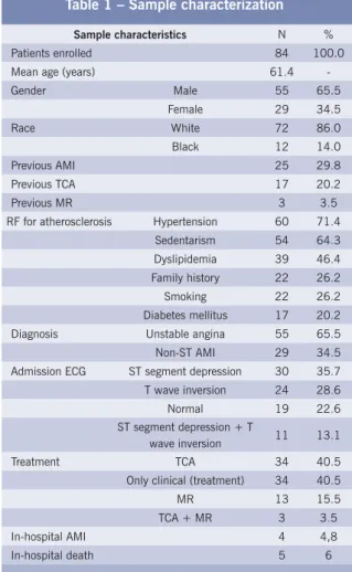
Table 1 – Sample characterization
Sample characteristics N %
Patients enrolled 84 100.0
Mean age (years) 61.4
-Gender Male 55 65.5
Female 29 34.5
Race White 72 86.0
Black 12 14.0
Previous AMI 25 29.8
Previous TCA 17 20.2
Previous MR 3 3.5
RF for atherosclerosis Hypertension 60 71.4
Sedentarism 54 64.3
Dyslipidemia 39 46.4
Family history 22 26.2
Smoking 22 26.2
Diabetes mellitus 17 20.2
Diagnosis Unstable angina 55 65.5
Non-ST AMI 29 34.5
Admission ECG ST segment depression 30 35.7
T wave inversion 24 28.6
Normal 19 22.6
ST segment depression + T
wave inversion 11 13.1
Treatment TCA 34 40.5
Only clinical (treatment) 34 40.5
MR 13 15.5
TCA + MR 3 3.5
In-hospital AMI 4 4,8
In-hospital death 5 6
RF – Risk factors; ECG – Electrocardiogram; TCA - Transluminal coronary angioplasty; MR – Myocardial revascularizarion; AMI – Acute myocardial infarct
presence of an unstable atherosclerotic lesion; number of arteries with an occlusion greater than 75% of the vessel lumen; and classifi cation of the lesion responsible for the ischemia, according to Ambrose et al3.
For Group II, the number of arteries with an occlusion greater than 75% of the vessel lumen and the presence of an unstable atherosclerotic lesion were analyzed.
TN-I, CRP and fi brinogen serum values were measured for the three angiographic groups, and the laboratorial defi nition for infarction was characterized by myoglobin > 70 ng/mL and/or CK-MB mass > 5.3 ng/mL at hospital admission or at the second measurement (on average 8 hours after admission). It is worth mentioning that for this paper the laboratory defi nition of acute myocardial infarction based solely on the increase of serum troponin levels was not used, as patients started to be enrolled before the American and European Heart Associations had published their agreement about the redefi nition of the disease. We chose not to alter the diagnoses of patients whose changes in enzyme levels were restricted exclusively to TN-I > 0.5 ng/mL, in order to preserve the original methodology.
For the IRA-identifi ed group (Group I), values for TN-I, CRP and fi brinogen correlated with the type of ischemia-related artery, TIMI fl ow, presence of thrombus and/or occlusion in the ischemia-related artery, presence of an unstable atherosclerotic lesion and the number of vessels with lesions > 75% of the lumen.
Finally, a multivariate analysis was conducted aiming to investigate the three biochemical markers (TN-I, CRP and fi brinogen) as independent predictors of unstable atherosclerotic lesions.
As such, Group III patients (normal coronaries or atherosclerotic occlusion < 50% of the vessel lumen) were excluded, and patients belonging to Groups I and II were analyzed as a whole and subdivided into two subgroups according to whether or not they had unstable atherosclerotic lesions.
It should be mentioned that, for the purpose of this analysis, positive TN-I values were considered as those greater than 0.5 ng/mL, and negative TN-I values were those smaller than or equal to 0.5 ng/mL.
The statistical analysis was done according to the following design: 1) Sample description with presentation of categorical and continuous variables distribution by means of central tendency and dispersion measurements. 2) Graphic representation of the continuous variables and their distribution in the groups using box plots. 3) Univariate analysis using Mann–Whitney and Kruskal– Wallis tests, as needed. 4) A generalized linear model with binomial distribution and a logit link function was used for the multivariate evaluation of the three markers, with cross validation and forward stepwise regression for the selection of variables according to the likelihood ratio.
5) The whole statistical analysis was planned with a 5% signifi cance level and 80% statistical power.
R
ESULTS
This study was carried out with eighty-four patients. Table 1 shows the characteristics of the patients enrolled in the study.
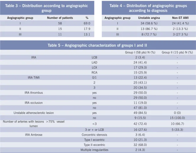
Table 2 shows the distribution of the biochemical markers analyzed. Theischemia-related artery (IRA) was identifi ed in 69% of the patients analyzed. Table 3 shows that fi fteen patients had coronary occlusion greater than 50% of the vessel lumen but it was not possible to identify the IRA, and eleven patients had normal coronaries.
In all three angiographic groups the main diagnosis was unstable angina and Group I had the largest proportion of non-ST AMI patients, as shown in Table 4. Table 5 displays the angiographic characteristics of Groups I and II.
Table 2 – Biochemical markers
Marker Mean Median First quartile Third quartile Standard deviation
Troponin I (ng/mL) 3.6 0.7 0.5 2.4 7.4
CRP (mg/dL) 1.5 0.8 0.5 1.9 2.5
Fibrinogen (mg/dL) 347.5 339.5 259.5 438.0 133.9
Table 3 – Distribution according to angiographic group
Angiographic group Number of patients %
I 58 69.0
II 15 17.9
III 11 13.1
Table 4 – Distribution of angiographic groups according to diagnosis
Angiographic group Unstable angina Non-ST AMI
I 34 (58.6 %) 24 (41.4 %)
II 13 (86.7 %) 2 (13.3 %)
III 8 (72.7 %) 3 (27.3 %)
Table 5 – Angiographic characterization of groups I and II
Group I (58 pts) N (%) Group II (15 pts) N (%)
IRA LCB 2 (3.4)
-LAD 24 (41.4)
-LCX 17 (29.3)
-RCA 15 (25.9)
-IRA TIMI 0/1 13 (22.4)
-2 25 (43.1)
-3 20 (34.5)
-IRA thrombus yes 29 (50.0)
-no 29 (50.0)
-IRA occlusion yes 11 (19.0)
-no 47 (81.0)
-Unstable atherosclerotic lesion yes 49 (84.5) 0 (0)
no 9 (15.5) 15 (100.0)
Number of arteries with lesions >75% vessel
lumen <3 42 (72.4) 10 (66.7)
3 or + or LCB 16 (27.6) 5 (33.3)
IRA Ambrose Concentric stenosis 3 (6.4)
-Type I eccentric 10 (21.3)
-Type II eccentric 32 (68.0)
-Multiple irregularities 2 (4.3)
-IRA – ischemia-related artery; GI – angiographic group I; GII – angiographic group II; LCB – left coronary branch; DA – LAD; LCX – circumfl ex artery; RCA – right coronary artery
to note that Group I had a mean TN-I value greater than the other two groups (p = 0.03). Moreover, Groups II and III had similar mean values (p = 0.47). CRP values appeared in decreasing order in Groups I, II and III, respectively, with statistical signifi cance (p = 0.001), and the same decreasing distribution was observed in the analysis of fi brinogen values among the three groups, also statistically signifi cant (p < 0.0001).
The analysis of the statistical correlation between angiographic variables and serum values of biochemical markers in Group I showed the following results: there was no correlation between the type of ischemia-related artery and the serum values of the biochemical markers analyzed, not even when patients were allocated to only two subgroups, right and left coronaries (p = 0.36; p = 0.10 and p = 0.10 for TN-I, CRP and fi brinogen, respectively).
When Group I patients were subdivided into two subgroups according to TIMI fl ow in the ischemia-related artery (TIMI 3 or TIMI <3), signifi cantly greater TN-1 values were observed in the subgroup with IRA TIMI fl ow < 3 (p = 0.006). Such a difference was not observed for CRP and fi brinogen values (p = 0.09 and p = 0.20, respectively).
Considering the presence or absence of a thrombus in the ischemia-related artery, no statistically signifi cant difference was observed for TN-I, CRP and fi brinogen (p = 0.08; p = 0.20 and p = 0.08, respectively).
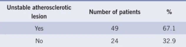
Table 6 – Breakdown of patients from Groups I and II according to the presence of unstable
atherosclerotic lesions
Unstable atherosclerotic
lesion Number of patients %
Yes 49 67.1
No 24 32.9
In the subgroup of patients with unstable atherosclerotic lesions, a signifi cant difference for TN-I and fi brinogen values (greater values) was observed as compared to patients in Group I with no unstable lesion (p = 0.02 and p = 0.01, respectively). However, no signifi cant difference was observed among the subgroups as to CRP values (p = 0.38).
Regarding the presence of multiarterial lesions (three or more arteries or LCB with lesions > 75% of vessel lumen), the CRP values observed were signifi cantly greater than those for the subgroup of patients with less than three arteries with lesions > 75% of the vessel lumen (p = 0.0005). For TN-I and fi brinogen, there was no signifi cant difference between these two subgroups (p = 0.8176 and p = 0.6911, respectively).
Group I and II patients were pooled and classifi ed according to the presence of an unstable atherosclerotic lesion, and 67.1% of them were found to have such a lesion instability. Group III patients were not included in this analysis since they had normal coronaries (Table 6).
It should be mentioned that the group with no unstable atherosclerotic lesions consisted of the fi fteen Group II patients and nine Group I patients who did not have unstable atherosclerotic lesions. The 49 patients with unstable atherosclerotic lesions were part of the group with an identifi ed IRA.
Multivariate analysis was used to examine CRP, fi brinogen and TN-I in order to identify which were prognostic markers of unstable atherosclerotic lesions.
Thismultivariate analysis showed that serum levels of CRP and fi brinogen, and whether or not TNI is positive, are independent factors of atherosclerotic lesion, i.e., the greater the levels of CRP and fi brinogen, the greater the probability of unstable atherosclerotic lesions; and when TN-I is positive (> 0.5 ng/mL), there is a greater probability of unstable atherosclerotic lesions as compared to anegativeTN-I (less than or equal to 0.5 ng/mL) (p = 0.002; 0.003, and 0.007, respectively).
D
ISCUSSION
In this paper, the diagnosis of unstable angina (UA) at hospital admission was more frequent than that of non-ST elevation acute myocardial infarction (non-ST AMI); this data are consistent with the fi ndings of a study by Zebrack et al4, who observed 58% of patients
with UA and 42% with non-ST AMI from a total of
1,360 patients with non-ST ACS. Such percentages are similar, since the cases described are prior to the year 2000 when acute myocardial infarction was redefi ned, and patients with TN-I values greater than the reference value, formerly seen as high-risk UA patients, started to be diagnosed as having a non-ST AMI, making this diagnosis more prevalent5,6.
Berk et al7, observed in their study that 90% of 37
patients with UA had CRP levels greater than 0.6 mg/dL at hospital admission. Another author conducting a study of patients with UA also described that 73% of them had serum CRP levels greater than 0.3 mg/dL7. In the present
study, 53 patients (63%) had serum levels of this marker greater than 0.5 mg/dL. The mean and median values for CRP and fi brinogen were also similar to the results observed by other authors in their analyses of patients with non-ST ACS4,7,8,9,10,11.
In the coronary angiographic analysis, specifi cally the percentage of patients with an identifi able IRA, there were differences between this study and the results observed by Benamer et al6 and Ambrose et al3, whose
percentages were 53 % and 60 %, respectively. However, those authors enrolled only patients with unstable angina which would explain the greater value observed in the present paper (69%)that alsoenrolled patients diagnosed with non-ST AMI, an illness with a higher prevalence of identifi cation ofthe ischemia-related artery12,13.
Fifty percent of Group I patients had an intracoronary thrombus, a percentage higher than that observed by Benamer et al6 and Dangas et al14 (18% and 14%,
respectively). As mentioned earlier, those authors analyzed only UA patients, whereas in this study, 34.5% of the sample consisted of patients diagnosed with non-ST AMI. Literature reports that angiographic fi ndings of intracoronary thrombus are more frequent among non-ST AMI patients, compared to UA, as well as in early coronary angiographic studies performed within the fi rst 24 hours of hospitalization. This would explain the high percentage found in the cases of patients here analyzed, as coronary angiography was performed on average within 24 hours after hospital admission15. In the cases described
by Benamer et al6, all coronary angiographies were
performed after 24 hours of hospitalization (on average, fi ve days post-admission), which might explain the 18% percentage of intracoronary thrombus observed.
Heeschen et al16, when analyzing 853 patients
with refractory UA by coronary angiography within the fi rst 24 hours after treatment onset, observed a higher prevalence of intracoronary thrombus (14.6%) in patients with a serum T troponin level (TN-T) greater than 0.1 mcg/L defi ned as TN-T positive (30.9% of the sample). In the present study, the high percentage of thrombi (50%) observed seems to be also associated with the high prevalence of TN-I positive patients (51%) in the sample.
these cases was based on the presence of an intracoronary thrombus, Ambrose II type eccentric lesion or IRA TIMI fl ow < 3. Other authors have also taken into consideration data such as the presence of a thrombus, coronary occlusion, TIMI fl ow and type II lesion eccentric as per Ambrose’s classifi cation to characterize atherosclerotic lesion instability3,6,8,17. It is worth mentioning that the
distribution of the patients analyzed in the present study according to the type of stenosis based on such a classifi cation followed a distribution similar to that described by the authors in 1985, when 54% of the stenoses were Ambrose type II eccentric lesions in groups of patients with unstable angina11. Another author
described a 66% percentage of Ambrose type II eccentric stenoses among 88 patients with UA, which is a value similar to the one found here (68%)18.
In 1996, Chen et al19, investigated the presence and
progression of angiographically complex lesions (irregular borders, ulcerated lesions and presence of a thrombus) in patients with stable and unstable angina pectoris. These authors observed 64% and 32% of complex lesions in the two groups, respectively. Moreover, they observed that the progression of these lesions had a greater prevalence in the unstable angina group, within a period of eight months. Medical literature reports that the complex stenoses observed in patients with stable angina, although angiographically similar to those of the unstable angina group, seem to differ in “activity”, according to a multifactor parameter that includes infl ammation, thrombogenic activity and vessel-reactivity20, corroborating
the conclusions of Chen et al19.
Other authors report the presence of unstable lesions in more than 70% of patients with acute coronary syndromes21, reaching 80% in the study by Benamer et
al6. Such data are consistent with those described here:
84.5% of unstable atherosclerotic lesions in the group with ischemia-related artery identifi ed, and in 67.1% of Groups I and II combined, i.e., in those patients with a coronaropathy defi ned as an occlusion greater than 50% of the vessel lumen. It should be mentioned that no patient in Group II had an unstable atherosclerotic lesion, since this characteristic was one of the criteria used to identify the IRA.
In the present sample, the TN-I highest mean value was observed in Group I, as compared to the other groups. Such data reinforce the results by Benamer et al6, in which serum TN-I elevation within the fi rst
twelve hours of hospital admission was an independent predictor of IRA identifi cation on coronary angiography. Moreover, patients with high TN-I levels had a higher prevalence of complex coronary stenoses (presence of thrombi or vessel occlusion) as compared to those with normal serum TN-I levels22,23,24.
CRP values were also signifi cantly higher in Group I patients compared to the other groups. In the study by Benamer et al6 there was no signifi cant difference in CRP
values among the groups with and without an identifi ed IRA on coronary angiography; however, the authors did not include in their analysis patients whose coronaries had no signifi cant atherosclerotic lesions and, consequently, with a smaller potential for coronary infl ammation.
Fibrinogen values were also signifi cantly higher in Group I patients. Such fi nding may be correlated with the high prevalence of intracoronary thrombi in this group (50%), as this biochemical marker is associated with inflammatory-thrombotic phenomena11. Also,
as mentioned previously, Group I consists of a higher percentage of patients with non-ST AMI as compared to the other two groups in which serum fi brinogen values are greater than those of UA patients, as shown in the TIMI IIIb study11.
No statistically significant correlation was found between the TNI, CRP and fi brinogen values and the type of artery responsible for the ischemia in Group I. This fi nding is consistent with two articles published that analyzed patients with non-ST ACS6,11. Likewise,
there was no signifi cant correlation between biochemical markers and TIMI fl ow, except the TN-I values that were greater in the subgroup with a TIMI of less than three, reinforcing the concept that the serum elevation of this enzyme is related to an arterial fl ow not suffi cient enough to prevent myocardial injury6,23,25. In a group of 1,161
patients with non-ST ACS, Lindhal et al24 demonstrated
an inverse relation between TN-T levels and IRA TIMI fl ow, i.e., the highest TN-T levels were detected in those patients with the smallest IRA TIMI fl ow.
No significant correlation was observed between biochemical markers and the presence of thrombi in the IRA. However, when this variable was analyzed together with others that were part of the defi nition of an unstable atherosclerotic lesion, such as type II Ambrose eccentric lesion and TIMI fl ow < 3, a signifi cant association with TN-I and fi brinogen serum levels was observed in the univariate analysis. It is possible that, with the inclusion of a greater number of patients, this variable may have isolated signifi cance.
Furthermore, in a study conducted with 1,150 non-ST UA and AMI patients during the period from October 1989 to June 1992, Antman et al26, did not detect a correlation
between the presence of a coronary thrombus and TN-I serum elevation either.
Lindhal et al24, however, demonstrated a direct
relationship between TN-I serum elevation and the presence of intracoronary thrombi, angiographically complex lesions and unfavorable TIMI fl ow in non-ST ACS patients.
coronary level6. In a study conducted with one hundred
patients with UA, no association was found between systemic infl ammation (elevation of plasma CRP) and the anatomical complexity of an atherosclerotic lesion in the IRA6.
Similarly, clinical studies with non-ST ACS patients suggest a connection between serum fi brinogen level and ischemic cardiovascular events since this marker is directly involved in the thrombotic process. However, no relationship between elevation of plasma fi brinogen and the presence of intracoronary thrombus has yet been demonstrated by coronary angiography11.
In the present study, no correlation was found between TN-I, CRP, and fi brinogen values and the presence of occlusion in the IRA. Benamer et al6 did not observe either
any association between TN-I elevation and occlusion of the vessel mentioned. As mentioned before, these authors have not demonstrated a correlation between serum CRP elevation and occlusion of the IRA either. As to fi brinogen, a marker for recent thrombosis, the lack of correlation may be associated with the presence of collateral circulation or non-thrombotic occlusion.
Correia et al10 described greater values of CRP in
patients with multiarterial coronariopathy hospitalized with diagnoses of UA or non-ST AMI. This fi nding is consistent with the results of this study in which merely the CRP value was signifi cantly higher in Group I patients with multiarterial coronariopathy, supporting the concept that TN-I is a marker of the presence of a vessel responsible for the ischemia and not of the number of vessels with atherosclerotic disease observed on coronary angiography6.
In the present study, patients with unstable atherosclerotic lesion were analyzed according to two different approaches.
Univariate analysis evaluated the correlation between serum levels of biochemical markers and the presence of lesion instability only for the group with identifi ed IRA (Group I). The analysis detected signifi cantly higher values of TN-I and fi brinogen in patients with lesion instability, a result similar to that of other authors for whom the elevation of these two biochemical markers was also associated with high angiographic complexity and signs of instability in the IRA (based on the Ambrose classifi cation, in the presence of thrombi and with abnormal TIMI fl ow)6,21,25. The analysis of CRP values, however, did
not show any difference between the values detected in groups with and without unstable lesions.
The exact relationship between infl ammation and atherosclerosis has yet to be fully defi ned, and there is not enough information available to determine if unstable coronary disease is due to a single vulnerable plaque or to diffuse vascular infl ammation. The recent work by Buffon et al27 suggests that individuals at increased risk of
acute coronary events, indicated by serum CRP elevation, supposedly have multiple vulnerable lesions in their coronary anatomy, i.e., vulnerable lesions in arteries other than those responsible for the ischemia, confi rming the association between the serum levels of this infl ammation marker and diffuse coronariopathy9,10.
Multivariate analysis investigated the relationship between the serum level of the three biochemical markers and the presence of atherosclerotic lesion instability in patients from Groups I and II combined, i.e., in all patients with obstructive coronariopathy with a block greater than 50% of the vessel lumen, subdivided according to the presence or absence of unstable lesions. In this analysis, besides TN-I and fi brinogen, CRP was also an independent predictor of the presence of unstable atherosclerotic lesions.
In 2000, Goldstein et al28, analyzed 253 patients
diagnosed with acute myocardial infarction who had undergone coronary angiography, and observed that 60.5% of them had one single complex atherosclerotic plaque (with thrombus, ulceration, irregular surface, insuffi cient fl ow and an obstruction of more than 50% of the vessel lumen), whereas 39.5% had multiple complex plaques, supporting the recent concept that ACS patients have diffuse coronary infl ammation that generates higher blood CRP levels. At the European Congress in 2003, Arroyo et al29 described a correlation
between the serum level of CRP measured at admission and the number of complex coronary stenoses in 125 UA patients. Thus, the association found in this study between the serum CRP level and the presence of unstable lesions can be justifi ed by the existence of more than one complex plaque, added to an extensive and diffuse coronary infl ammatory process in the group of patients with unstable lesions in the IRA, since none of the patients from Group II had unstable lesions and only nine patients (15.5%) from Group I did not have unstable lesions.
Sano et al30 recently analyzed ninety patients with
infarction who had undergone coronary intravascular ultrasound during the fi rst six hours of the onset of symptoms, and suggested that the elevation of serum CRP may be linked with atherosclerotic plaque rupture when there is intense infl ammatory activity. This conclusion also justifi es the greater CRP values in the group of patients with unstable lesions.
R
EFERENCES
1. Braunwald, E. Heart Disease A textbook of Cardiovascular Medicine. 5ª ed. Philadelphia: W. B. Saunders Company, 1997, 1996.
2. Standarts NCFCI. Procedures for the collection of diagnostic blood specimens by venipucture. 3rd ed. NCCLS Document H3 – A3. 1991.
3. Ambrose JA, Winters SL, Stern A, Teicholz LE, Gorlin R, Fuster V. Angiographic morphology and the pathogenesis of unstable angina pectoris. J Am Coll Cardiol 1985; 5: 609-16.
4. Zebrack JS, Anderson JL, Maycock CA, Horne BD, Bair TL. Usefulness of high sensivity C – reactive protein in predicting long-term risk of death or acute myocardial infarction in patients with unstable or stable angina pectoris or acute myocardial infarction. Am J Cardiol 2002; 15; 89(2): 145-9.
5. The Joint European Society of Cardiology/American College of Cardiology Committee. Myocardial infarction redefi ned. Eur Heart J 2000; 21: 1502-13.
6. Benamer H, Steg P, Benessiano J. Elevated cardiac troponin I predicts a high risk angiographic anatomy of the culprit lesion in unstable angina. Am Heart J 1999; 137: 815-20.
7. Berk BC, Weintraub WS, Alexander RW. Elevation of C reactive protein in “active” coronary artery disease. Am J Cardiol 1990; 65: 168-72.
8. Liuzzo G, Biasucci LM, Rebuzzi AG et al. Plasma protein acute-phase response in unstable angina is not induced by ischemic injury. Circulation 1996; 94: 2373-80.
9. Lindhal B, Toss H, Siegbahn A, Vengi P, Wallentin L. Markers of myocardial damage and inflammation in relation to long., term mortality in unstable coronary artery disease. N Engl J Med 2000; 343: 1139-47.
10. Correia LCL, Lima JC, Gerstenblith G, Magalhães LP et al. Correlação entre medidas de Proteína C Reativa pelos métodos de nefelometria e turbidimetria em pacientes com angina instável ou infarto agudo do miocárdio sem supra desnível do segmento ST. Arq Bras Cardiol 2003; 81: 129-32.
11. Becker RC, Cannon CP, Bovil EG et al. for the TIMI III Investigators. Prognostic value of plasma fi brinogen concentration in patients with unstable angina and non Q wave myocardial infarction (TIMI III B Trial). Am J Cardiol 1996; 78: 142-7.
12. Ambrose JA, Winters SL, Arora RR et al. Coronary angiographic morphology in myocardial infatction: a link between the Pathogenesis of unstable angina and myocardial infarction. J Am Coll Cardiol 1985; 6: 1233-8.
13. Campos A, Amino JG, Magalhães CK, Nagib E, Tura B. Graus crescentes de complexidade angiográfica correlacionam-se com formas progressivamente mais graves de síndrome isquêmica aguda sem supradesnível de segmento ST. Arq Bras Cardiol 2003; 81(supl. III): 28.
14. Dangas G, Mehran R, Wallenstein S, Kakarala V, Ambrose JA. Correlation of angiographic morphology and clinical presentation in unstable angina. J Am Coll Cardiol 1997; 29: 519-25.
15. ACC/AHA Guidelines for the management of patients with Unstable Angina and Non ST segment elevation myocardial infarction.J Am Coll Cardiol 2000; 36: 970- 1056.
16. Heeschen C, Van der Brand MJ, Hamm CW, Simoons ML. Angiographic findings in patients with refractory unstable angina according to Troponin T status. Circulation 1999; 104:1509-14.
17. Ambrose JA, Israel DH. Angiography in unstable angina. Am J Cardiol 1991; 68: 78B-84B.
18. Bugiardini R, Pozzati A, Borghi A et al. Angiographic morphology in unstable angina and its relation to transient myocardial ischemia and hospital outcome. Am J Cardiol 1991; 67: 460-4.
19. Chen L, Chester MR, Crook R, Kaski JC. Differential progression of complex culprit stenoses in patients with stable and unstable angina pectoris. I Am Coll Cardiol 1996; 28: 597-603.
20. Keaney JF, Vita JA. The value of infl ammation for predicting Unstable Angina. N Engl J Med 2002; 347: 55-7.
21. Zaacks S, Liebson PR, Calvin JE. Unstable Angina and non – Q wave infarction. Does the clinical diagnosis have therapeutic implications? J. Am. Coll. Cardiol, 1999; 33: 107-18.
22. Kodama K, Sakura M, Ueda Y, Yamaguchi O, Hirayama A. The role of plaque rupture in the development of acute coronary syndrome evaluated by the coronary angioscopy. Intern Med 2000; 39: 333-5.
23. Galvani M, Ottani F, Ferrini D, Ladenson JM, Destro A, Baccos D. Prognostic infl uence of elevated values of cardiac troponin I in patients with unstable angina. Circulation 1997; 95 (8): 2053-8.
24. Lindhal B, Venge P, Wallentin L. for the FRISC study group. Relation between troponin T and risk of subsequent cardiac events in unstable coronary artery disease. Circulation 1996; 93:1651-7.
25. Magalhães CK, Amino JG, Campos A, Ferreira E, Tura B. Correlação entre o fl uxo distal à lesão e marcadores não-invasivos de mau prognóstico nas síndromes coronarianas isquêmicas agudas sem supradesnível de segmento ST. Arq Bras Cardiol 2002; 79 (supl III): 92.
26. Antman EM, Tanasijevic MJ, Thompson B et al. Cardiac-specific troponin I levels to predict the risk of mortality in patients with acute coronary syndromes. N Engl J Med 1996; 335 (18): 1342-9.
27. Buffon A, Biasucci LM, Liuzzo G et al.Widespread Coronar y Infl ammation in Unstable Angina.N Engl J Med 2002; 347: 5-12.
28. Goldstein JA, Demetriou D, Grines CL, Pica M, Shoukfeh M, O’Neill WW. Multiple complex coronary plaques in patientes with acute myocardial infarction. N Engl J Med 2000; 343: 915-22.
29. Arroyo R, Avanzas P, Sales JC, Vasquez E, Gimeno J. C- reactive protein, had a high prevalence of three-artery disease (34%), and
none of which had an obstruction of less than 75% of the vessel lumen.
On the other hand, in patients from Group II (comprising 62.5% of the subgroup without an unstable lesion in the multivariate analysis), the infl ammatory process at the coronary level was not as exuberant, since 60% of patients in this group had merely moderate atherosclerotic occlusions (between 50% and 75% of the vessel lumen) and none of them had unstable lesions, which explained the smaller serum CRP level.
In 1999, Lagrand et al31 described CRP as a marker
that refl ects infl ammation associated not only with the extension but also with the severity of the atherosclerotic occlusion, corroborating the results reported here.
coronary disease activity and severity of coronary atherosclerosis in patients with angina pectoris.In: ESC Congress 2003, Austria. Eur Heart J 2003; 24(abstract suppl): 493.
30. Sano T, Kawarabayashi T, Tanaka A, Nishida Y, Shimada K, Yoshikawa J. C- reactive protein and lesion morphology in patients with acute
myocardial infarction. In: ESC Congress 2003, Austria. Eur Heart J 2003; 24(abstract suppl): 495.