Applicability of Longitudinal Strain of Left Ventricle in Unstable Angina
Natasha Soares Simões dos Santos, Andrea de Andrade Vilela, Rodrigo Bellio de Mattos Barretto, Marcela
Paganelli do Vale, Mariana Oliveira Rezende, Murilo Castro Ferreira, Alexandre José Aguiar Andrade,
Nelson Henrique Goes Scorsioni, Olívia Ximenes de Queiroga, David Le Bihan
Instituto Dante Pazzanese de Cardiologia, São Paulo, SP – Brazil
Mailing Address: Natasha Soares Simões dos Santos •
Av. Dante Pazzanese, 500. Postal Code 04012-909, Vila Mariana, São Paulo, SP – Brazil
E-mail: [email protected], [email protected] Manuscript received January 31, 2017, revised manuscript November 13, 2017, accepted November 22, 2017
DOI: 10.5935/abc.20180062
Abstract
Background: Unstable angina (UA) is a common cause of hospital admission; risk stratification helps determine strategies for treatment.
Objective: To determine the applicability of two-dimensional longitudinal strain (SL2D) for the identification of myocardial ischemia in patients with UA.
Methods: Cross-sectional, descriptive, observational study lasting 60 days. The sample consisted of 78 patients, of which fifteen (19.2%) were eligible for longitudinal strain analysis. The value of p < 0.05 was considered significant.
Results:The group of ineligible patients presented: a lower proportion of women, a higher prevalence of diabetes mellitus (DM), use of ASA, statins and beta-blockers and larger cavity diameters. The main causes of non-applicability were: presence of previous infarction (56.4%), previous CTA (22.1%), previous MRI (11.5%) or both (16.7%) and the presence of specific electrocardiographic abnormalities (12.8%). SL2D assessment revealed a lower global strain value in those with stenosis greater than 70% in some epicardial coronary arteries (17.1 [3.1] versus 20.2 [6.7], with p = 0.014). Segmental strain assessment showed an association between severe CX and RD lesions with longitudinal strain reduction of lateral and inferior walls basal segments; (14 [5] versus 21 [10], with p = 0.04) and (12.5 [6] versus 19 [8], respectively).
Conclusion: There was very low SL2D applicability to assess ischemia in the studied population. However, the global strain showed a correlation with the presence of significant coronary lesion, which could be included in the UA diagnostic arsenal in the future. (Arq Bras Cardiol. 2018; 110(4):354-361)
Keywords: Angina, unstable / physiopathology; Ventricular Dysfunction, Left; Myocardial Ischemia / physiopathology; Strain; Echocardiography / methods.
Introduction
In United States, unstable angina (UA) is the most common cardiovascular cause of hospitalization and is responsible for most hospitalizations in coronary units.1 The diagnosis of UA
is performed by clinical criteria based on angina duration and intensity.2 UA patient has a variable prognosis for unfavorable
events such as acute myocardial infarction (AMI), recurrence of angina, necrosis biomarkers, ventricular function and need for myocardial revascularization.3
Speckle tracking (ST) is a technology introduced in the 1980s that allows the quantification of global and regional myocardial deformity by tracking the natural heart muscle "acoustic marks" by ultrasound, presenting reduced values in presence of myocardial ischemia.4,5 ST allows myocardial strain calculation and has shown
great utility in the identification of subendocardial ischemia as
in unstable angina, with greater sensitivity and specificity than two-dimensional echocardiogram.6
However, for ST to track adequately speckles, there are some variables that may interfere in deformity analysis, so when present, they may give erroneous results or even impede myocardial strain analysis. In addition, for myocardial ischemia identification in UA patients, infarction previous myocardial presence or other myocardial injury (such as significant valvar heart disease) may alter myocardial deformity and cause an incorrect analysis of deformity decrease true cause. These are the variables that interfere in myocardial deformity correct analysis, and for that reason, they are considered exclusion criteria in the majority of published studies (aimed at analyzing acute ischemia): previous infarction, atrial fibrillation, left bundle branch block, ventricular arrhythmia aortic and/or mitral valvar disease, previous cardiac surgery, ventricular hypertrophy, cardiac pacemaker and inadequate acoustic window.7
The main objective is to study the applicability prevalence of two-dimensional longitudinal strain (SL2D) in all hospitalized patients diagnosed with UA during the 60-day observation period.
Methods
This is a cross-sectional, descriptive, performed at the Emergency Room (ER) and Coronary Unit (CU) of Dante Pazzanese Institute of Cardiology (IDPC).
Inclusion criteria were: hospitalized patients of both sexes, age greater than 18 years and clinical diagnosis of UA who were admitted to IDPC service during the study period and who accepted the participation in the study, having signed informed consent form. We emphasize that there was no calculation of sample size. A census was carried out of all patients who had inclusion criteria. Patient arrival to IDPC service was by convenience.
Exclusion criterion was the change in diagnosis during hospitalization. These cases occurred in patients who entered the service with initial chest pain and after propaedeutic and complementary exams, diagnosis of UA was ruled out. In case of differential diagnosis and closing as final diagnosis: acute myocardial infarction (AMI) with supra or no supra-ST segment, aortic dissection, pulmonary embolism and aortic stenosis.
We a n a l y z e d c l i n i c a l - e p i d e m i o l o g i c a l a n d electrocardiographic characteristics, as well as tests collection for troponin I and creatinine.
Risk stratification was done using GRACE risk score.8.9
Electrocardiographic analysis was performed by two experienced cardiologists; in case of disagreement regarding the diagnosis, tracing would be analyzed by a specialized service in the electrocardiographic reports of the institution where the research was carried out. Transthoracic echocardiography was performed within 48 hours of patient precordial pain last episode in ER or CU. The equipment for conducting examination was GE® Vivid E9 (General Electric Medical System, Norway) with transducer "array in phase" with 3.5 megahertz emission frequency. Images obtained during the examination were acquired with harmonic, in a repetition of frames between 50 and 70 frames/second, in digital clips form ( three consecutive cycles average) and recorded in CDs for later analysis in workstation EchoPAC PC version 6.0.1® (GE VingmedUltrasound).
According to American Society of Echocardiography and European Society of Echocardiography committee guidelines, which standardized the acquisition of tomographic sections obtained during echocardiographic examinations, with the patient in left lateral decubitus and electrocardiogram monitored, we acquired transthoracicly echocardiographic images by the Spectral Doppler (pulsatile and continuous) Doppler and flow mapping in color.10
Measures acquired:
• Two-dimensional: diastolic and systolic left ventricular (LV) diameter, left atrium anteroposterior diameter (LA), aortic root diameter, interventricular septum and posterior wall thickness. LV diastolic and systolic volume. Calculation of LV ejection fraction (EF) by the modified biplanar Simpson method.
• Doppler and Color Flow Mapping: Mitral flow with spectral Doppler (pulsatile and continuous) for diastolic
function analysis and mitral valvopathy quantification, when present. Aortic flow with spectral Doppler (pulsatile and continuous), to determine aortic valve opening and closing (to mark the systolic event), and aortic valvopathy quantification, when present. Valve lesions diagnosis and quantification followed American Society of Echocardiography recommendations.11
The technique to obtain longitudinal tension was done as follows:
• Marking systolic event with aortic flow pulsating Doppler. • Determination of three points of endocardial border
in each of the following images: apical 3 chambers (at anterosseptal wall base, at inferolateral wall base and at apex), apical 4 chambers (at septum base, at lateral wall base and at apex) and apical 2 chambers (at inferior wall base, at anterior wall base and at apex).
• Through the Automatic Function Imaging® (AFI) tool, the deformation of each of the 17 myocardial segments was automatically calculated, providing posteriorly left ventricle global deformation (analyzed segments mean). The program provides SL2D curves and polar map with longitudinal strain values in each segment.
The maximum absolute value of the two-dimensional strain curve was defined as the systolic peak. Adjacent myocardial segments with altered strain value were defined as ischemic territory, correlating them with coronary irrigation, according to polar map shown in Figure 1.
According to the literature,7,12,13 the situations mentioned
below may lead to a change in myocardial deformity, or to a real deformity impairment, or by limitation of the software to identify acoustic marks during the cardiac cycle:
• Concentric ventricular hypertrophy (LVH);
• Aortic and/or mitral valvar diseases greater than moderate degree;
• Pacemaker pace;
• At least one of the following electrocardiographic changes: left bundle branch block (LBBB), atrial fibrillation (AF) rhythm and complex ventricular arrhythmia; • S e c o n d a r y u n s t a b l e a n g i n a ( a c u t e a n e m i a ,
tachyarrhythmia and infection);
• Prior AMI or prior myocardial (percutaneous or surgical) revascularization procedure and
• Inadequate acoustic window.
Based on the above, we hypothesized that the presence of one of these alterations may impair SL2D analysis in severe coronary disease identification in UA patients. These concepts has fundamental importance for the knowledge of SL2D real applicability in this population, when the examination purpose is to evaluate coronary disease responsible for the acute condition.
Figure 1 – Polar map with coronary irrigation correlation. AD: anterior descending coronary artery; CX: circumflex coronary artery; RD: right coronary artery. Ante
rosept
al Ante
rior
La
te
ra
l
Inferior
Inferol
ater al
In
fe
ro
se
pt
al
2 1
7 8
6 12 16
11 5
3 9
4 10
15 17 14
13 AD
CX RD
Results of cardiac catheterization (CC) and Coronary Angiography by Computed Tomography (CACT) exams were also analyzed. Stenosis greater than or equal to 70% in epicardial coronary arteries or stenosis greater than or equal to 50% in left main coronary artery (LMCA) was considered.
The sample was divided into two groups: Group A - patients in whom it was possible to analyze by SL2D and Group B - patients in whom analysis by SL2D was not possible.
The research protocol was submitted and approved by the institution's Ethics and Research Committee.
There was no interference in individual medical conduct due to participation in the study. Such conduct was based on ER and CU routine that corresponds to US and national guidelines3,14 for UA patients treatment patients.
Statistical analysis
Statistical analysis was performed with Statistical Package for Social Sciences (SPSS), version 19.0.
Kolmogorov-Smirnov and Shapiro-Wilk tests were performed to verify our sample normal distribution. As the normality hypothesis was rejected, we used nonparametric tests for analysis.
Groups were compared using Mann-Whitney test and Fisher exact test as appropriate.
Continuous variables were presented as median and interquartile range, and categorical variables were expressed as percentage (%).
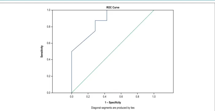
ROC curve was used to evaluate SL2D discriminative
power in severe coronary stenosis identification (≥ 70%)
in UA patients.
Level of significance was 5%.
Results
We evaluated 93 patients diagnosed with UA at admission to ER; however, fifteen (16.2%) patients were excluded from the study due to diagnosis change during hospitalization, 13 (14%) cases with non-ST-segment AMI, one (1.1%) with UA post-MI and one (1.1%) with type A aortic dissection.
At the end, 78 UA patients were investigated, of which fifteen (19.2%) were eligible for longitudinal strain analysis.
Main population clinical characteristics are summarized in Table 1.
About 70% of sample had no change in QRS complex duration or morphology complex; more than half (60.3%) showed no change in ventricular repolarization. Five patients (6.4%) presented ST segment depression on admission. Main electrocardiographic changes are detailed in Table 2.
Of the 63 patients in whom the longitudinal strain was not applied, 40 (63.5%) performed two-dimensional echocardiography during ER stay. Main echocardiographic findings of this population, including the 15 patients submitted to SL2D, are shown in Table 3.
In total, 50 patients completed the investigation with CC and five with CACT. In the first exam, three patients presented LMCA severe lesions (3.9%), 22 (28.2%) anterior descending coronary artery lesions (AD), 21 (26.9%) in right coronary artery, 2%) in circumflex coronary artery (CX). In patients submitted to CACT, one presented AD severe damage (1.3%) and one in RD (1.3%).
During hospitalization, 23 patients (29.5%) were submitted to intervention. Coronary transluminal angioplasty (CTA) was the main revascularization therapy. In three cases (3.8%), revascularization was surgical.
Comparing patients eligible for longitudinal strain analysis (group A) to those not eligible (group B), we found that group B had a lower proportion of women, a higher prevalence of diabetes, left cavities larger dimensions, larger root aorta diameter and lower systolic function on two-dimensional echocardiography; in addition to a higher ASA use rate, statins and beta-blockers, according to the data in Table 4.
Main causes to strain non-applicability were presence of prior infarction (56.4%), previous CTA (22.1%), prior surgical (CTA) revascularization (MRI), MRI and previous CTA (16, 7%), and presence of the following electrocardiographic alterations: LBBB, AF, pathological Q wave and pacemaker pace (12.8%).
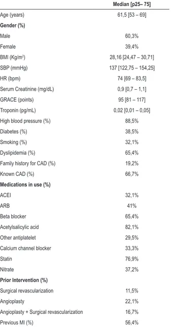
Table 1 – Clinical characteristics of studied population (n = 78)
Median [p25– 75]
Age (years) 61,5 [53 – 69]
Gender (%)
Male 60,3%
Female 39,4%
BMI (Kg/m2) 28,16 [24,47 – 30,71]
SBP (mmHg) 137 [122,75 – 154,25]
HR (bpm) 74 [69 – 83,5]
Serum Creatinine (mg/dL) 0,9 [0,7 – 1,1]
GRACE (points) 95 [81 – 117]
Troponin (pg/mL) 0,02 [0,01 – 0,05]
High blood pressure (%) 88,5%
Diabetes (%) 38,5%
Smoking (%) 32,1%
Dyslipidemia (%) 65,4%
Family history for CAD (%) 19,2%
Known CAD (%) 66,7%
Medications in use (%)
ACEI 32,1%
ARB 41%
Beta blocker 65,4%
Acetylsalicylic acid 82,1%
Other antiplatelet 29,5%
Calcium channel blocker 33,3%
Statin 76,9%
Nitrate 37,2%
Prior Intervention (%)
Surgical revascularization 11,5%
Angioplasty 22,1%
Angioplasty + Surgical revascularization 16,7%
Previous MI (%) 56,4%
BMI: body mass index; SBP: systolic blood pressure; HR: heart rate; CAD: coronary artery disease; AMI: acute myocardial infarction; ACEI: angiotensinogen converting enzyme inhibitor; ARB: angiotensin receptor AT-2 blocker.
Coronary anatomy evaluation revealed a severe lesion in LMCA in 1 case (6.7%). Number of patients with severe lesions in coronary arteries AD, CX and RD was 2 (13.3%), 4 (26.7%) and 4 (26.7%), respectively.
SL2D evaluation revealed a reduced global strain value in those who had severe lesion in any epicardial coronary artery (17.1 [3.1] versus 20.2 [6.7] with p = 0.014), area under the ROC curve 0.875, as shown in Figures 2 and 3.
Segmental strain assessment showed an association between severe CX lesion and longitudinal strain reduction of lateral wall basal segment (14 [5] versus 21 [10] with p = 0.04
and area under ROC curve = 0.864), and (12.5 [6] versus 19 [8] with p = 0.026 and area under ROC curve = 0.86).
Discussion
Acquisition of images by ST with longitudinal strain determination allows a more complete myocardial function assessment and can detect subtle alterations in segmental contractility in ischemic heart disease patients, with good inter and intraobserver reproducibility.7,12 Thus, this method
has been gaining more space in coronary artery disease evaluation, with a large number of studies produced in recent years.15-17
Table 2 – Electrocardiographic findings (n = 78)
Change Frequency (%)
LBBCD 10,3%
RBBB 3,8%
LASDB 2,6%
RBBB + LASDB 2,6%
LBBB 3,8%
Q wave pathological 3,8%
Pacemaker pace 3,8%
High response AF 1,3%
CVR anterosseptal 5,1%
Previous CVR 5,1%
Lower CVR 9%
Side CVR 7,7%
Diffuse CVR 11,5%
Infra/ST > 0,5 mm 1,3%
LBBCD: left bundle branch conduction disorder; RBBB: right bundle branch block; LASDB: left anterior superior divisional block; CVR: change in ventricular repolarization; LBBB: left bundle branch block; AF: atrial fibrillation.
Table 3 – Echocardiographic findings (n = 55)
Variable Median [p25 – p75]
LVEF Simpson 0,59 [0,5 – 0,65]
LA (mm) 39 [36 – 42]
LVFDD (mm) 51 [48 – 56]
LVFSD (mm) 32 [30 – 37,75]
Septum (mm) 10 [9 – 11]
Posterior wall (mm) 9 [9 – 11]
Mass index (g/m2) 124,5 [110 – 153,5]
PASP (mmHg) 32 [31 – 36]
Aorta root (mm) 34 [31 – 36]
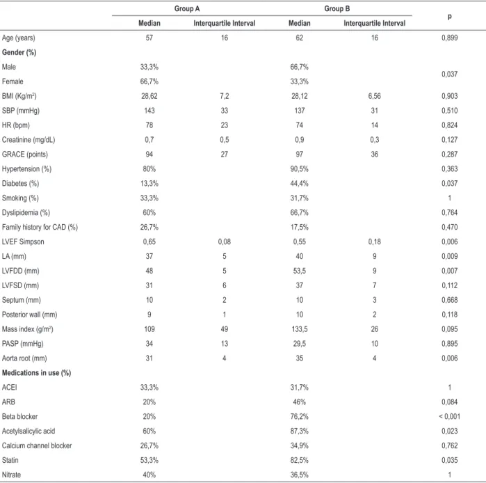
Table 4 – Clinical and echocardiographic characteristics of patients undergoing longitudinal strain analysis (group A, n = 15) compared to non-submitted patients (group B, n = 63)
Group A Group B
p Median Interquartile Interval Median Interquartile Interval
Age (years) 57 16 62 16 0,899
Gender (%)
Male 33,3% 66,7%
0,037
Female 66,7% 33,3%
BMI (Kg/m2) 28,62 7,2 28,12 6,56 0,903
SBP (mmHg) 143 33 137 31 0,510
HR (bpm) 78 23 74 14 0,824
Creatinine (mg/dL) 0,7 0,5 0,9 0,3 0,127
GRACE (points) 94 27 97 36 0,287
Hypertension (%) 80% 90,5% 0,363
Diabetes (%) 13,3% 44,4% 0,037
Smoking (%) 33,3% 31,7% 1
Dyslipidemia (%) 60% 66,7% 0,764
Family history for CAD (%) 26,7% 17,5% 0,470
LVEF Simpson 0,65 0,08 0,55 0,18 0,006
LA (mm) 37 5 40 9 0,009
LVFDD (mm) 48 5 53,5 9 0,007
LVFSD (mm) 31 6 37 7 0,112
Septum (mm) 10 2 10 3 0,668
Posterior wall (mm) 9 1 10 2 0,118
Mass index (g/m2) 109 49 133,5 26 0,095
PASP (mmHg) 34 13 29,5 10 0,895
Aorta root (mm) 31 4 35 4 0,006
Medications in use (%)
ACEI 33,3% 31,7% 1
ARB 20% 46% 0,084
Beta blocker 20% 76,2% < 0,001
Acetylsalicylic acid 60% 87,3% 0,023
Calcium channel blocker 26,7% 34,9% 0,762
Statin 53,3% 82,5% 0,035
Nitrate 40% 36,5% 1
BMI: body mass index; SBP: systolic blood pressure; HR: heart rate; LVEF: left ventricular ejection fraction; LA: measurement of the left atrium; LVFDD: left ventricular final diastolic diameter; LVFSD: left ventricular final systolic diameter; PASP: pulmonary artery systolic pressure. ACEI: angiotensinogen converting enzyme inhibitor; ARB: angiotensin receptor AT-2 blocker. Mann-Whitney was used for continuous variables (expressed in median and interquartile range) and Fisher’s exact test for categorical variables (expressed as percentage).
Table 5 – Risk score of patients submitted to longitudinal strain analysis
Score Frequency (%)
GRACE
≤ 108 points 86,7%
109-139 points 13,3%
≥ 140 points 0%
GRACE - Low risk - ≤ 108, moderate risk - 109 to 139, ≥ 140 - high risk
The present study is one of the pioneers in longitudinal strain applicability evaluation in UA patients attended at the Emergency Room of a Tertiary-level Cardiology Hospital.
due to ischemia. We emphasize that in the study population, 56.1% had previous infarction and 44.6% had previous cardiac procedure (CTA, MRI or both).
Shimoni et al.,18 evaluated SL2D in 97 hospitalized
patients with angina and normal ventricular function; of these, 69 patients had major coronary disease. Global strain analysis was -17.3 ± 2.4 with an area under the ROC curve (AUC) of 0.80 to identify significant CAD in patients with angina; in the subgroup of patients with unstable angina the global strain
also demonstrated good accuracy in predicting angiographic obstructive CAD (AUC = 0.86).18 Findings of this study are
similar to those found in relation to strain diagnostic accuracy to identify significant CAD in angina , however, there was no reference as to method applicability to the sample.
We verified in the present study a statistically significant association between reduced global strain values and the presence of anatomically severe CAD, and similar accuracy to data available in the literature.19 When we analyzed Figure 2 – ROC curve to evaluate ability of global strain to identify severe lesion (> 70%) in any epicardial coronary artery. Area under the ROC curve 0.875, with p < 0.014.
1.0
1.0 0.8
0.8 0.6
0.6 0.4
0.4 0.2
0.2 0.0
0.0
1 – Specificity ROC Curve
Sensitivity
Diagonal segments are produced by ties
1. Effects of tissue plasminogen activator and a comparison of early invasive and conservative strategies in unstable angina and non-Q-wave myocardial infarction. Results of the TIMI IIIB Trial. Thrombolysis in Myocardial Ischemia. Circulation. 1994;89(4):1545-56. doi: https://doi. org/10.1161/01.CIR.89.4.1545.
2. Braunwald E. Unstable angina: a classification. Circulation. 1989;80(2):410-4. https://doi.org/10.1161/01.CIR.80.2.410.
3. Anderson JL, Adams CD, Antman EM, Bridges CR, Califf RM, Casey DE, et al. 2012 ACCF/AHA 2007 guidelines for the management of patients with unstable angina/non-ST-elevation myocardial infarction: a report of the American College of Cardiology Foundation/American Heart Association Task Force on Practice Guidelines. J Am Coll Cardiol. 2013;61(23):e179-347. doi: 10.1016/j.jacc.2013.01.014. Erratum in: J Am Coll Cardiol. 2013;62(11):1040-1.
4. Amundsen BH, Helle-Valle T, Edvardsen T, Torp H, Crosby J, Lyseggen E, et al. Noninvasive myocardial strain measurement by speckle tracking echocardiography; validation against sonomicrometry and tagged magnetic resonance imaging. J Am Coll Cardiol. 2006;47(4):789-93. doi: https://doi. org/10.1016/j.jacc.2005.10.040.
5. Choi JO, Cho SW, Song YB, Cho SJ, Song BG, Lee SC, et al. Longitudinal 2D strain at rest predicts the presence of left main and three vessel coronary artery disease in patients without regional wall motion abnormality. Eur J Echocardiogr. 2009;10(5):695-701. doi: 10.1093/ejechocard/jep041.
6. Perk G, Tunick PA, Kronzon I. Non-Doppler two-dimensional strain imaging by echocardiography–from technical considerations to clinical applications. J Am Soc Echocardiogr. 2007;20(3):234-43. doi: http://dx.doi.org/10.1016/j. echo.2006.08.023.
7. Geyer H, Caracciolo G, Abe H, Wilansky S, Carerj S, Gentile F, et al. Assessment of myocardial mechanics using speckle tracking echocardiography: fundamentals and clinical applications. J Am Soc Echocardiogr. 2010; 23(4):351-69. doi: 10.1016/j.echo.2010.02.015. Erratum in: J Am Soc Echocardiogr. 2010;23(7):734.
8. Granger CB, Goldberg RJ, Dabbous O, Pieper KS, Eagle KA, Cannon CP, et al; Global Registry of Acute Coronary Events Investigators. Predictors of hospital mortality in the global registry of acute coronary events. Arch Intern Med. 2003;163 (19): 2345-53. doi: 10.1001/archinte.163.19.2345.
9. Eagle KA, Lim MJ, Dabbous OH, Pieper KS, Goldberg RJ, Van de Werf F, et al; Grace Investigators. A validated prediction model for all forms of acute coronary syndrome: estimating the risk of 6-month postdischarge death in an international registry. JAMA. 2004;291(22):2727-33. doi: 10.1001/jama.291.22.2727.
10. Lang RM, Bierig M, Devereux RB, Flachskampf FA, Foster E, Pellikka PA, et al; Chamber Quantification Writing Group; American Society of Echocardiography’s Guidelines and Standards Committee; European Association of Echocardiography. Recommendations for chamber quantification: a report from the American Society of Echocardiography’s Guidelines and Standards Committee and the Chamber Quantification
References
segmental strain, we found a significant association only in basal segment deformity reduction of lateral and inferior
walls, with stenosis ≥ 70% in CX and RD coronaries,
respectively. We believe that segmental strain findings would be more robust if the sample was larger.
In a meta-analysis published in 2016 with 1385 patients included in 10 studies, global longitudinal strain demonstrated good accuracy in detecting moderate to severe CAD in symptomatic patients with AUC of 0.81, sensitivity of 74.4% and specificity of 72.1%.19
Despite the low SL2D applicability in ER and CU, most probably due to patients profile that our institution attends, current evidence and our findings indicate that this method may be a complementary exam in diagnostic algorithm of CAD and useful tool in early ischemia evaluation.
Conclusion
In 80.8% of the cases, it was not possible to apply longitudinal strain, mainly due to the following criteria: presence of previous infarction or prior revascularization (percutaneous or surgical). We believe that the method applicability in a profile of patients with less clinical complexity would be greater, due to the method technical limitations.
In spite of this limitation, we can observe that the global strain showed a correlation with the presence of anatomically severe coronary lesion. In this way, SL2D could be included in the diagnostic arsenal of UA, in emergency units, since it is a noninvasive examination with diagnostic information available in a short period.
Author contributions
Conception and design of the research: Santos NSS, Vilela AA, Barretto RBM, Rezende MO, Ferreira MC, Andrade AJA, Scorsioni NHG, Queiroga OX, Le Bihan D; Acquisition of data: Santos NSS, Vilela AA, Vale MP, Rezende MO, Ferreira MC, Andrade AJA, Scorsioni NHG, Queiroga OX; Analysis and interpretation of the data: Santos NSS, Vilela AA, Barretto RBM, Vale MP, Rezende MO, Ferreira MC, Andrade AJA, Scorsioni NHG, Queiroga OX, Le Bihan D; Statistical analysis and Critical revision of the manuscript for intellectual content: Santos NSS, Vilela AA, Rezende MO, Ferreira MC, Andrade AJA, Scorsioni NHG, Queiroga OX; Obtaining financing and Writing of the manuscript: Vilela AA.
Potential Conflict of Interest
No potential conflict of interest relevant to this article was reported.
Sources of Funding
There were no external funding sources for this study.
Study Association
This study is not associated with any thesis or dissertation work.
Ethics approval and consent to participate
Writing Group, developed in conjunction with the European Association of Echocardiography, a branch of the European Society of Cardiology. J Am Soc Echocardiogr. 2005;18(12):1440-1463. doi: 10.1016/j.echo.2005.10.005.
11. Nishimura RA, Otto CM, Bonow RO, Carabello BA, Erwin JP 3rd, Fleisher LA, et al. 2017 AHA/ACC Focused Update of the 2014 AHA/ACC Guideline for the Management of Patients With Valvular Heart Disease. A Report of the American College of Cardiology/American Heart Association Task Force on Clinical Practice Guidelines. J Am Coll Cardiol. 2017;70(2):252-89. doi: 10.1016/j.jacc.2017.03.011.
12. Del Castillo JM, Herszkowicz N, Ferreira C. Speckle tracking–a contratilidade miocárdica em sintonia fina. Rev bras ecocardiogr imagem cardiovasc. 2010;23(3):46-54.
13. Leitman M, Lysyansky P, Sidenko S, Shir V, Peleg E, Binenbaum M, et al. Two dimensional strain: a novel software for real-time quantitative echocardiographic assessment of myocardial function. J Am Soc Echocardiogr. 2004;17(10):1021-9. doi: 10.1016/j.echo.2004.06.019.
14. Nicolau JC, Timerman A, Marin-Neto JA, Piegas LS, Barbosa CJ, Franci A, et al; Sociedade Brasileira de Cardiologia. [Guidelines of Sociedade Brasileira de Cardiologia for unstable angina and non-ST-segment elevation myocardial infarction (II edition, 2007) 2013-2014 update]. Arq Bras Cardiol. 2014;102(3 Suppl 1):1-61. doi: http://dx.doi.org/10.5935/ abc.2014S001.
15. Eek C, Grenne B, Brunvand H, Aakhus S, Endresen K, Smiseth AO, et al. Strain echocardiography predicts acute coronary occlusion in patients with non-ST-segment elevation acute coronary syndrome. Eur J Echocardiogr. 2010;11(6):501-8. doi: 10.1093/ejechocard/jeq008.
16. Grenne B, Eek C, Sjoli B, Dahlslett T, Uchto M, Hol PK, et al. Acute coronary occlusion in non-ST-elevation acute coronary syndrome: outcome and early identification by strain echocardiography. Heart. 2010;96(19):1550-6. doi: 10.1136/hrt.2009.188391.
17. Dahlslett T, Karlsen S, Grenne S, Eek C, Sjoli B, Skulstad H, et al. Early assessment of strain echocardiography can accurately exclude significant coronary artery stenosis in suspect non-ST-segment elevation acute coronary syndrome. J Am Soc Echocardiogr. 2014;27(5):512-9. doi: 10.1016/j. echo.2014.01.019.
18. Shimoni S, Gendelman G, Ayzenberg O, Smirin N, Lysyansky P, Edri O, et al. Differential effects of coronary artery stenosis on myocardial function: the value of myocardial strain analysis for the detection of coronary artery disease. J Am Soc Echocardiogr. 2011;24(7):748-57. doi: 10.1016/j.echo.2011.03.007.
19. Liou K, Negishi K, Ho S, Russell EA, Cranney G, Ooi SY. Detection of obstructive coronary artery disease using peak systolic global longitudinal strain derived by two-dimensional speckle-tracking: a systematic review and meta-analysis. J Am Soc Echocardiogr. 2016;29(8):724-35.e4. doi: 10.1016/j.echo.2016.03.002.