Pedro Miguel Gomes Gião Vicente
Degree in Cell and Molecular Biology
Disclosing CCBE1 role in Cardiac
Differentiation of Human Pluripotent
Stem Cells
Dissertation to obtain Master Degree in
Biotechnology
Supervisor: Dr. Margarida Serra, Senior Scientist, IBET/ITQB-NOVA
Jury:
President: Prof. Doctor Susana Filipe Barreiros Arguer: Doctor José Manuel Café Inácio
Supervisor: Doctor Maria Margarida de Carvalho Negrão Serra
September 2018
Disc los ing CCB E 1 ro le in Car d ia c Diff e rent iatio n of Hu man P lu ripo tent S tem Cells P ed ro V ic en te 2018
Pedro Miguel Gomes Gião Vicente
Degree in Cell and Molecular Biology
Disclosing CCBE1 role in Cardiac Differentiation of
Human Pluripotent Stem Cells
Dissertation to obtain Master Degree in
Biotechnology
Supervisor: Dr. Margarida Serra, Senior Scientist, IBET/ITQB-NOVA
Jury:
President: Prof. Doctor Susana Filipe Barreiros Arguer: Doctor José Manuel Café Inácio
Supervisor: Doctor Maria Margarida de Carvalho Negrão Serra
Disclosing CCBE1 role in Cardiac Differentiation of Human Pluripotent Stem Cells
Copyright © Pedro Miguel Gomes Gião Vicente, Faculdade de Ciências e Tecnologia, Universidade Nova de Lisboa
A Faculdade de Ciências e Tecnologia e a Universidade Nova de Lisboa têm o direito, perpétuo e sem limites geográficos, de arquivar e publicar esta dissertação através de exemplares impressos reproduzidos em papel ou de forma digital, ou por qualquer outro meio conhecido ou que venha a ser inventado, e de a divulgar através de repositórios científicos e de admitir a sua cópia e distribuição com objetivos educacionais ou de investigação, não comerciais, desde que seja dado crédito ao autor e editor.
Acknowledgements
Disclosing CCBE1 role in Cardiac Differentiation of Human Pluripotent Stem Cells V
Acknowledgments
I would like to express my gratitude to all the people directly or indirectly involved in this thesis. To my supervisor Dr. Margarida Serra, for her guidance, helpful discussions and critical suggestions. Thank you for giving me the opportunity to join the Stem Cell Bioprocessing group and for helping me to grow as a scientist.
To Dr. Patrícia Gomes-Alves, for her guidance, constant encouragement and support during this thesis. For the optimism and always being there to help.
To Dr. Paula Alves, for giving me the opportunity to do my master thesis at Animal Cell Technology Unit at ITQB-NOVA/IBET, for the good working conditions offered and for being a strong example of leadership.
To all my colleagues of the Animal Cell Technology Unit, for the good working environment, friendship and help during this year, especially those from the stem cells group. To Marta Silva, Maria João and Bernardo for the scientific and technical training given and fruitful discussions. To Marta Paiva for the critical opinions and constant support.
A special thanks to Marta Silva, to whom I owe most of the skills and knowledge acquired during my master thesis. Thank you for having the patience to teach me every technique used in this project, for the encouragement, and guidance, for being there for me during all time and for being a strong example of a scientist. I learned a lot with you this last year.
Aos meus colegas de mestrado, especialmente ao João e ao Miguel, mas também ao Ivo, Nuno e Rita pela amizade e todos os momentos bem passados. Um obrigado especial por estarem sempre disponíveis para ajudar e apoiar ao longo deste último ano.
A todos os restantes colegas da FCT, em especial à Francisca pela amizade partilhada ao longo dos últimos 5 anos. Por me fazeres sempre acreditar em mim, pela compreensão e apoio incondicional em todos as ocasiões. Por todos os momentos, conversas e até os grupos de estudo superprodutivos.
Aos meus amigos de longa data, por todas as ocasiões e experiencias vividas em conjunto e por me fazerem sempre rir em qualquer altura.
À minha família, em especial aos meus pais e à minha irmã por todo apoio incondicional em todos os momentos e circunstâncias. Por me terem proporcionado todas as condições para que fosse possível atingir cada etapa da minha vida, obrigado.
Preface
Disclosing CCBE1 role in Cardiac Differentiation of Human Pluripotent Stem Cells VII
Preface
This work was performed at the Animal Cell Technology Unit, IBET and ITQB-NOVA, under the scope of the projects CardioRegen (HMSP-ICT/0039/2013) and iNOVA4Health - UID/Multi/04462/2013, financially supported by the Fundação para Ciência e Tecnologia (FCT), Portugal and co-funded by FEDER under the PT2020 Partnership Agreement.
Part of the work presented in this thesis has been included in poster communications and a manuscript is under preparation.
Poster Communications:
• Silva MM, Vicente P et al. CCBE1 role along cardiac commitment of human pluripotent stem cells: exploring gene editing tools on loss-of-function studies. Frontiers in Cardiovascular Biology, April 20th – 22nd, 2018, Vienna, Austria.
• Silva MM, Vicente P et al. CCBE1-induced cardiac commitment on human pluripotent stem cells: exploring gene editing tools on loss-of-function studies. Workshop on Cardiovascular Research @ Bayer, May 31st – June 3rd, 2018, Cape Cod, USA.
Conference proceeding:
• Silva MM, Vicente P et al. (2018) CCBE1 role along cardiac commitment of human pluripotent stem cells: exploring gene editing tools on loss-of-function studies. Cardiovascular Research. 114. S20-S20. 10.1093/cvr/cvy060.038.
Manuscript:
• Silva MM, Vicente P et al. (2018) Unveiling CCBE1 role as a modulator of human pluripotent stem cells cardiomyocyte differentiation. In preparation.
Abstract
Disclosing CCBE1 role in Cardiac Differentiation of Human Pluripotent Stem Cells IX
Abstract
Cardiovascular diseases (CVD) are the leading cause of death worldwide. Within CVDs, myocardial infarction (MI) is associated with a massive and irreversible loss of cardiomyocytes (CM). An in-depth comprehension of key cellular mechanisms and molecules involved in cardiogenesis is fundamental to improve cardiac therapies by exposing novel therapeutic targets. CCBE1, a collagen and calcium-EGF biding domain 1 protein, was identified to be expressed in mouse heart precursors. Mutations in CCBE1 have been associated with Hennekam syndrome, which is characterized by abnormal lymphatic system and congenital heart defects. However, the CCBE1 functional role in cardiac specification is still unknown. Therefore, the main aim of this thesis was to unveil CCBE1 role in CM and Endothelial cells (EC) specification. For this purpose, a modified hiPSC line displaying the CRISPR interference technology (CRISPRi) was used to selectively knockdown (KD) CCBE1 gene expression along CM and EC differentiation.
We showed that CCBE1 downregulation did not affect hiPSCs growth, morphology and stemness. Nonetheless, a significant reduction on gene expression of cardiac troponin T2 gene (TNNT2) and lower gene expression ratios of cardiac troponin I isoforms (TNNI3:TNNI1) and myosin heavy chains (MYH7:MYH6) were detected in CMs derived from CRISPRi-CCBE1 KD cell line at day 15. Ultrastructural changes were also observed in this condition, CMs presented lower sarcomere length and alignment, indicating a more immature state. In contrast, EC differentiation was not affected by CCBE1 KD, with no impact on EC morphology or gene expression levels. Therefore, CCBE1 seems to have a key role on CM specification and maturation. Moreover, we successfully selected hiPSC clonal populations with higher level of CCBE1 KD for future studies. This work may contribute with new insights towards the development of CCBE1-mediated therapeutic strategies for cardiac regenerative medicine.
Keywords: Cardiovascular Disease; human induced Pluripotent Stem Cells (hiPSCs); CCBE1;
Resumo
Disclosing CCBE1 role in Cardiac Differentiation of Human Pluripotent Stem Cells XI
Resumo
As doenças cardiovasculares são das principais causas de morte, sendo o enfarte do miocárdio associado à perda excessiva e permanente de cardiomiócitos (CM). Uma melhor compreensão dos mecanismos celulares que desempenham um papel crucial na cardiogénese é fundamental para identificar novos alvos terapêuticos e melhorar as terapias cardiovasculares. A expressão da proteína CCBE1 (collagen and calcium-EGF binding domain 1) foi detetada nos percursores cardíacos durante o desenvolvimento embrionário em ratinhos. Para além disso, mutações nesta proteína estão associadas ao síndrome de Hennekam, apresentando deficiências no sistema linfático e defeitos congénitos no coração. Dado que, o papel funcional da CCBE1 na diferenciação cardíaca não é conhecido, o principal objetivo desta tese foi investigar a função desta proteína, utilizando células estaminais pluripotentes induzidas (hiPSC) e métodos de modificação génica (tecnologia de interferência CRISPR) para induzir a perda de função do gene em causa durante a diferenciação em CM e células endoteliais (CE).
A Inibição da expressão da CCBE1 não afetou o crescimento, morfologia nem a pluripotência das hiPSC. Por outro lado, durante a diferenciação em CMs observou-se um decréscimo da expressão do marcador cardíaco TNNT2 e diminuição dos rácios de expressão das isoformas de troponina I (TNNI3:TNNI1) e das cadeias pesadas de miosina (MYH7:MYH6). Estes CMs também apresentavam alterações ultraestruturais, nomeadamente no tamanho e alinhamento dos sarcómeros, indicadores de um fenótipo mais imaturo comparativamente com a cultura com níveis de CCBE1 normais. No entanto, durante a diferenciação em CE não foi observado qualquer efeito da diminuição da expressão da CCBE1, tanto a nível morfológico como na expressão génica. Estes dados sugerem que esta proteína pode ter uma função importante no fenótipo dos CM. Adicionalmente, foram selecionados clones com maior nível de inibição da expressão da CCBE1 para estudos futuros. Este trabalho poderá contribuir para o desenvolvimento de novas terapias cardíacas baseadas no papel da CCBE1.
Palavras-chave: Doenças Cardiovasculares; Células estaminais pluripotentes induzidas
List of Contents
Disclosing CCBE1 role in Cardiac Differentiation of Human Pluripotent Stem Cells XIII
List of Contents
Acknowledgments ... V
Preface ... VII
Abstract ... IX
Resumo ... XI
List of Contents ... XIII
Figure Index ... XV
Table Index ... XVII
Abbreviations... XIX
1. Introduction... 1
1.1. Cardiovascular Diseases: Prevalence & Treatment ... 1
1.2. Novel Therapies for heart regeneration and repair ... 2
1.2.1. Stem cell-based therapy for Myocardial Regeneration ... 2
1.2.1.1. Understanding the human Cardiogenesis ... 5
1.2.2. Cell-Free Approaches for Myocardial Regeneration ... 7
1.2.2.1. Neovascularization and Lymphangiogenesis to augment heart repair ... 8
1.2.3. The Role of CCBE1 as a potential modulator of cardiac function ... 9
1.3. Gene editing tools ... 12
2. Aim of the thesis ... 17
3. Material & Methods ... 19
3.1. hiPSC culture & differentiation ... 19
3.1.1. hiPSC lines ... 19
3.1.2. hiPSC expansion ... 19
3.1.3. hiPSC cardiac differentiation ... 19
3.1.4. hiPSC endothelial differentiation ... 19
3.2. CCBE1 knockdown: gRNA design, cell electroporation and selection ... 20
3.3. hiPSC Characterization ... 21
3.3.1. Cell concentration and viability determination ... 21
3.3.2. Cell proliferation ... 21
List of Contents
Disclosing CCBE1 role in Cardiac Differentiation of Human Pluripotent Stem Cells XIV
3.3.4. Flow Cytometry ... 22
3.3.5. mRNA Extraction and RT-qPCR ... 22
3.3.6. Transmission electron microscopy (TEM) ... 23
3.4. Statistical Analysis ... 24
4. Results and Discussion ... 25
4.1. CCBE1 expression is transiently upregulated at early stages of cardiomyocyte differentiation ... 25
4.2. Generation of CCBE1 knockdown in iPSC cell line ... 28
4.3. CCBE1 knockdown impacts cardiomyocytes differentiation ... 30
4.4. CCBE1 knockdown has no impact on endothelial expression markers ... 35
4.5. CCBE1 knockdown is more prominent in the selected clones compared to the polyclonal populations ... 36
5. Conclusions and Future Perspectives ... 39
Figure Index
Disclosing CCBE1 role in Cardiac Differentiation of Human Pluripotent Stem Cells XV
Figure Index
Figure 1.1 – Death causes of European population in 2016 ... 1
Figure 1.2 – Schematic representation of the expressed transcriptional factors along iPSCs cardiac differentiation. ... 5
Figure 1.3 – Schematic representation of CCBE1 and its key roles. ... 10
Figure 1.4 – Lymphangiogenesis: VEGF-C activation by CCBE1 and ADAMTS3 complex and its role in myocardial infarction (MI). ... 11
Figure 1.5 – Differences in action method of Streptococcus pyogenes Cas9 and dCas9. ... 14
Figure 1.6 – Potential of CRISPRi hiPSC lines developed by Mandegar and colleagues. ... 15
Figure 2.1 – Schematic representation of the major aims of this thesis and outlined strategy. .. 17
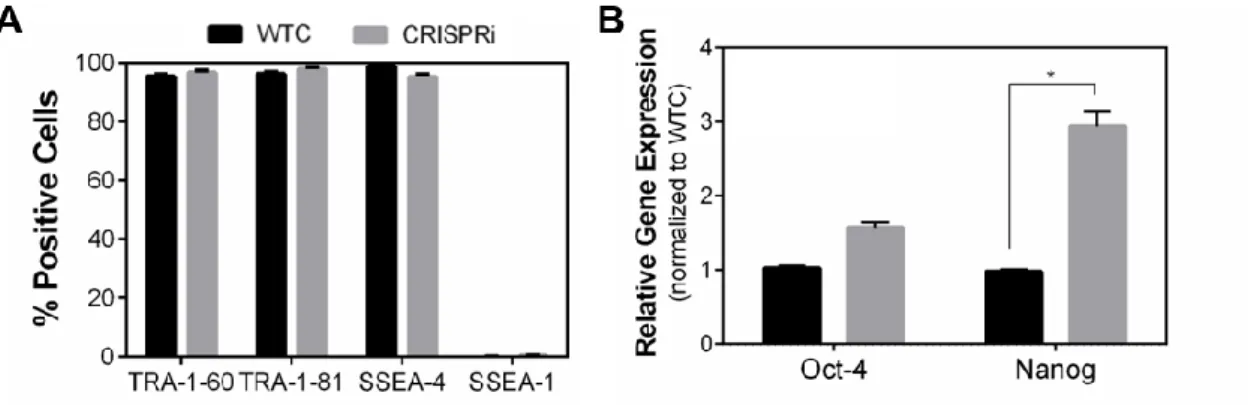
Figure 4.1 – Pluripotency of WTC and CRISPRi hiPSC lines. ... 25
Figure 4.2 – human induced pluripotent stem cells efficiently differentiate into cardiomyocytes. ... 27
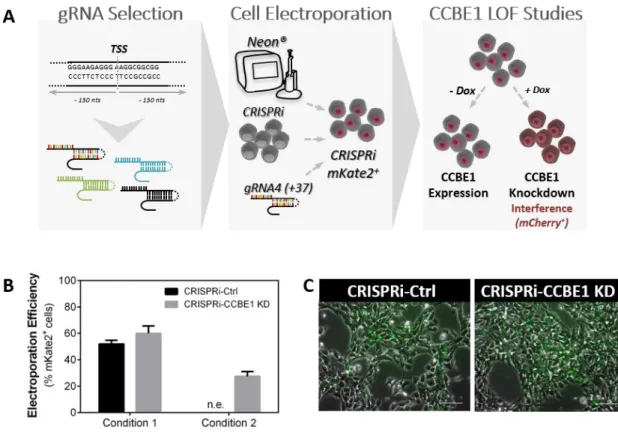
Figure 4.3 – CRISPRi technology for CCBE1 knockdown. ... 28
Figure 4.4 – Efficient CCBE1 knockdown with the selected gRNA in hiPSCs. ... 30
Figure 4.5 – CCBE1 knockdown during cardiomyocyte differentiation. ... 32
Figure 4.6 – CCBE1 knockdown affects other cardiac specific markers gene expression. ... 33
Figure 4.7 – Impact of CCBE1 knockdown on cardiomyocyte differentiation/maturation. ... 34
Figure 4.8 – Effect of CCBE1 knockdown on cardiac endothelial differentiation. ... 35
Table Index
Disclosing CCBE1 role in Cardiac Differentiation of Human Pluripotent Stem Cells XVII
Table Index
Table 1.1 – Structural, gene expression and energy related characteristics in adult and
immature-like CMs. ... 6 Table 1.2 – Preclinical results from cardiac regeneration therapies for ischemic heart diseases. 7 Table 1.3 – Comparison of the major gene editing tools. ... 12 Table 3.1 – List of gRNA oligo sequences. ... 20 Table 3.2 – List of all antibodies and dilutions used for immunocytochemistry and flow
cytometry analysis. ... 23 Table 3.3 – List of all Primers used in RT-qPCR. ... 24
Abbreviations
Disclosing CCBE1 role in Cardiac Differentiation of Human Pluripotent Stem Cells XIX
Abbreviations
ADAMTS3 A Disintegrin and Metalloproteinase with Thrombospondin Motifs-3
BM Bone marrow
BMP Bone morphogenic protein
BSA Bovine Serum Albumin
Cas9 CRISPR associated protein 9
CCBE1 Collagen and calcium-binding EGF domain-1
CMs Cardiomyocytes
CO2 Carbon dioxide
CPC Cardiac Progenitor Cell
CRISPR Clustered Regularly Interspaced Short Palindromic Repeats
CSC Cardiac Stem Cell
cTnI Cardiac muscle troponin I
cTnT Cardiac muscle troponin T
CVD Cardiovascular diseases
DMEM Dulbecco’s modified Eagle medium DMSO Dimethyl sulfoxide
Dox Doxycycline
DSB Double strand break
EC Endothelial Cell
EdU 5-ethynyl-2´-deoxyuridine
Endo Endocardium
Epi Epicardium
FBS Fetal bovine serum
FDR False discovery rate
FGF Fibroblast growth factor
FHF First Heart Field
GAPDH Glyceraldehyde-3-Phosphate Dehydrogenase
GATA4 Transcription factor GATA-4
gRNA guideRNA
hESC Human embryonic stem cells
HF Heart Failure
hiPSC Human induced pluripotent stem cells
IgG Immunoglobulin G
IgM Immunoglobulin M
IWR Inhibitors of Wnt response
KD Knockdown
KDR Kinase insert domain receptor
Abbreviations
Disclosing CCBE1 role in Cardiac Differentiation of Human Pluripotent Stem Cells XX
LVEF Left ventricular ejection fraction
MESP1 Mesoderm Posterior BHLH Transcription Factor 1
MI Myocardial infarction
MSC Mesenchymal stem cells
MYH6 Myosin heavy chain 6, α isoform protein MYH7 Myosin heavy chain 7, β isoform protein
MYL2 Myosin light chain 2, ventricular/cardiac muscle isoform (MLC2v)
MYL7 Myosin light chain 7, atrial isoform (MLC2a)
Nanog Homeobox Protein Nanog
Nkx2-5 NK2 Homeobox 5
Oct4 Transcription factor octamer 4
PAM Protospacer-adjacent motif
PBS Phosphate-buffered saline
PFA Paraformaldehyde
PSC Pluripotent stem cells
RNAi RNA-mediated interference
RPLP0 Ribosomal Protein Lateral Stalk Subunit P0
RT-qPCR Reverse transcriptase quantitative polymerase chain reaction
SHF Second Heart Field
SIRPα/β Signal-regulatory protein alpha/betta
SMs Skeletal myoblasts
SSEA-1 Stage-specific embryonic antigen-1
SSEA-4 Stage-specific embryonic antigen-4
TALENs Transcription activator-like effector nucleases
TNNI1 Troponin I1, Slow Skeletal Type
TNNI3 Troponin I3, Cardiac Type
TNNT2 Cardiac muscle troponin T
TRA-1-60 Human embryonal carcinoma marker antigen 60
TRA-1-81 Human embryonal carcinoma marker antigen 81
VCAM1 Vascular cell adhesion molecule 1
VE-cadherin Vascular endothelial cadherin, CD144
VEGF Vascular endothelial growth factor
WNT Wingless-related integration site
Introduction
Disclosing CCBE1 role in Cardiac Differentiation of Human Pluripotent Stem Cells 1
1.Introduction
1.1.Cardiovascular Diseases: Prevalence & Treatment
Cardiovascular diseases (CVD), a group of disorders of the heart and blood vessels, persist as the leading cause of death worldwide, accounting for 17.7 million deaths per year, a number that is expected to grow even further, to 23.6 million deaths by 2030 [1]. In Europe CVDs were responsible for almost 45% of all deaths in 2016 (Figure 1.1). In particular, myocardial infarction (MI) or heart attack results in cardiac muscle loss, due to cardiomyocyte (CM) death either by apoptosis or necrosis. Moreover, the limited capability of the heart tissue to regenerate makes this loss largely irreversible and a scar tissue constituted by fibroblasts is formed, resulting in loss of contractility and decreased heart function. This consequently leads to the development of heart failure (HF) [2]. Although adult CMs do not proliferate, evidences of a resident cardiac progenitors’ cells (CPC) population able to differentiate into CMs may provide some endogenous regenerative capacity in the adult heart, however at insufficient rates to compensate for the massive cell loss caused by MI [3].
Figure 1.1 – Death causes of European population in 2016. Adapted from [4].
Heart transplantation remains the best long-term solution for end-stage HF, however the limited number of donors available, the high costs and possible tissue rejection, makes it unrealistic to be considered a standard therapy. Current treatments can improve patient’s survival and well-being, yet they fail to regenerate or repair the damaged heart. For example, pharmacological approaches, such as inhibitors of renin-angiotensin system, aim to reduce myocardial fibrosis, hypertrophy and incidence of heart failure [5]. In addition, revascularization
Cardiovascular diseases (44,5%) Cancer (22%) Respiratory diseases (6,5%) Injuries and poisoning (6,5%)
All other causes (20,5%)
Introduction
Disclosing CCBE1 role in Cardiac Differentiation of Human Pluripotent Stem Cells 2 approaches have also been widely used to improve blood flow after MI [6]. Even though these methods have successfully reduced the mortality rates, they are intrinsically non-curative and hence novel approaches capable to promote heart regeneration and repair are still required.
1.2.Novel Therapies for heart regeneration and repair
1.2.1.Stem cell-based therapy for Myocardial Regeneration
Regeneration of the injured heart by replacing the lost CM population, is an attractive approach to repair the heart and avoid future HF. To date, clinical efforts towards cardiac repair and regeneration have largely focus on stem based therapies. The first generation of cell-based therapies used for this purpose included the transplantation of noncardiac stem cells, such as, mesenchymal stem cells (MSCs), bone marrow (BM)-derived cells and myoblast cells, as they were more easily obtained [7]. One of the first cell types to be tested, with the goal of remuscularization in mind, were the skeletal myoblasts (SMs), although they did not yield any improvements in the randomized phase-II MAGIC trial [8]. In addition, concerns about their arrhythmogenic potential led to the end of further development of cell therapies based on this cell type [9].
Other cell types used in cardiac regeneration trials were BM-derived cells, due to their safety, easy isolation and encouraging initial results [10]. Early clinical trials, namely the BOOST [11] and REPAIR-AMI [12], have shown some beneficial effects in patients with MI through improvement of the ejection fraction in cell-treated groups compared to placebo. Nevertheless, other clinical trials with a wider number of patients, didn’t display any beneficial outcomes [13, 14]. The ongoing investigations with these cells aim to end these controversies and to draw a conclusion about their beneficial effects for patients.
The last major noncardiac cell source studied in regenerative therapies are the multipotent adult MSCs, which demonstrated a great potential for cardiac regeneration in preclinical studies, displaying an inherent capacity for self-renew and differentiation into adipocytes, chondrocytes, hepatocytes, osteoblasts, neurons and skeletal muscle cells [15]. When co-cultured with primary CMs or in the presence of the DNA methyltransferase inhibitor 5-azacytidine these cells are also able to differentiate into CMs in vitro [16, 17]. Human MSCs can be isolated primarily from bone marrow, but also from other adult and fetal tissues, including adipose tissue, cord and peripheral blood, placental and umbilical tissues [18]. Their availability and high expansion rate, combined with successful cryopreservation and strong paracrine effects, namely their angiogenic, anti-inflammatory and immunomodulatory properties, makes them a very attractive source in autologous or allogenic therapies for heart regeneration [18]. However, in clinical trials such as POSEIDON [19] and MSC-HF [20], transplantation of MSCs resulted only in modest improvements for patients with ischemic heart failure.
In conclusion, the transplantation of noncardiac stem cells has not shown consistent positive results in the treatment of heart diseases yet, and the few favorable effects were likely
Introduction
Disclosing CCBE1 role in Cardiac Differentiation of Human Pluripotent Stem Cells 3 due to paracrine mechanisms such as neovascularization and remodeling of the scar, rather than the formation of new CMs and the direct regeneration of the heart [21]. These heterogeneous outcomes could be due to low cell engraftment and limited differentiation potential, therefore, the research and clinical focus shifted to the second-generation of stem cells, comprising cardiac progenitors/stem cells (CPC/CSC) and pluripotent stem cells (PSC).
CPCs/CSCs are a resident population of the heart with multipotent, self-renewal and clonogenic capacity, possessing the ability to differentiate into multiple lineages of the heart: CMs, smooth muscle cells and endothelial cells, without the aptitude for teratoma formation, as observed in PSC [22]. Thus, they offer an appealing alternative for cell transplantation therapies since they can be widely propagated in vitro, transplanted into the diseased heart and then differentiated into cardiovascular cells. Furthermore, these CPC/CSC are considered to stimulate the regenerative capacity of the heart through secretion of growth factors involved in signaling pathways, activating the endogenous cardiac cells and/or paracrine mechanisms. The SCIPIO clinical trial, which was the pioneer in the treatment of ischemic cardiomyopathy using CSCs demonstrated an increased recovery of left ventricular (LV) function and decreased infarct size [23, 24]. A second clinical trial, CADUCEUS, also showed a reduction in infarct size, but failed to show an improvement in LV function [25]. However, the low engraftment rates shown in the preclinical trials and the questionable capacity to form functional CMs were still a major concern. Moreover, the recent CAREMI clinical trial demonstrated that allogenic CSCs can be safely administrated in patients with MI, with no deaths or adverse cardiac events reported. The absence of immune rejection events was also described in this study, with no differences found in terms of LV remodeling and infarct size reduction, between CSCs and placebo-treated groups [26].
Pluripotent stem cells, including embryonic stem cells (ESC) and induced pluripotent stem cells (iPSC) are non-specialized cells with the capacity to proliferate continuously and give rise to differentiated cells, under the presence or absence of specific signals [27], making them an extremely attractive cell source for cellular therapy, drug discovery and disease modeling [28]. A major breakthrough in the stem cell research field arose when mouse ESC were isolated from the inner cell mass of the blastocysts in 1981 [29], followed by isolation of their human counterparts in 1998 [30]. Despite the pluripotent potential of these cells (i.e. ability to differentiate into cells derived from the 3 germ layers: ectoderm, endoderm and mesoderm) and their high self-renewal capacity (i.e. they can proliferate continuously and give rise to undifferentiated cells), there are still major issues preventing the fulfilment of their great potential, namely the ethical issues due to the manipulation of embryos, immunological incompatibility and propension to teratoma formation [28, 31]. These drawbacks in ESCs urged the discovery of new cell alternatives with similar pluripotent phenotype. Thus, in 2006, Takashi and Yamanaka showed that reprogramming adult mouse fibroblasts, forcing the expression of four recombinant factors (OCT4, SOX2, KLF4 and C-MYC), was enough to convert these cells into embryonic-like state and named them iPSCs [32]. A year later they were able to reproduce this accomplishment with human somatic cells [33].
Introduction
Disclosing CCBE1 role in Cardiac Differentiation of Human Pluripotent Stem Cells 4 The first reprogramming protocols relied on the use of retroviral vectors to efficiently produce iPSCs. However, random transgene insertion could lead to insertional mutations, interrupting important genes which might result in tumorigenesis and disturb the pluripotent state of the cells. To overcome these risks, safer methods for reprogramming somatic cells have emerged, using non-integrative virus (e.g. adenoviruses, Sendai virus) or virus-free approaches (e.g. piggyBac transposon, microRNAs, plasmids, episomal and minicircle vectors) [34]. Nonetheless, a lower reprogramming efficiency is still observed when compared to the use of integrative vectors. Thus, new and more efficient approaches are still needed to promote the use of these cells in a safer and more efficient way in regenerative medicine.
iPSCs are remarkably like ESC in many key aspects critical for their application in regenerative medicine. Nevertheless, differences in gene expression profile can be found, like distinct microRNA (miRNA) expression and epigenetic markers [35]. When reprogramming happens, a global epigenetic remodeling occurs which is necessary for the successfulness of the reprogramming, but genetic aberrations can also arise during this process [27]. Both of these PSCs are easily expandable and can be differentiated into functional CMs in vitro offering the opportunity to obtain sufficient number of CMs for transplantation to the damaged heart. However, preclinical trials were not able to draw a conclusion yet about the efficacy of PSCs-derived CM for heart regeneration showing mixed results depending on the animal model [36–38]. Even though in some of these cases there was an improvement in LV function and remuscularization of the heart, their ability to form teratomas and induce arrhythmias remains a major problem [39]. The recent ESCORT trial, testing hESC-derived CPC is an attempt to set aside these concerns with promising preliminary outcomes [40]. In the end iPSC still hold great potential not only for clinical applications and personalized medicine but for cell biology research too. Being ethically less controversial, avoiding the need of embryo use and presenting a lower probability of immune rejection when compared with ESCs, since iPSCs can carry the genome of the patient from whom it was derived [31, 41]. Therefore, they can turn out to be the “Gold Standard” for regenerative medicine in the future.
To date the disappointing results obtained in the cell therapy clinical trials are assumed to be due to the poor engraftment an inadequate dosage of the cells regardless of the cell type used [42]. An emerging alternative approach for cardiac treatment is the repeated dose cell therapy, which already showed promising outcomes in rodent models, where one single dose of CPC was less efficient when giving the same number of cells divided into three smaller doses a few weeks apart [42, 43]. Nevertheless, new studies and clinical trials that incorporate these repeated treatments are still essential to test their truly safety and efficacy. Another emerging strategy is to develop genetically engineered cells to optimize their quality, functionality and performance, to serve as enhanced therapeutic agents for heart regeneration and repair [44]. However, the heart is one of the most challenging organs to repair, so an in-depth comprehension of how the heart develops and a better understanding of heart regeneration processes could improve the efficiency of therapies targeting cardiovascular regeneration after MI.
Introduction
Disclosing CCBE1 role in Cardiac Differentiation of Human Pluripotent Stem Cells 5
1.2.1.1.Understanding the human Cardiogenesis
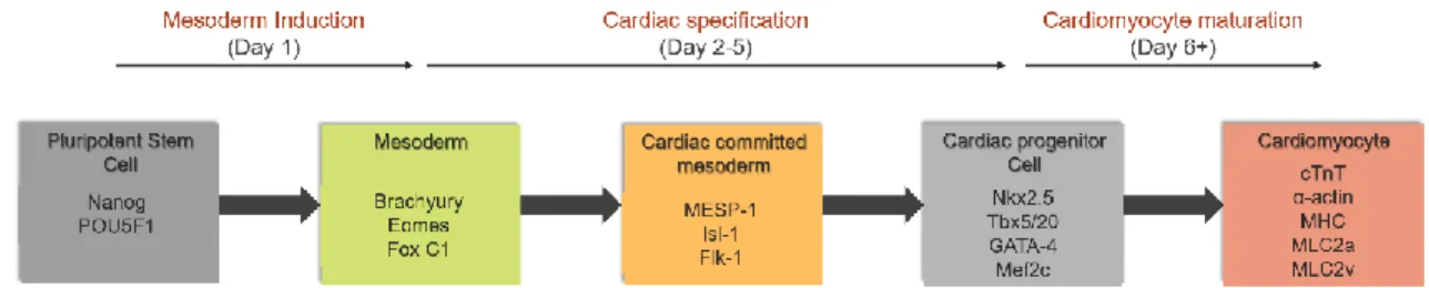
While human cardiogenesis is still not fully understood, cardiac development in animal models (e.g. mouse) has provided sufficient insights that allowed the improvement of CMs differentiation protocols from hiPSCs. To efficiently differentiate hiPSC into CMs, the most common and reproducible strategies involve replicating in culture the key steps along their natural development path in vivo (Figure 1.2), which require stage-specific activation and inhibition of different signaling pathways, such as Wingless/INT (WNTs), Nodal, bone morphogenic protein (BMP) and fibroblast growth factor (FGF) .Therefore, an in-depth knowledge of these pathways is crucial, as different differentiation protocols rely on their modulation by exposing hiPSC to several growth factors at specific times points and in precise doses to guide them towards cardiac fate. [45].
Figure 1.2 – Schematic representation of the expressed transcriptional factors along iPSCs cardiac differentiation. The five major stages in the differentiation of iPSC to cardiomyocytes: pluripotent stem cell,
mesoderm, cardiac committed mesoderm, cardiac progenitor cells and cardiomyocyte, are characterized by the distinct expression of different transcription factors. Adapted from. [46].
Cardiac specification begins with Nodal signaling and gastrulation that prompt mesoderm formation. The Nodal signal also upregulates BMP4 expression, which in turn induces WNT3 expression. Then WNT promotes the expression of mesoderm markers such as T (brachyury) and Eomes (eomesodermin) with the consequent activation of MESP1, the “master regulator” of cardiac progenitors specification [47, 48]. In this phase, cardiac differentiation will proceed with the inhibition of WNT/ β-catenin signaling. This pathway has a biphasic role during cardiogenesis, being important at the beginning to induce the primitive streak formation and after this stage, inhibition of this pathway is crucial to direct the progenitor cells into cardiac fate. At this point the cardiac mesoderm is formed and several transcription factors begin to be expressed, such as ISL1, GATA4, TBX5 and NKX2-5. This cardiac mesoderm gives rise to the endocardium, the first heart field (FHF) and the second heart field (SHF). While the FHF forms the left ventricle, the majority of the atria and part of the right ventricle, the SHF forms the majority of the right ventricle, outflow tract and part of the atria [49]. The three major cellular lineages that compose the heart tissue, CMs, endothelial cell (EC) and vascular smooth muscle cells (SMC) are all derived from the mesoderm phase [50].
To obtain functional CMs from hPSCs, the cardiac progenitors still need to differentiate into beating CMs, which are identified by the expression of certain proteins involved in
Introduction
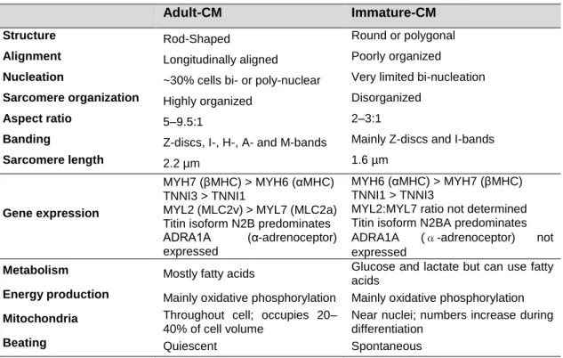
Disclosing CCBE1 role in Cardiac Differentiation of Human Pluripotent Stem Cells 6 morphogenic events leading to the formation of the heart, such as α-actinin, α-myosin or the cardiac isoform of Troponin-T (cTnT) [51]. However, generated CMs are still immature, to the point where their phenotype resembles more the fetal CMs in vivo than the adult CMs [52]. This lack of maturity in hPSC-derived CMs can be assessed by analyzing the structure, energy related characteristics and gene expression of cardiac specific markers (Table 1.1), like the myosin heavy (MYH6, MYH7) and light chains (MYL2, MYL7). For example, while the MYH7 and MYL2 are predominant in adult CMs, their isoforms, MYH6 and MYL7 respectively, predominate in immature related-CMs [53].
Table 1.1 – Structural, gene expression and energy related characteristics in adult and immature-like CMs. Adapted from [53].
Adult-CM Immature-CM
Structure Rod-Shaped Round or polygonal
Alignment Longitudinally aligned Poorly organized
Nucleation ~30% cells bi- or poly-nuclear Very limited bi-nucleation
Sarcomere organization Highly organized Disorganized
Aspect ratio 5–9.5:1 2–3:1
Banding Z-discs, I-, H-, A- and M-bands Mainly Z-discs and I-bands
Sarcomere length 2.2 µm 1.6 µm
Gene expression
MYH7 (βMHC) > MYH6 (αMHC) TNNI3 > TNNI1
MYL2 (MLC2v) > MYL7 (MLC2a) Titin isoform N2B predominates ADRA1A (α-adrenoceptor) expressed
MYH6 (αMHC) > MYH7 (βMHC) TNNI1 > TNNI3
MYL2:MYL7 ratio not determined Titin isoform N2BA predominates ADRA1A (α-adrenoceptor) not expressed
Metabolism Mostly fatty acids Glucose and lactate but can use fatty
acids
Energy production Mainly oxidative phosphorylation Mainly oxidative phosphorylation Mitochondria Throughout cell; occupies 20–
40% of cell volume
Near nuclei; numbers increase during differentiation
Beating Quiescent Spontaneous
Additionally, CMs populations derived from hiPSC are still heterogeneous. These populations include myocytes with nodal, atrial and ventricular properties [54]. This variability poses a challenge for the potential application of these cells in transplantation therapies, as it potentiate the risk of arrhythmia [55]. New approaches to generate homogenous populations of CMs from hiPSC in a reproducible way are still necessary. Alternatively, investigating how to efficiently purify a mixture of cells to select the desirable cell type could also be a promising approach. The uncertain outcome in cell-based therapies urged the search for new methods to improve cell differentiation, retention, survival and coupling, by using miRNAs [56], biomaterials [57], 3D cell constructs [58], bispecific antibodies [59],and cytokines [60].
Introduction
Disclosing CCBE1 role in Cardiac Differentiation of Human Pluripotent Stem Cells 7
1.2.2.Cell-Free Approaches for Myocardial Regeneration
Based on the hypothesis that the small improvements observed in cell therapy were due to paracrine mechanisms, new strategies bypassing the use of cells as the transplanted agents have emerged. For example, the direct administration of paracrine factors, such as growth factors, non-coding RNAs and extracellular vesicles or direct reprogramming of fibroblast into CMs (Table
1.2) [61, 62].
Table 1.2 – Preclinical results from cardiac regeneration therapies for ischemic heart diseases.
Adapted from [63, 64]
Therapy Mechanism Disease Model
Delivery
method Outcome Comments Ref Allogenic iPSC-CMs Direct replacement of CMs IHF Surgical intramyocardial injection LVEF improvement (≈ 10%) at 12 weeks Ventricular arrhythmias in all cell transplanted animals [65] hECS-CMs No significative changes in LVEF [37] Microparticles loaded with FGF-1/NRG1 Angiogenesis and Reversal of fibrosis IHF Intramyocardial injection LVEF improvement (≈ 9%) at 3 months Reduction in ventricular remodeling and increase in vascularization [66] miR-199a-3p miR-590-3p Promote endogenous CMs proliferation IHF MI Intramyocardial or with cationic lipid formulations LVEF improvement (≈ 10-20%) at 8 weeks Higher number of positive CMs for the DNA synthesis marker EdU [67] MR-409 (GHRH agonist) Pleiotropic effects and activation of GHRH IHF Subcutaneous injection Reduction in the scar size observed after 4 weeks Failed to improve the cardiac function [68] Recombinant FSTL1 in patch Pleiotropic effects Stimulation of CMs proliferation and arteriogenesis IHF Surgical implantation of a patch LVEF improvement (≈ 10%) [69] Retroviral GHMT Direct reprogramming of human fibroblasts towards the cardiac fate IHF MI Intramyocardial LVEF improvement (≈ 25%) after 12 weeks Reduction in the scar size
[70]
IHF, ischemic heart failure; FGF-1, fibroblast growth factor 1; FSTL1, follistatin-related protein 1; GHRH, growth hormone-releasing hormone; LVEF, left ventricular ejection fraction; MI, myocardial infarction; NRG1, neuregulin 1.
Although these preclinical studies showed some level of LVEF enhancement, they are still far away from complete regeneration of the infarcted heart. These marginal improvements (9%-25%), in Table 1.2, help to establish the therapeutic value of these approaches, paving the way for other therapeutic targets. Namely, restauration of coronary vasculature after MI, to
Introduction
Disclosing CCBE1 role in Cardiac Differentiation of Human Pluripotent Stem Cells 8 improve heart repair and increase the chance of survival. Nevertheless, this field is still recent and major issues and optimizations still need to be addressed to draw a concrete conclusion about their efficacy in the clinics.
1.2.2.1.Neovascularization and Lymphangiogenesis to augment heart
repair
Treatment of ischaemic heart disease has focused on protecting the heart from progression to HF. Even though coronary intervention can restore coronary blood flow after MI, microvasculature obstruction still persists, due to endothelial cell death, inflammation and thrombotic and plaque debris. This leads to poor wound healing and ventricular remodeling as well as increase HF events, diminishing the patients’ chances of survival [71]. Therefore, the regeneration of coronary microcirculation is essential for effective heart repair. To achieve this goal, a better understanding of how coronary vasculature is formed during the heart development is crucial. These vessels are essentially composed by vascular endothelium, smooth muscle and fibroblasts, while endothelial cells arise primarily from the sinus venosus (SV) and endocardium (Endo), the epicardium (Epi) acts as a source of trophic factors and progenitors’ cells, which ultimately give rise to the smooth muscle cells and fibroblast [72, 73]. This different coronary progenitors’ populations can compensate for each other if one exhibits defects, providing robustness in heart development. This compensatory mechanism was recently reported demonstrating the regulation of distinct coronary progenitor pools by both genetic timing (ELABELA-APJ signaling) and the microenvironment (hypoxia) to ensure the establishment of the proper vasculature needed for heart physiology [74].
A crosstalk between the Epi and myocardium is vital for coronary vessel formation during heart development. Epicardial cells secrete essential growth factors that support the developing myocardium, which in turn secretes angiogenic factors to promote vasculogenesis in the developing heart. For example, through fibroblast growth factor (FGF) and vascular endothelial growth factor (VEGF) signaling pathways [75]. FGF, secreted by epicardium, is essential for normal formation of the coronary vasculature and stimulates the secretion of A and VEGF-B by the myocardium. Moreover, myocardial factor thymosin β4 promotes EC migration, proliferation and initiates the epicardial progenitors cells activation [75]. After MI, quiescent epicardial cells reactivate to support heart repair and neovascularization [76]. One way to improve this neovascularization, is priming the epicardium before the injury with thymosin β4 [77]. Still, the precise mechanisms of coronary revascularization upon injury are uncertain and a better understanding of how epicardial response is modulated along the regeneration process would lead to more effective therapeutic approaches.
Besides CM replacement discussed above (in section 1.2.1), other therapies are being studied to promote heart regeneration, namely the stimulation of CM proliferation, the activation of lymphangiogenesis and angiogenesis, immunomodulation and reversal and/or inhibition of fibrosis [64]. For example, the delivery of specific recombinant proteins, such as VEGF-A was shown to improve neovascularization in animal models, although it failed to show beneficial
Introduction
Disclosing CCBE1 role in Cardiac Differentiation of Human Pluripotent Stem Cells 9 effects in clinical trials (EUROINJECT-ONE and NORTHERN) [78]. However, other growth factors, such as follistatin-related protein 1 (FSTL1), were shown to improve myocardium regeneration after MI [69]. Nevertheless, by changing the strategy of delivery, resorting to intramyocardial injection of synthetic modified RNA encoding human VEGF-A in a mouse MI model, led to an improvement of heart function [79]. Thus, the poor outcomes observed in the growth factor-based approaches in clinical trials might be due to inappropriate dosages, inadequate delivery strategy and/or lack of organ selectivity.
Another emerging strategy is the stimulation of lymphangiogenesis, which is required for clearance of edema and to reduce inflammation. One way to do this is through the stimulation of VEGF-C signaling [80], that is the key mediator of lymphangiogenesis during development and required for SV sprouting through its binding to VEGF receptor 3 (VEGFR3) following maturation process [81]. The vascular system consists of blood and lymphatic vessels; these lymphatic vessels are essential for tissue and body fluid homeostasis with vital role for the transport of macromolecules, immune system cells and absorption of lipids from the digestive system. During the differentiation process, lymphatic endothelial cells differ from the others due to the collective action of different transcriptional factors, thereby any defective mutation of these proteins may cause primary lymphedema [82, 83].
Genetic studies in zebrafish and mice models lacking lymphatic vessels development, as well as in rare individuals with Hennekam syndrome, have contributed to the identification of a secreted protein collagen and calcium-binding EGF domain 1 (CCBE1), that is required for proper lymphatic vascular development (Figure 1.3 A) [84, 85]. Moreover, over the last years reports suggested the requirement of this particular protein and a metalloproteinase (ADAMTS3) for the activation of VEGF-C [86].
1.2.3.The Role of CCBE1 as a potential modulator of cardiac function
CCBE1 was identified to be expressed in heart precursors in mouse embryos from embryonic day (E)7.0 to (E)9.5 [87], particularly near the developing lymphatic vessels and in the developing heart [88]. Also, it was identified in early cardiac progenitors in chick embryos and in heart precursors of first and second heart field [87]. In humans, mutations on this protein were found to be associated with Hennekam syndrome, which is an autosomal recessive lymphatic disorder where about 25% of the patients exhibit mutations in CCBE1 [89]. This rare disease displays diverse pathological features like lymphedema, lymphangiectasias and intellectual disability [84]. Moreover, EGF protein family, which includes CCBE1 was identified in embryonic cardiac fibroblasts as a paracrine factor involved in the regulation of CMs proliferation [90]. Also, there are several reports suggesting the importance of this protein on cancer context, such as tumor suppressor gene in ovarian cancer and as a potential biomarker in the detection of lung [91] and gastrointestinal stromal tumors (Figure 1.3 B) [92].Introduction
Disclosing CCBE1 role in Cardiac Differentiation of Human Pluripotent Stem Cells 10 Figure 1.3 – Schematic representation of CCBE1 and its key roles. (A) CCBE1 protein domains. SP, signal peptide; EGF, epidermal growth factor domain; Ca-EGF, calcium binding EGF domain; ColA, collagen repeat A; ColB, collagen repeat B. (B) CCBE1 key roles suggested so far. Adapted from [93].
As previously mentioned, CCBE1 is involved in the activation of the major lymphangiogenic growth factor VEGF-C, which is crucial for lymphatic development in mouse embryos and for the major part of the lymphangiogenesis process in adults [93, 94]. However, VEGF-C is synthesized as a precursor molecule, the pro-VEGF-C, and needs to be further activated to play its key role [95]. For this purpose, the ADAMTS3 (A Disintegrin And Metalloproteinase with Thrombospondin Motifs-3) protease in a complex with CCBE1, is responsible for the cleavage of pro-VEGF-C to generate a mature and activated version of this factor, which binds to and further activates the VEGFR-3 receptor (Figure 1.4 B) [82, 86]. The C-terminal of CCBE1 is essential for an effective activation of VEGF-C, whereas the N-C-terminal is important for the colocalization of pro-VEGF-C with CCBE1 and ADAMTS3 on the endothelial cell surface which may be necessary for an efficient cleavage of this factor by ADAMTS3, contributing to the VEGFR-3 signaling increase [93, 96].
While mutations in CCBE1 C-terminal domain have shown to result in the absence of lymphatic structures in mice, mutations in the N-terminal resulted in incomplete and disorganized lymphatic vessels, suggesting a role in the organization and migration of lymphatic endothelial cells [93]. Recent studies also demonstrated the importance of CCBE1 in coronary vasculature development through the activation of VEGF-C during the embryonic development [74, 97]. In these studies, CCBE1 knockout in mouse models displayed similar heart defects as in VEGF-C mutants, exhibiting a stunted angiogenesis compared to the wildtype. Therefore, CCBE1 could also be used as a therapeutic factor to stimulate neovascularization after MI (Figure 1.4 B). However, obtaining sufficient amounts of stable recombinant full-length CCBE1 protein (49 kDa) remains a major hurdle, being an issue for functional and therapeutic studies using this protein [86, 98]. Alternatively, the use of precise gene editing tools (e.g. CRISPR/Cas9) in combination
Introduction
Disclosing CCBE1 role in Cardiac Differentiation of Human Pluripotent Stem Cells 11 Figure 1.4 – Lymphangiogenesis: VEGF-C activation by CCBE1 and ADAMTS3 complex and its role in myocardial infarction (MI). (A) MI is followed by adverse remodeling of epicardial collector lymphatics,
with subsequent edema, severe inflammation and fibrosis. A therapeutic approach is based on VEGF-C administration to increase lymph flow and resolves inflammation, improving the cardiac function. (B) CCBE1 secretion at sites of lymphatic vessel growth promotes the proteolytic cleavage of pro-VEGF-C form by the disintegrin/metalloprotease ADAMTS3. The mature form of VEGF-C can further activate VEGFR-3. Most of the VEGF-C cleavage may occur on lymphatic endothelial cell (LEC) surface mediated by CCBE1 and ADAMTS3. Adapted from [99, 100].
Introduction
Disclosing CCBE1 role in Cardiac Differentiation of Human Pluripotent Stem Cells 12 with hiPSCs would greatly contribute for in vitro studies helping to uncover CCBE1 role in human cardiac repair.
1.3.Gene editing tools
To better understand the regulatory networks that drive specific cellular activities, in healthy or disease conditions, one can use precise and effective tools for gene manipulation. The capacity to manipulate the expression of desirable genes either by repression or activation facilitates the understanding of pathophysiological mechanisms in cardiovascular diseases. This manipulation can now be achieved with the emergence of genomic-editing systems which are getting more advanced, efficient and simpler to use. Advantages and disadvantages of the currently major gene editing tools are summarized in Table 1.3.
Table 1.3 – Comparison of the major gene editing tools. Adapted from [101]
Gene editing tool ZFN TALENs CRISPR/cas9
Source Bacteria, Eukaryotes Bacteria
(Xanthamonas sp.)
Bacteria (Streptococcus sp).
Easy of design Difficult Moderate Easy
Specificity High High High
Efficiency Low High High
Multiplexing Low Moderately High High
Sequence limitations
Non-guanosine rich sequence hard to
target
5′targeted base must be thymine for
each TALEN monomer
PAM sequence must follow target site
Cost High Moderate Cheap
Nowadays, different systems for genome manipulation have already been described, such as RNA-mediated interference (RNAi) and customized classes of DNA binding-chimeric proteins for instance zinc-finger proteins (ZFs), Transcription activator-like effector nucleases (TALENs) and more recently, the promising guide RNA (gRNA)-driven Cas9 (CRISPR) system. These tools offer a great prospect for the future of cardiovascular field and for in vivo genome-editing therapies. The RNAi was the first tool being explored [102]. In this system small interfering RNAs (siRNAs) or short hairpin RNAs (shRNAs) bind to the target endogenous mRNAs transcripts promoting their cleavage [102]. However, its low reported efficacy and non-specificity has limited its wide application [103, 104]. For precise genetic modifications, custom engineered and site-specific endonucleases were successfully developed, namely the ZF and TALENs.
The ZFs consist of programable DNA-binding domains fused to a functional domain, which allows the manipulation of gene expression levels in a modular way by recruiting effectors into transcriptional sites of the target genes. For a better genomic specificity, a combination of at
Introduction
Disclosing CCBE1 role in Cardiac Differentiation of Human Pluripotent Stem Cells 13 least six ZFs in the DNA binding domain is recommended, as each one can recognize approximately only three bp of DNA [105]. This tool has already been applied for the genome-editing of several living organisms [106–109] and to different cell lines [110, 111] in a successful way.
TALENs system is similar to the ZFs in a way that both have a DNA-binding domain fused with a functional domain. However in this system the binding protein consists of highly conserved TALEN tandem repeats of 33-35 amino acids, that can be easily designed and with the potential to target any sequence with a high success rate [112].
An emerging alternative based on RNA-guided nuclease overcome the above limitations. The type II Clustered Regularly Interspaced Short Palindromic Repeats (CRISPR), derives from the adaptive immune system of Streptococcus pyogenes, which protects the bacteria from exogenous DNA-containing phages and plasmids, thanks to the combination of CRISPR loci and a Cas9 nuclease [113]. This loci together with short spacer sequences derived from past virus infections, are transcribed into long RNAs (crRNAs), which forms a complex with a small transactivating CRISPR RNA (tracrRNA), called guide RNA (gRNA) that will provide the capacity to search and guide the Cas9 nuclease to cleave the target viral DNA by creating a double strand break (DSB) [113]. The Cas9 has two nuclease like-domains, HNH and RuvC that will promote a DSB in the target sequence after binding to a short DNA sequence, named protospacer-adjacent motif (PAM) which flanks the RNA-binding site [114]. This DSB triggers the DNA repair mechanisms that sporadically introduces indel mutations in the target sequence, allowing the introduction, in a simple and easy manner, any desirable mutations in target genes [115, 116] One of the major drawbacks for the application of this technology is the off-target effects. It has already been reported that the Cas9 binds to off-target sites and although only a small subset of those are cleaved efficiently, they still represent a major concern, since other genes could be mutated with serious damage potential [117, 118].
In addition to gene editing, CRISPR technology can be used for regulation of gene expression, without cleaving the target site. For this purpose, a nuclease-deficient Cas9, labeled as dead Cas9 (dCas9) has been developed by inserting mutations into the two nuclease domains of Cas9, the HNH and RuvC. The CRISPR-dCa9, also called CRISPR interference (CRISPRi) is still capable to specifically bind to the target sequence when guided by the gRNA either in the promotor or regulatory sequences and manipulate its transcription process without changing the genomic sequence (Figure 1.5) [114, 119]. It can prevent the transcription initiation or elongation by blocking the binding of important transcription factors in the transcription site or the RNA polymerase II [114, 119, 120]. Furthermore, the genetic regulation using CRISPR-dCas9 is not permanent and can be easily reversed. Moreover, grouping of dCas9 with a transcriptional repressor, like the Krüppel-associated box (KRAB) [121] or four concatenated mSin3 interaction domains (SID4X) [122] can improve the repression of endogenous genes, this was already demonstrated in eukaryotic models [121] and more recently in hiPSC [123]. In addition, coupling
Introduction
Disclosing CCBE1 role in Cardiac Differentiation of Human Pluripotent Stem Cells 14 the dCas9 with a transcriptional activation domain, such as VP64 or p65, termed CRISPR activation (CRISPRa) can increase the expression of endogenous genes [124, 125].
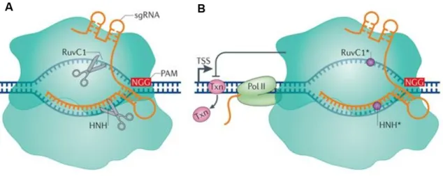
Figure 1.5 – Differences in action method of Streptococcus pyogenes Cas9 and dCas9. (A) The single
guide RNA (sgRNA) leads Cas9 to the target DNA sequences. This targeting is dependent on the presence of a 5′ protospacer-adjacent motif (PAM) in the DNA, which in S. pyogenes is usually NGG. After binding the two nuclease domains (RuvC1 and HNH) cleave the target sequence. (B) Mutations in the two nuclease domains deactivate the Cas9 protein (dCas9), inactivating its nuclease function (circles), but still retains the capacity to target specific sequences through sgRNA guidance and PAM. dCas9 binds near the transcription start site (TSS) and blocks transcription elongation by obstructing RNA polymerase II (Poll II) or blocking the binding of important transcription factors (Txn). Adapted from [126]
In fact, genome editing has already been used to create more reliable cardiac disease models or correct genetic mutations in IPSC-derived CMs by introducing genetic alterations [127]. One example, was a disease model for Barth syndrome (mitochondrial disorder caused by mutation of the gene encoding tafazzin) developed by Wang and colleagues, which combined patient-derived IPSC and genome editing tools, like CRISPR/cas9 to mimic the pathophysiology of this specific disorder in vitro [128].
For loss-of function studies, Mandegar and colleagues developed a versatile CRISPRi-dCas9 inducible system for hiPSC lines. This repression system enables precise control of single or multiplexed gene expression upon doxycycline addition, making this CRISPRi-hiPSCs lines an attractive tool for identification of novel factors involved in cell differentiation and maturation. Moreover, this system was validated for multiple genes along cardiac differentiation, in hiPSC, CPC and CMs, demonstrating its efficacy and reliability (Figure 1.6) [123].
These current advances in gene editing tools and in iPSC technology offers a great opportunity to better understand the pathophysiological mechanism of cardiac diseases and to develop reliable differentiation protocols resulting in more homogenous cardiac populations and trustworthy cell models suitable to improve disease understanding and to propose novel cell therapies.
Introduction
Disclosing CCBE1 role in Cardiac Differentiation of Human Pluripotent Stem Cells 15 Figure 1.6 – Potential of CRISPRi hiPSC lines developed by Mandegar and colleagues. GEN1C cell
line was generated by integration of CRISPRi construct into the AAVS1 locus of WTC. Posteriorly, a gRNA or gRNAs are selected and introduced in the cell line to specifically target a gene or genes of interest. After that, the targeted gene expression will be repressed upon Dox induction. This system is also validated for multiple genes specific in different stages of CM differentiation. Adapted from [123].
Aim
Disclosing CCBE1 role in Cardiac Differentiation of Human Pluripotent Stem Cells 17
2.Aim of the thesis
CCBE1 has been studied as an important protein for lymphatic vessels development and despite the increasing evidence of a potential involvement in cardiac commitment, further investigation is still needed to validate this hypothesis. Therefore, the aim of this thesis was to unveil the role of CCBE1 on cardiovascular development exploring gene editing tools (Figure
2.1). In this context, we performed CCBE1 loss-of-function studies using the CRISPRi technology
in hiPSC to knockdown CCBE1 gene expression during CM and EC differentiation process. The first objective consisted on evaluating the CCBE1 knockdown efficiency and its impact on hiPSCs pluripotent phenotype. To accomplish this, CCBE1 knockdown hiPSC lines were generated. Additionally, their self-renewal capacity, differentiation potential and CCBE1 gene expression were assessed. The second objective was to evaluate CCBE1 knockdown impact along hiPSC differentiation into CM and EC. We performed a detailed characterization of hiPSC-derived CM/EC phenotype in CCBE1 knockdown cell line.
Overall this work provides new insights on CCBE1 role in cardiac development. The knowledge here described would help to identify CCBE1-modulatory pathways and explore CCBE1 as a therapeutic molecule for cardiovascular regenerative medicine.
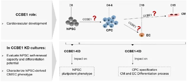
Figure 2.1 – Schematic representation of the major aims of this thesis and outlined strategy. The
main aim of this thesis was to unveil the role of CCBE1 on cardiovascular commitment. hiPSC- human induced pluripotent stem cells; CCBE1- collagen and calcium-EGF biding domain; CPC- Cardiac Progenitor Cell; CM- Cardiomyocytes; EC- Endothelial Cells; KD- knockdown.
Material & Methods
Disclosing CCBE1 role in Cardiac Differentiation of Human Pluripotent Stem Cells 19
3.Material & Methods
3.1.hiPSC culture & differentiation
3.1.1.hiPSC lines
In this study two human iPSCs lines with the same genetic background were used. Wild-type C (hereafter designated as hiPSC-WT) and a modified cell line CRISPRi Gen1C (hereafter designated as hiPSC-CRISPRi), integrating a Tet-On inducible system that modulate the expression of a deactivated Cas9 (dCas9) fused with the repressor KRAB domain. mCherry reporter gene is under the control of the same inducible promoter (at downstream of dCas9-KRAB, separated by p2A). These cell lines were derived by Mandegar MA and colleagues [123] and provided by The J. David Gladstone Institutes under a Material Transfer Agreement.
3.1.2.hiPSC expansion
hiPSC lines were routinely propagated in static culture systems, 6-well plates (Falcon™), coated with growth factor reduced (GFR) Matrigel®, Phenol Red Free (BD Biosciences) using mTeSR1TM media (STEMCELL Technologies), according to the protocol described by Mandegar and colleagues [123]. Cells were maintained under humidified atmosphere with 5% CO2 at 37ºC.
3.1.3.hiPSC cardiac differentiation
hiPSC were differentiated into CM in monolayer culture systems, according to the recently published protocol [129]. hiPSC single cell suspensions were prepared by incubation with Accutase (STEMCELL Technologies) for 3 min at 37ºC and seeded at a density of 7-9 × 10⁴ cell/cm2 in Matrigel coated 6-well plates (Falcon™) or μ-Slide 4 well formats (ibidi®). Two days after cell seeding, the differentiation was induced by replacing the expansion media with RPMI 1640 medium (Gibco®) supplemented with 2% (v/v) B27 minus insulin (Invitrogen), 12 µM CHIR99021 (Biogen Cientifica S.L), 80 ng/mL Activin A (PeproTech) and 50 µg/mL ascorbic acid (Sigma-Aldrich). Twenty-four hours later (day 1 of differentiation) the media was replaced by RPMI supplemented with 2% (v/v) B27 minus insulin, 5 µM IWR-1 (Sigma-Aldrich) and 50 µg/mL ascorbic acid. At day 3 of differentiation, cells were incubated with RPMI supplemented with 2% (v/v) B27 minus insulin, 5 µM IWR-1. From day 6 until day 15, the medium was changed 3 times per week with the RPMI supplemented with 2% (v/v) B27 minus insulin [129]. Cells were maintained under humified atmosphere with 5% CO2 at 37ºC.
3.1.4.hiPSC endothelial differentiation
hiPSC were differentiated into endothelial cells according to the protocol described by Giacomelli and colleagues [130]. Cells were seeded at 1.25 × 10⁴ cell/cm2 in 6-well plates coated in Matrigel 24 hours prior initiation of differentiation process. At day 0 of differentiation, the media was replaced by APEL-Li (STEMCELL Technologies) supplemented with Activin A (20 ng/ mL), BMP4 (20 ng/mL) and CHIR99021 (1.5 µM). Then medium was replaced every three days by APEL-Li supplemented with VEGF (50 ng/mL) until day 10 of differentiation. Cells were
Material & Methods
Disclosing CCBE1 role in Cardiac Differentiation of Human Pluripotent Stem Cells 20 maintained under humified atmosphere with 5% CO2 at 37ºC. All supplements were supplied by Peprotech.
3.2. CCBE1 knockdown: gRNA design, cell electroporation and selection
For CCBE1 knockdown, four gRNAs were designed to target near the transcription start site (TSS) of CCBE1 (between 150 bp upstream and 150 bp downstream). All gRNAs were phosphorylated, annealed and cloned into the pgRNA-CKB vector (kindly provided by Bruce Conklin; Addgene plasmid # 73501) at BsmBI restriction site. All the cloning steps were performed as described elsewhere [123]. gRNA oligo sequences are listed in Table 3.1.The pgRNA-CKB expression vector, containing mKate2 as reporter gene and blasticidin as antibiotic selection marker (mKate2-T2A-Bsd), was transfected into CRISPRi cells using the Neon Transfection System (Thermo Fisher Scientific) according to manufacturer’s instructions. Two conditions were tested (condition 1: 1400 V, 20 ms, 2 pulses and condition 2: 1100 V, 30ms, 1 pulse). CRISPRi cells (2×10⁶ cells) were transfected with 5 μg of vector carrying a CCBE1-specific gRNA generating the CRISPRi-CCBE1 KD cell line or with empty pgRNA-CKB vector, without a gRNA to generate the CRISPRi-Ctrl cell line (control condition).
Table 3.1 – List of gRNA oligo sequences. Each gRNA indicates the binding relative to the transcription
start site (TSS) of CCBE1 gene, and whether they target the template (T) and non-template (NT) strand. Forward and reverse primers for cloning into the pgRNACKB gRNA-expression vector are listed from 5’ to 3’. gRNA Name (Targeting Strand) Oligo Sequences 5’ – Forward Primer – 3’ 5’ – Reverse Primer – 3’ CCBE1 g-145 (NT) TTGGAAGGGGGTACCTGCGGTGTC AAACGACACCGCAGGTACCCCCTT CCBE1 g-82 (NT) TTGGCAGGGGTCCGGAATATTATG AAACCATAATATTCCGGACCCCTG CCBE1 g+22 (T) TTGGAGCAGGACGCTTGGTCCGGA AAACTCCGGACCAAGCGTCCTGCT CCBE1 g+37 (NT) TTGGTCCCAGCGCCGAGCTCCGTC AAACGACGGAGCTCGGCGCTGGGA
Twenty-four hours post transfection, blasticidin selection was applied by culturing the cells in mTeSR1 supplemented with Y-27632 (10 μM) and blasticidin (10 µg/mL). Stable colonies were pooled and passaged five times to enrich for cells with integration into sites of active transcription. The percentage of nucleofected cells was evaluated by mKate2 expression using an inverted fluorescence microscope (Leica Microsystems GmbH).
CRISPRi mediated gene knockdown studies, using CCBE1 g+37 (Table 3.1), were performed by supplementing the media with doxycycline (Dox; 2 µM). To allow CCBE1 expression, cells were cultured in the absence of Dox. The gene knockdown efficiency was examined by RT-qPCR.
![Figure 1.1 – Death causes of European population in 2016. Adapted from [4].](https://thumb-eu.123doks.com/thumbv2/123dok_br/19237322.970080/23.892.181.699.567.888/figure-death-causes-european-population-adapted.webp)

![Table 1.3 – Comparison of the major gene editing tools. Adapted from [101]](https://thumb-eu.123doks.com/thumbv2/123dok_br/19237322.970080/34.892.125.766.459.753/table-comparison-major-gene-editing-tools-adapted.webp)