Mahder Seifu Manenda
Graduated in
Erasmus Mundus Masters In Membrane Engineering
Title
Separation of Bioactive Peptides by
Electrodialysis with Ultrafiltration
Membranes: Membrane Characteristics,
Ex-situ and In-Ex-situ Digestion and Their Impact
on Peptide Migration
Dissertation for obtaining the Master degree in Membrane Engineering
Erasmus Mundus Master in Membrane Engineering
Advisor: Laurent Bazinet, Professor, Université Laval
Co-advisor(s): Isabel Coelhoso, Professor, FCT-UNL
João G. Crespo, Professor, FCT-UNL
Jury:
President: João Crespo, Professor, FCT-UNL Examiner(s): S. Velizarov, Professor, FCT-UNL
Member(s): Karel Bouzek, Professor, ICTP André Ayral, Professor, UM2
MAHDER SEIFU MANENDA
Graduated in
ERASMUS MUNDUS MASTERS IN MEMBRANE ENGINEERING
TITLE
Separation of Bioactive Peptides by
Electrodialysis with Ultrafiltration
Membranes: Membrane Characteristics,
Ex-situ and In-Ex-situ Digestion and Their Impact
on Peptide Migration
Dissertation presented to Faculdade de Ciências e Tecnologia, Universidade Nova de Lisboa for obtaining the master degree in Membrane Engineering
TITLE: Separation of Bioactive Peptides by Electrodialysis with Ultrafiltration Membranes: Membrane Characteristics, Ex-situ and In-situ Digestion and Their Impact on Peptide Migration
The EM3E Master is an Education Programme supported by the European Commission, the European Membrane Society (EMS), the European Membrane House (EMH), and a large international network of industrial companies, research centres and universities (http://www.em3e.eu).
Copyright @ Manenda, FCT/UNL
A Faculdade de Ciências e Tecnologia e a Universidade Nova de Lisboa têm o direito, perpétuo e sem limites geográficos, de arquivar e publicar esta dissertação através de exemplares impressos reproduzidos em papel ou de forma digital, ou por qualquer outro meio conhecido ou que venha a ser inventado, e de a divulgar através de repositórios científicos e de admitir a sua cópia e distribuição com objectivos educacionais ou de investigação, não comerciais, desde que seja dado crédito ao autor e editor.
ACKNOWLEDGMENTS
“In questions of science, the authority of a thousand is not worth the humble reasoning of a single individual.” Galileo Galilei
First and foremost I would like to thank my family for their understanding and patience during my endless travels while on this program. Their encouragement and expectations keep my
dreams alive.
I am deeply grateful to my supervisor Prof. Laurent Bazinet for his continuous supervision, encouragement and motivation throughout my research work at Université Laval. This manuscript would not be complete without his diligent attention to details and continuous
constructive feedback.
The contributions of Elodie Rozoy, Shyam Suwal, Dr. Cyril Roblet, Dr. Alain Doyen and Daine
Gagnon in the form of discussions and suggestions are recognized.
I thank program coordinators Prof André Ayral, Prof. Isabel Coelho, Prof. João Crespo and Dr Elena Vallejo for their support in organizing my internship and for the continuous guidance they offered during the masters program.
The financial support received from the European Union in the form of scholarship throughout
the masters program is appreciated.
ABSTRACT
Trypsic hydrolysis of whey protein isolate was performed simultaneously with (in-situ) and before (ex-(in-situ) fractionation by electrodialysis with ultrafiltration membrane (EDUF) to obtain bioactive peptides. Two ultrafiltration membrane (UFM) materials, PES and PVDF, were used for a 120 minute EDUF fractionation of the hydrolysate. The two membranes showed similar zeta potential measurements at the pH of operation, 7.8, but had significantly different conductivity which decreased significantly after use in EDUF. Peptide migration to anionic (A
-RC) and cationic (C+RC)
peptide recovery compartment was strongly dependent on the electrical conductivity of the UFMs than their types or the digestion strategy used. For UFMs with close values of conductivity, peptide migration to the A
-RC was observed to be higher with
in-situ digestion while peptide migration to the C+
RC was higher in an ex-situ digestion.
When the two membrane types, PES and PVDF, have closer values of conductivities, PVDF was observed to exhibit more migration than PES. Peptide migration to the C+
RC
varied from 16.56 ± 5.36 µg/mL to 103.10 ± 2.76 µg/mL for PVDF membrane with significantly different conductivities: 2.73 ± 0.32 mS/cm and 5.47 ± 0.56 mS/cm, respectively. Peptide migration to the A
-RC varied from 4.41 ± 0.86 µg/mL to 49.65 ±
6.13 µg/ml for PVDF membrane with significantly different conductivities: 3.163 ± 0.12 mS/cm and 5.23 ± 0.04 mS/cm, respectively. HPLC-MS studies showed 23 major peaks that were generated on whey protein isolate digestion by trypsin. 9 of these peaks migrated to the A
-RC while 3 migrated to the C+RC. Among the anionic peptides 3
peptides are known to have hypocholesterolemic effect, 1 is antibacterial and 1 is antihypertensive. Among the cationic peptides 1 is antihypertensive. EDUF appears to be a powerful new technique that can fractionate bioactive peptides in terms of mass and charge.
Index of Figures
Figure 2.1 EDUF cell configuration for the hydrolysis of BiPro protein and recovery of anionic and cationic peptides
Figure 4.1: Evolution of Zeta potentials of PES and PVDF UFMs as a function of pH
Figure 4.2: Evolution of feed temperature as a function of time during 120 minutes of EDUF
Figure 4.3: Evolution of system resistance as a function of time during 120 minutes EDUF
Figure 4.4: Evolution of conductivity in the anionic peptide recovery compartment (A
-RC) for
in-situ and ex-in-situ experiments with PES and PVDF membranes
Figure 4.5: Evolution of conductivity in the cationic peptide recovery compartment (C+ RC) for
in-situ and ex-situ experiments with PES and PVDF membranes
Figure 4.6: Evolution of conductivity in the feed recirculation compartment for PES in-situ, PES ex-situ, PVDF in-situ and PVDF ex-situ EDUF experiments
Figure 4.7: Total peptide migration into the A
-RC as a function of time for PES and PVDF
membranes with in-situ and ex-situ digestion
Figure 4.8: Total peptide migration into the C+
RC as a function of time for PES and PVDF
membranes with in-situ and ex-situ digestion
Figure 4.9: Zeta Potential of UFM-I and UFM-II (after use in EDUF) and the original PVDF membrane before use in EDUF
Figure 4.10: Evolution of system temperature and resistance as a function of time for ex-situ and in-situ experiments
Figure 4.11: Evolution of conductivity as a function of time in the A
-RC for ex-situ and in-situ
experiments
Figure 4.12: Evolution of conductivity as a function of time in the C+
RC for ex-situ and in-situ
experiments
Figure 4.13: Evolution of conductivity as a function of time in the feed compartment for ex-situ and in-situ experiments
Figure 4.14: Evolution of total peptide concentration in the A
-RC and C+RC as a function of time
for ex-situ and in-situ experiments with PVDF
Figure 4.15: Chromatogram of initial BiPro solution before digestion by trypsin
Figure 4.16: Chromatograms of BiPro A) after 120 minutes of ex-situ digestion, B) after 120 minutes of EDUF after an ex-situ digestion and C) after 120 minutes of simultaneous (in-situ)
digestion and EDUF treatment
Figure 4.17: Chromatograms of peptides obtained in the A
-RC after 120 minutes EDUF of A)
ex-situ digested BiPro and B) in-ex-situ digested BiPro
Figure 4.18: Chromatograms of peptides obtained in the C+
RC after 120 minutes EDUF of A)
Index of Tables
Table 1: Thickness (mm) and conductivity (mS/cm) of UFMs and IEMs used in EDUF experiments
Table 2: Thickness (mm) and conductivity (mS/cm) of PVDF and IEMs before and after use in EDUF
Abbreviations
EDUF: electrodialysis with ultrafiltration membranes ED: electrodialysis
UFM: Ultrafiltration Membrane UF: Ultrafiltration, NF: Nanofiltration PVDF: Polyvinylidiene Floride PES: Polyether Sulfone
MWCO: Molecular Weight Cutoff MW: Molecular Weight
ACE: Angiotensin Converting Enzyme BCA: Bicinchoninic Acid
IEM: Ion-Exchange Membrane CEM: Cation-Exchange Membrane. AEM: Anion-Exchange Membrane
A-RC: Anionic Peptides Recovery Compartment;
C+RC: Cationic Peptides Recovery Compartment
TABLE OF CONTENTS
Acknowledgements ... I
Abstract ... II
Index of Figures ... III
Index of Tables ... IV
Abbreviations ... V
1 Introduction... 1
1.1 Background and motivation ... 1
1.2 Objectives ... 2
2 Literature review ... 3
3 Materials and methods ... 12
4 Results and discussion ... 16
5 Conclusions and future perspectives ... 49
1.
Introduction
1.1. Background and Motivation
As the knowledge of molecules contained in food sources deepen, scientists have long started looking at food as more than just a source of calories and nutrients. The presence of bioactive molecules contained in the complex food matrix and other natural sources has attracted much attention from food scientists and the food industry. Bioactive molecules are molecules obtained from natural sources that can exert physiological changes in microbes or in humans. They can exist as polysaccharides or oligosaccharides, polyphenolic compounds, conjugated linoleic acids, peptides etc. The existence of such molecules in everyday food sources, making them functional foods, or the isolation of such molecules from food sources and their preparation into specialized products (nutraceuticals) can be a cost effective alternative to the increasingly very expensive drug based health care (Health Canada, 2013).
Bioactive peptides have long been known to exert a range of physiological effect in humans and other organisms acting as antihypertensive, antithrombotic, antimicrobial, antioxidative, immunomodulatory and opioid molecules (Bazinet L & Firdaous L, 2009). The bioactivity of these peptides is inherent in their amino acid sequence that sometimes leads to multi-functionality. However due to their low concentrations in nature and their interactions with other molecules that can potentially lead to loss of bioactivity, bioactive compounds have limited bioavailability. These led to several attempts to either directly synthesize the peptide sequences or isolate them from their natural sources efficiently. Direct synthesis by chemical method or using recombinant DNA technology are, as of now, costly for large peptides and difficult to streamline while isolation from natural sources including everyday food materials by hydrolysis and then isolation presents a cost effective and easy to scale up alternative.
fractionate cationic and anionic peptide which may have close molecular mass but completely different bioactivity (Yuanhui et al.2007).
Electromembrane processes; including electrically-enhanced membrane filtration systems, forced flow membrane electrophoresis and electrodialysis with ultrafiltration (EDUF) have the ability to fractionate cationic and anionic peptides even when the peptides have very close molecular weights. Moreover EDUF have the general advantage of lower fouling compared to purely pressure driven process (Langevin M., et al 2012). EDUF has now been used to isolate
bioactive compounds from multitude of sources. Labbé et al (2005) used EDUF system to
obtain a migration of 50% catechins (antioxidant molecules from a green tea infusion). Firdaous
et al (2010) reported the isolation of an angiotensin converting enzyme (ACE) inhibitor peptide
fraction from alfalfa white protein and Doyen et al (2012) used EDUF to recover and
concentrate the active antibacterial fraction from a snow crab by-products hydrolysate.
In a recent study Doyen et al (2011) compared polyether sulfone (PES) and cellulose acetate
(CA) membranes and reported no significant difference in total peptide migrations. The two membranes were reported to show a slight difference in the way their conductivities changed after being used in EDUF: PES membranes had reduced conductivity while CA did not. The authors reported no migration to the cationic recirculation compartment (C
-RC) for both
membranes until after 180 minutes of EDUF. This could be due to the 20kDa MWCO of UFM used and also due to a relatively smaller electric field strength applied: 2V/cm. Hence we propose to apply larger electric field strength and use UFMs with a larger MWCO to enhance peptide migration and further study the role of UFM material using PES and PVDF membrane materials. Moreover the effect of digestion within the EDUF setup or outside of it has not been studied before. PES and PVDF membranes have desirable physical and chemical properties that make them popular for ultrafiltration and other applications (Rong G. et al. 2005, Zhang Q. et
al. 2002, & Huyen T. et al.2006). Their differences in surface properties and electrical
properties resulting from their chemical makeup could be important factors in EDUF.
1.2. Objectives
In light of the background presented above, the aim of the present work is investigate the role of digestion techniques and membranes in the separation of bioactive peptides from hydrolysate of whey protein isolate digested by trypsin using EDUF. The specific objectives include:
i. to study the effect of UFM material on EDUF isolation of bioactive peptides ii. to study the effect of in-situ and ex-situ digestion on EDUF isolation of
bioactive peptides
2.
Literature Review
2.1. Bioactive Peptides
2.1.1. Definitions
Bioactive peptides are generally defined as specific protein fragments that have a positive impact on body functions and conditions and may ultimately influence health (Bazinet L & Firdaous L, 2009). Even though the terms “functional foods” and nutraceuticals have now
become part of the scientific lexicon in food science literature, a universal definition for both is difficult to find. However, functional foods can be defined as products that are consumed as food and not in dosage form and have beneficial effect on the health of the consumer. Nutraceutical is defined, in Canadian law, as referring to "a product isolated or purified from foods that is generally sold in medicinal forms not usually associated with food. A nutraceuticals are demonstrated to have physiological benefits or provide protection against chronic diseases” (Health Canada, 2013).
2.1.2. Bioactive Peptides from Milk
Bioactive molecules occur naturally in plant and animal products, usually at very low concentrations. Several bioactive peptides that are present in many food proteins have long been demonstrated to posses physiological benefits (Kitts D. & Weiler K., 2003) apart from the provision of basic nutrition as there is more to food than just calories and nutrients. Given their
benefits it is not surprising to see the number of publication regarding functional foods and nutraceuticals from protein sources increasing by the year and the global functional foods and
nutraceutical market growing at a rate that is outpacing the processed food market (Korhonen H. & Pihlanto A., 2003).
improve the human health, reduce health care costs and support economic development in rural communities (Korhonen H. & Pihlanto A., 2003).
Milk proteins are considered the most important source of bioactive peptides. The activity of these peptides is based on their inherent amino acid composition and sequence found buried in the native protein. The size of active sequences in peptides may vary from 2 – 20 amino acid residues, and many peptides are known to reveal multifunctional properties (Kitts D. & Weiler K., 2003). Over the last decade a great number of peptide sequences with different bioactivities have been identified in various milk proteins. The best characterized sequences include antihypertensive, antithrombotic, antimicrobial, antioxidative, immunomodulatory, and opioid peptides (Korhonen H. & Pihlanto A., 2003). These peptides have been found in enzymatic protein hydrolysates and fermented dairy products, but they can also be released during gastrointestinal digestion of proteins, as reviewed in many articles (Mullaly M. et al 19975, Vandekerckhove J et al &7). For instance trypsic digestion can release peptides β-LG f15– 20, β-LG f102–105 and β-LG f142–148 all of which are identified as having ACE inhibitory activity (Mullaly, M. et al 1978, Fitzgerald R. & Meisel, H., 2009). Trypsin also releases
bactericidal peptides β-LG f15–20 and β-LG f92–100 (Pellegrini, A. et al, 2000), and
hypocholesterolemic peptide β-LG f71–75 (Nagaoka, S, et al 2001), and the opioid peptide
from β-LG f102–105 (Meisel, H. & Schlimme E., 1995).
2.2. Methods of Bioactive Peptide Production and Separation
2.2.1. Production of Bioactive Peptides
2.2.1.1. Direct Peptide synthesis and Recombinant DNA Technology
Due to their low concentrations in nature and their interactions with other molecules that can potentially lead to loss of bioactivity, bioactive compounds have limited bioavailability. This challenge on bioavailability has encouraged scientists and industries to investigate possible synthesis, isolation, concentration and delivery of specific bioactive peptides for increased efficacy in their role as modulators of physiological functions. Direct peptide synthesis of specific sequences is one of the methods being investigated to produce bioactive peptides of desired amino acid sequence in the laboratory. This can be done by a liquid-phase or a solid-phase synthesis. The liquid-solid-phase synthesis has been used for the synthesis of small peptides and its main advantage is that the intermediate products can be isolated and purified after each step of synthesis and recombined later to obtain larger peptides of the desired sequence. But the productivity of these methods is very limited except for relatively shorter sequences (8-10 amino acid) (Nishiuchi et al. 1998) Solid-phase peptide synthesis consists in the elongation of a
linked by amide (peptide) bond formation between the carboxyl group of the incoming amino acid and the amino group of the amino acid previously bound to the matrix, until the peptide of the desired sequence and length has been synthesized (Nilsson et al. 2005). Despite their wide
spread use in the pharmaceutical industry application of solid phase peptide synthesis for peptides of more than 10 residues are challenging and technically complicated (Patarroyo et al.,
1988).
Recombinant DNA technology is also another method of producing desired peptides and if the system is established, peptides can be obtained in large quantities from very inexpensive starting materials via fermentation but this requires a long and expensive research and development phase before starting to produce the desired peptide (Korhonen , H. & Pihlanto A., 2003). In fact Meister et al (2003) produced β-Casomorphins (an opiate peptide from milk),
from both human and bovine milk, by genetically engineering the genes for the peptide and cloning it into a plasmid of E. coli but the concentrations they obtained were very low (38.22
nmol/L). Hence the direct synthesis of peptides, chemically (for longer sequences than 8-10 amino acids) or by recombinant DNA technology, is both costly and technically complicated for scale up.
2.2.1.2. Microbial Fermentation
Microbial fermentation of food matrices and/or biomass can be a source of several bioactive peptides. A mixture of system of lactic acid bacteria, such as Lactococcus lactis, Lactobacillus
helveticus and Lactobacillus delbrueckii var. bulgaricus, (Hartmann R. & Meisel H., 2007)
have the enzymatic machinery to systematically cleave large proteins into oligopeptides and then into amino acids and also into bioactive peptides which can be extracted from the fermentation broth by various techniques. The enzymes involved in milk protein degradation can either be cell-envelope proteinases (CEP) or intracellular peptidases (Mullaly M. & Meisel H., 1997). Longer oligopeptides which are not transported into the cells can be sources for the liberation of bioactive peptides in fermented milk products when further degraded by intracellular peptidases after bacterial cell lysis or in the digestive system of humans. Yamamoto et al. (1994) reported that casein hydrolyzed by the cell wall-associated proteinase
from L. helveticus CP790 showed antihypertensive activity in rats. Several ACE inhibitory and
one antihypertensive peptide were isolated from the hydrolysate. Using the same proteinase, Maeno et al. (1996) identified a -casein-derived antihypertensive peptide from the casein
2.2.1.3. Enzymatic Digestion
Enzymatic digestion of peptides involves the release of peptide ad amino acids by hydrolysis of the peptide bond to obtain a hydrolysate. Hydrolysate’s peptide composition, and consequently their properties, is dependent of protein and enzyme used, as well as, on hydrolysis conditions (temperature, pH, enzyme to substrate ratio and reaction time). Hydrolysates can be characterized according to several molecular characterization methods, including HPLC-MS, which reflect their molecular properties.
Enzymatic digestion of proteins by a digestive enzyme, or a combination of them, is the most common way of producing bioactive peptides from food materials. Enzymes like trypsin, chymotrypsin, alcatase, pepsin etc have been used to generate bioactive peptides from proteins. Amongst the enzymes, trypsin is the most commonly used mainly because of its tendency to generate more bioactive peptides (specially angiotensin-converting enzyme (ACE) inhibitor peptides) without needing to be combined with other enzymes (Meister W. et al., 1994).
Trypsin digests proteins from milk that remain soluble at pH 4.6 which are commonly known as whey protein isolates (WPI). These proteins are commonly isolated by ion exchange chromatography or microfiltration and mainly contain β-lactoglobulin and α-lactalbumin both of which have high nutritional value. WPI also contain relatively high branched chain amino acids (leucine, isoleucine, valine) that contribute to their important role in muscle growth and repair. They are also an important source for bioactive peptides (Clare & Swaisgood, 2000) after trypsic digestion. However after digestion the peptides have to be separated in order to enhance their biological activity.
2.2.2. Separation of Bioactive Peptides
Membrane processes are the most commonly used processes used in a large scale separation of bioactive peptides.
2.2.2.1. Pressure Driven Processes in Bioactive Peptide Isolation
advantage of recycling the enzyme and continuous exposure of the protein to the digestive enzyme leading to an improved production of the peptides. Fractions of peptides with a wide range of molecular size and sequence can be obtained depending on the molecular cutoff of the ultrafiltration membrane used (Langevin M. et al. 2012). Ultrafiltration membranes have also
been applied in a pressure drive process without involving immobilized enzymes to separate peptides from hydrolysates obtained from a separate digestion step.
In an interesting application of ultrafiltration membranes for the fractionation of hydrolysate obtained from whey proteins, Turgeon et al (Turgeon L. et al. 1990) used a stepped
ultrafiltration procedure to limit the range of peptides obtained in different fractions to a narrow range. In their experiments they used ultrafiltration membranes with MWCO 1 and 30 kDa to
enrich ACE inhibitory peptides from purified α- lactoglobulin and β-lactoglobulin and obtained
two fractions: one rich in higher molecular weight peptides and the other rich in amino acids and low molecular weight peptides. Several studies have also reported the use of stepped ultrafiltration procedure to fractionate different hydrolysates in to different fractions which have different physiological effects depending upon the peptides they contain (Xie Z, et al., 2008,
Pihlanto A. et al., 2008). But such procedures are limited because they use only the size of the
peptides generated to fractionate the peptides as opposed to other emerging procedures which use a combination of size, shape and charge of peptides to fractionate them.
Several nanofiltration (NF) membranes were also used to separate peptides on the basis of charge interaction with the membranes since most peptides contain charged functional groups at a given pH. Tsuru et al. (1994) reported that separation of amino acids and
peptides with nanofiltration membranes having a MWCO around 2000–3000 Da was successful based on a charge effect mechanism while nanofiltration membranes having a MWCO below 300Da were not suitable for separation of amino acids or peptides. Charged amino acids and peptides were rejected in their fractionation while neutral amino acids and peptides permeated through the FilmTech, Toray and Nitto membranes they used. The separation of peptides having different isoelectric points with nanofiltration membranes was possible by adjusting the pH (Tsuru et al. 1994).
combination of ultrafiltration and nanofiltration to fractionate peptides contained in sweet whey. In this combination the first step consisted in the ultrafiltration of the sweet whey with a MWCO of 10 kDa to completely retain the whey proteins. The resulting permeate fractions were then subjected to a fractionation by nanofiltration with a MWCO of 1 kDa. Amongst the pressure-driven membrane techniques, UF and NF are preferred for the fractionation of protein hydrolysates due to the fact that the molecular weight of most bioactive peptides is within the normal range of the MWCO of these membranes.
Despite their wide use in the fractionation of protein hydrolysates pressure-driven processes like NF and UF lead to a significant accumulation of particles on membrane leading to fouling that causes modification of the membrane transport selectivity (Langevin M. et al., 2012) Moreover
both UF and NF lack the ability to fractionate peptides depend on the charge of the peptide. Though in nanofiltration the charge of the peptide does play a role in determining its interaction
with the membrane this role is limited as it doesn’t allow simultaneous separation of oppositely
charged peptides.
2.2.2.2. Electromembrane Processes in Bioactive Peptide Isolation
The challenge of significant fouling and the fractionation of peptides only by their molecular weight (not considering their charge and its change with pH) have made scientists to look for other options of bioactive peptide isolation. Electromembrane processes are among the options investigated for possible improvement of peptide isolation. These processes involve the coupling of the transport of electrical charges and a mass transfer across ion-exchange, bipolar, filtration etc membranes. These processes can be used together with pressure driven processes to reap the benefits of both types of process and improve process productivity.
In an interesting approach of combining pressure driven process with electromembrane processes Huotari et al. (1999) superimposed an electrical field to a conventional membrane
filtration unit effectively forming an electrically-enhanced membrane filtration system. The superimposition can be either in a parallel orientation with flat sheet filtration membranes or the electric field can be applied between the inner rode and the membrane in a tubular membrane module. Loose nanofiltration and ultrafiltration membranes have been reported to be used in this configuration (Lapointe J. et al., 2006). But this configuration lacks a distinct electrolyte
In the recent years Electrodialysis with Ultrafiltration Membranes (EDUF) stacked has
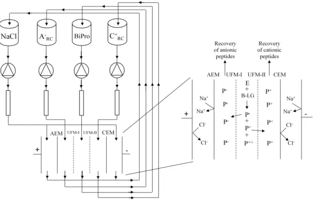
emerged to be capable of separating peptides not only on the basis of their molecular weight but also on the basis of their charge. In a configuration patented by Bazinet et al. (2005) EDUF
consists of four recirculation compartment for the feed, anionic peptide recirculation compartment (A
-RC), cationic peptide recirculation compartment C+RC, and the electrolyte
solution at the anode and cathode (Figure 1). The feed recirculation is separated from A -RC and
C+
RC by two ultrafiltration membranes (UFMs) which impart the configuration the ability to
screen peptides in terms of their molecular weights. Anion exchange membrane (AEM) and cation exchange membranes (CEM) separate the two peptide recovery compartments from the electrolyte recirculation compartment near the anode and cathode, respectively. The two UFMs can have the same or different MWCO, they can be of the same or different material, and interestingly as much as 7 UFMs have been placed in between the ion exchange membranes (IEMs) (Firdaous et al., 2010) giving an efficient functional unit. It is important to note that no
pressure difference is applied in between any compartment of the EDUF configuration.
Figure 2.1: EDUF cell configuration for the hydrolysis of BiPro protein and recovery of anionic and cationic peptides. AEM: anion-exchange membrane, CEM: cation exchange membrane,
UFM: ultrafiltration membrane, A
-RC: anionic peptide recovery compartment, C+RC: cationic
peptide recovery compartment, BiPro: whey protein extract, E: trypsin enzyme, P+: cationic
peptide, P-: anionic peptide (Adapted from Doyen
EDUF technology showed several potential applications for the food industry, notably for the separation and recovery of bioactive compounds from diverse raw matrices. Doyen et al.,
(2012) used EDUF to recover and concentrate the active antibacterial fraction from a snow crab by-products hydrolysate. They used two ultrafiltration membranes with different MWCO (20 kDa and 50 kDa) and two electrical field strengths (2 and 14 V/cm) and reported 94% abundance of peptides with molecular weight range 300–600 Da peptide in the recovery compartments. In a different report (Doyen et al., 2011) the same researchers identified an
anticancer peptide fraction from snow crab byproduct hydrolysate after a selective separation by EDUF with 20 kDa MWCO cellulose acetate (CA) UFM stacked in the system.
Firdaous et al., (2010) reported the isolation of an angiotensin converting enzyme (ACE)
inhibitor peptide fraction from alfalfa white protein hydrolysate by stacking a polyether sulfone (PES) UFM with10 kDa MWCO in EDUF cell. Poulin et al. (2006) performed fractionation of a β-lactoglobulin hydrolysate and demonstrated the simultaneous separation of acid and basic bioactive peptides is possible by stacking 20 kDa MWCO cellulose acetate (CA) UFMs. Roblet
et al., (2013) found out that pH modulation appeared to be an efficient way to concentrate the
low molecular weight peptides (400 Da) in the cationic peptides recovery compartment C+ RC
and to limit the diversity of peptides recovered in the A
-RC in purification of soy peptides from a
complex matrix.
It is important to note that EDUF can indeed be used in isolation of bioactive molecules not only from protein sources but also from other several types of sources. Labbé et al. (2005)
demonstrated that catechins (antioxidant molecules from a green tea infusion) can migrate at a high rate through an EDUF system. They reported that UFM with 1kDa can achieve migration as high as 50%. In a more recent study, Bazinet et al., (2009) reported an 18% increase of the
antioxidant capacity of cranberry juice the enriched by EDUF treatments demonstrating that the EDUF process might be used for natural enrichment of cranberry juice with antioxidant phenolics (Bazinet L. et al. 2009).
Despite the growing interest in EDUF and the parameters that influence separation in EDUF the effect of ultrafiltration membrane (UFM) material have not been studied extensively. In a comparative study Doyen et al. (2011) compared two different types of membranes, polyether
sulfone (PES) and cellulose acetate (CA) for their performance when used in EDUF system. No significant difference in total peptide migration was reported between the two types of UFM material studied during EDUF separations. Total peptide migration depended only on the duration of EDUF and, for the C
-RC, the authors observed no migration for both membranes until
in EDUF when compared to their respective control membranes but no significant such difference was detected for CA (Doyen A. et al., 2011). In general the report indicates that CA
UFM allowed the recovery of high molecular weight molecules (900-20000 Da) in both recovery compartments.
Polyethersulfone (PES) is a heat-resistant, transparent, amber, non-crystalline engineering plastic while PVDF is a highly non-reactive and pure thermoplastic fluoropolymer produced by the polymerization of vinylidene fluoride. PVDF highly desirable insolubility and electrical properties result from the polarity of alternating CH2 and CF2 groups on the polymer chain. It
withstands exposure to harsh thermal, chemical, or ultraviolet conditions. PES, due to its non-crystalline nature, is attacked by highly polar solvents: such as esters, ketones and trichloroethylene while PVDF is resistant to most chemicals and solvents. PES membranes generally have lower roughness than PVDF membranes which could be an interesting difference with regards to peptide migration or peptide deposition onto the membranes surfaces (Rong G.
3.
Materials and Methods
3.1. Materials
BiPro protein was purchased from Davisco Foods International Inc. (Minnesota, USA). Bovine pancreatic trypsin was purchased from Sigma– Aldrich (St. Louis, MO, USA). PVDF and PES ultrafiltration (both with MWCO 50 kDa) membranes were purchased from Synder Filtration (California, USA). Ion exchange membranes were supplied by Eurodia Industries (PERTUIS, France). HCl and NaOH solutions were obtained from Fisher Scientific (Montreal, QC, Canada). NaCl and KCl were purchased from ACP Inc. (Montréal, QC, Canada).
3.2. Configuration of EDUF
The electrodialysis cell used for our experiment was MP type cell (100 cm2 of effective surface
area) manufactured by ElectroCell Systems AB Company (Täby, Sweden). The configuration was the same as the one used by Doyen et al. (2013) (Fig 2.1). Briefly, EDUF configuration
consisted of one Neosepta CMX-SB cationic membrane (Tokoyuma Soda Ltd, Tokyo Japon), one Neosepta AMX-SB anionic membrane (Tokoyuma Soda Ltd, Tokyo, Japan) and two polyether sulfone or two polyvinylidiene floride UFMs with a molecular weight cut-off (MWCO) of 50 kDa (Synder Filtration Inc. Vacaville, CA USA)
The configuration consisted of 4 compartments. Two of them, containing 2 L of aqueous KCl (2 g/L) were used for the recovery of recovery of peptides (anionic (A
-RC) and cationic (C+RC)
peptide recovery compartments): they were located near the anode and the cathode respectively. The third compartment contained the electrode NaCl rinsing solution (3 L, 20 g/L), and, the last compartment contained the feed solution (BiPro, 2 L) at 12.5 g/L. The solutions were circulated using four centrifugal pumps and the flow rates were controlled using flowmeters (figure 2.1). Permeate and feed solution flow rates were 1.5 L/min while the flow rate of the electrode solution was 2 L/min (figure 2.1).
3.3. Hydrolysis and Separation Procedures
BiPro solution was prepared by a overnight hydration of 25g of BiPro in 2L of distilled water (1.25% w/v) in a cold room of 40C.Trypsic hydrolysis of BiPro carried-out in two setups: one in
a beaker (ex-situ) with continuous stirring of the BiPro solution and the other in the EDUF system (in-situ) simultaneously with separation after the pH was adjusted to 7.8. Enzymatic hydrolysis was started by the addition of 10 mL trypsin solution (125 mg/L of trypsin (w/v)). In both in-situ and ex-situ digestions the hydrolysis was performed for 120 minutes after which the enzymatic reaction was stopped by raising the temperature of the solution to 800C for 30
A constant electric field of 8.22 V/cm was applied between EDUF electrodes. The hydrolysis and fractionation procedures were performed during 120 min. The system was started initially at room temperature and the EDUF parameters are recorded every 15 min during the 120 min experiment. During EDUF, the reaction was maintained at pH 7.8, corresponding to the optimum pH value of trypsin, with 0.5 M NaOH using a pH meter from Thermo Scientific Orion 9206BN probe (VWR International Inc., Mississauga, Ontario, Canada). The pH of recovery compartments was also maintained at 7.8 by a continuous addition of NaOH and HCl by using the same type of pH meter. 10mL of samples from the hydrolysate and each recovery compartment were collected before applying voltage and every 30 min during the treatment from the A
-RC, C+RC and the feed/hydrolysate compartments. Samples are heated to 800C to stop
the action of the enzyme. Following each EDUF treatment, the final volumes of A
-RC, C+RC and
the feed/hydrolysate compartments were recovered and freeze dried for storage. Finally a clean-in-place procedure for the EDUF cell was performed after every repeat of EDUF to ensure the recovery of the UFMs and IEMs performances.
Our experiment was performed in two parts. Part one involved EDUF experiments, in an in-situ and situ setup, with both PES and PVDF membranes. In the second part EDUF, in both ex-situ and in-ex-situ setups, was done only with only PVDF membranes and the recovered peptides were analyzed by HPLC-MS in only the second part.
3.4.
Analysis
3.4.1.
Membrane thickness
Membrane thickness was measured using a Mitutoyo Corp. digimatic indicator (model ID- 110 ME, Japan) and a digimatic mini-processor (model DP-1HS, Japan) specially designed for
plastic film thickness measurement. The resolution was of 1 μm and the range of 10 mm.
3.4.2.
Membrane Conductivity
The membrane electrical conductivity was measured according to the method of Bazinet and Araya-Farias M, using a specially designed clip from the Laboratoire des Matériaux Echangeurs
3.4.3.
Zeta potential measurements
A SurPASS electrokinetic analyzer (Anton Paar, Graz, Austria) equipped with a clamping cell was used to measure the zeta potential of UFMs. The determination of zeta potential with the SurPASS electrokinetic analyzer is based on the streaming current or streaming potential measurement created by the circulation of the electrolyte through a capillary system. The streaming current was measured in 1 mM KCl solution in a pH range of 2.5-11. The streaming channel of well-defined dimensions (25 mm in length and 5 mm in width) was formed by two identical flat membranes mounted opposite of each other and separated by one spacer. The streaming current was measured alternatively in the two flow directions by pressure ramps in the range 0 to 300 mbar. Two cycles of pressure ramps in each direction were conducted and measured average zeta potential values were computed using the Fairbrother-Mastin model.
3.4.4.Measurement of Solution Conductivities
Conductivities of A
-RC, C+RC and feed compartments were measured every 15 min during the
120 min of EDUF with a YSI conductivity meter (model 3100) equipped with a YSI immersion probe (model 3252, cell constant K = 1 cm-1, yellow Springs Instrument Co., Yellow springs,
OH, USA). The conductivities were measured in order to evaluate the mineralization or demineralization of the solutions during the process.
3.4.5. Total Peptide Determination in Different Compartments
Total peptide migrations to the A
-RC, C+RC and feed compartments were determined from
samples withdrawn every 30 min over a period of 120 min using the BCA protein assay (Pierce, Rockford, IL, USA). The microplate was first incubated 370C and then cooled to room
temperature and the absorbance was read at 562 nm on a microplate reader (THERMOmax, Molecular devices, Sunnyvale, CA). Concentration was determined with a standard curve in a range of 5–2000 µg/mL of bovine serum albumin (BSA).
3.4.6. Protein and Peptide Profile with HPLC
The peptide composition of the A
-RC, C+RC and hydrolysate solutions was determined by
RP-HPLC according to the method of Firdaous et al. (2010) adapted to the specific conditions of
wavelength was 214 nm which is typically used to monitor peptide bonds (Firdaous et al., 2009,
and Stachelhaus, T., et al. 1998).
3.4.7. Peptide Molecular Weight Determination
Protein and peptide molecular weights were determined by using MS. MS analysis were performed with a scan range of 300 - 2200 m/z in positive polarity, with an ESI ion source type, at a dry temperature of 350°C, a nebulizer at 30.00 psi and dry gas of 8.00 L/min.
The molecular weight (MW) of proteins and peptides in recovered samples were determined by mass spectrometry (LC-MS) analyses according using ion trap method. The system used was an Agilent 1100 series (Agilent Technologies, Palo Alto, CA, USA). Peptides were analyzed with the same method and the same column used for RP-HPLC analyses. To reduce the effect of TFA, mass-spectrometry was performed after infusing (10 µL/min) a mixture of 50% propionic acid and 50% isopropanol to the existing flow before the MS interface. Signals were recorded in positive mode using a 90-V fragmentation with a scan range of 300–3000 m/z (Firdaous
et al., 2009).
3.5. Statistical Analysis
4.
Result and Discussion
4.1. Studies with PES and PVDF Membranes
4.1.1. Membrane characterization
A. Membrane Thickness
Membrane thickness and conductivity were determined for every membrane used before and after EDUF experiments. Measurement of membrane thickness can be a rapid method to follow effects of electrodialysis (ED) or EDUF on membranes (Casademont C., et al., 2010). The
mean values together with the standard deviations of quadruplicate measurements are presented in Table 1.
ANOVA showed that membrane material, PES and PVDF, had a significant effect on membrane thickness (P<0.001) with averages for all conditions being of 0.194 ± 0.007 mm and 0.234 ± 0.016 mm, respectively (table 1). Especially for PES membrane, ANOVA showed that its thickness varied significantly before and after EDUF (P<0.002), with UFM number (UFM-I or UFM-II, P<0.001) and with ex-situ vs in-situ experiments (P<0.001). Moreover double interaction were also detected between ex-situ/in-situ experiments and before/after EDUF (P<0.022), ex-situ/in-situ experiments and UFM number (P<0.011) and before/after EDUF and UFM number (P<0.022). No triple interactions were detected. For PVDF membrane thickness, ANOVA indicated a significant difference only for ex-situ vs in-situ (P<0.001) and for UFM number (P<0.001). For IEMs, their thicknesses varied significantly with IEM type (AEM vs CEM, P<0.001) and UFM type with which it was used (PES vs PVDF, P<0.001). The control AEM and CEM had mean thicknesses of 0.140 ± 0.003 mm and 0.170 ± 0.003 mm, respectively.
PES UFMs showed significant change in thickness while PVDF indicated no significant change in thickness which may suggest a slight difference in susceptibility to surface deposition of foulants. Marginal differences in thickness of UFM-I and UFM-II of same membrane types could be due to differences in storage and position of folding during the rolling of membrane for shipment and storage. The other types of membranes; AEM and CEM showed no a significant change in thickness as a result of EDUF. Although change in thickness of AEM after use in ED has been reported before (Casademont C., et al., 2010), we observed no significant difference in
thickness of AEM after being used in our EDUF experiments. Due to the nature of the driving force involved, which is applied electric field, the possibility of having uncharged peptides depositing on the surface of UFMs is reduced compared to pressure driven processes (Poulin J.,
Table 1: Thickness (mm) and conductivity (mS/cm) of UFMs and IEMs used in EDUF experiments
Thickness [mm]
Conductivity [mS/cm]
UFM
Material
Digestion Strategy
Membrane Type
Before EDUF After EDUF Before EDUF After EDUF
PES
Ex-situ
UFM-I 0.193 ± 0.003a 0.189 ± 0.002b 6.48 ± 0.24a 6.04 ± 0.19b
UFM-II 0.200 ± 0.003c 0.206 ± 0.005d 6.68 ± 0.16c 6.31 ± 0.11d
AEM 0.142 ± 0.002a 0.140 ± 0.002a 9.09 ± 0.18a 7.55 ± 0.19b
CEM 0.176 ± 0.002c 0.176 ± 0.001c 6.26 ± 0.07c 3.28 ± 0.31d
In-situ
UFM-I 0.185 ± 0.003a 0.191 ± 0.001b 5.94 ± 0.19a 5.85 ± 0.54b
UFM-II 0.190 ± 0.003c 0.198 ± 0 .003d 4.98 ± 0.31c 4.00 ± 0.38d
AEM 0.140 ± 0.003a 0.144 ± 0.002a 9.13 ± 0.74a 6.61 ± 0.24b
CEM 0.175 ± 0.002c 0.173 ± 0.002c 10.76 ± 0.09a 10.3 ± 0.31c
PVDF
Ex-situ
UFM-I 0.238 ± 0.005a 0.242 ± 0.003a 3.13 ± 0.12a 1.59 ± 0.10b
UFM-II 0.212 ± 0.001c 0.215 ± 0.002c 2.73 ± 0.32c 1.58 ± 0.29d
AEM 0.139 ± 0.001a 0.139 ± 0.001a 9.12 ± 0.09a 7.6 ± 0.09b
CEM 0.171 ± 0.001c 0.171 ± 0.002c 11.19 ± 0.16a 8.64 ± 0.12c
In-situ UFM-I 0.255 ± 0.003
a 0.256 ± 0.002a 5.23 ± 0.04a 5.52 ± 0.20b
UFM-II 0.227 ± 0.001c 0.225 ± 0.005c 4.27 ± 0.18c 4.64 ± 0.22d
AEM 0.139 ± 0.002a 0.143 ± 0.003a 8.56 ± 1.14a 6.64 ± 0.13b
CEM 0.172 ± 0.003c 0.173 ± 0.002c 10.66 ± 0.16c 7.99 ± 0.15d
Different letters, in a row or a column, indicate statistically significant difference (P=0.005) for the parameter/membrane of interest.
B.
Membrane Conductivity
ANOVA also indicated a significant difference in conductivity of PVDF membranes used in ex-situ and in-ex-situ experiments (P<0.001), before and after EDUF (P<0.001) and UFM number (P<0.001). Within both PVDF UFM-I and UFM-II significant difference in conductivity was observed for ex-situ vs in-situ experiments (P<0.001, for both) (Table 1). Within PVDF in-situ experiments significant differences in conductivity are observed for UFM number (P<0.001) and significant differences were also seen within before EDUF (3.13 ± 0.12mS/cm and 5.23 ± 0.04 mS/cm, for UFM-I ex-situ and in-situ, respectively, P<0.001) and within after EDUF (P<0.001) for ex-situ vs in-situ experiments (Table 1).
Amongst the UFMs, PES UFM-II used in ex-situ experiment exhibited the highest conductivity at 6.68 ± 0.36 mS/cm while PVDF UFM-II showed the lowest conductivity at 2.73 ± 0.32 mS/cm. PVDF membranes used as both UFM-I and UFM-II particularly in the ex-situ experiment had significantly (P<0.0036) lower conductivities (3.13 ± 0.12 mS/cm and 2.73 ± 0.32 mS/cm, respectively) compared to other PVDF UFM-I and UFM-II membranes used in the in-situ experiment (5.23 ± 0.05 mS/cm and 4.27 ± 0.19 mS/cm, respectively).
Electrical conductivities of UFMs are not provided by manufacturers as the main market and researchers that use/study UFMs use them in pressure driven processes for which MWCO are more important than electrical conductivity. However Donose et al., (2011) reported that
conductivity can indeed be used to check the integrity of RO and NF membranes as a surrogate measurement for rejection of ions by the membranes. In the same manner the conductivity of UFMs can be used to check for relation to peptide migration across membranes in EDUF and check for structural integrity of membranes. Lower conductivities for the original membranes observed for UFM-I and UFM-II used in PVDF ex-situ experiment can, therefore, suggest certain structural anomalies that can lead to vulnerability to fouling which is indicated by the significant decrease, almost by half, of conductivity in these membranes after use in EDUF (table 1). In fact these aberrations were sometimes apparent in the different physical appearance of membranes obtained from the same roll/sheet and this could be the reason why membranes had such a significantly different conductivity before EDUF.
In a study reported by Doyen et al., (2011), the conductivity of PES membrane changed
For IEMs ANOVA showed that IEM material had no significant effect on the conductivity of IEMs while there was a significant decrease in IEM conductivity after use in EDUF (P<0.001). Marginal differences in IEM conductivity were detected for the two types of (PES and PVDF) UFMs the IEMs were used with (P=0.033). Double interactions were detected between UFM type and IEM type (P=0.012). ANOVA also showed a significant difference in IEM conductivity used in ex-situ and in-situ experiments (P=0.012) (Table 1).
The least IEM conductivity before EDUF was recorded for CEM membrane used with PES ex-situ experiment (6.26 ± 0.07 mS/cm) while the largest was recorded for CEM membrane used in PVDF ex-situ experiment (11.19 ± 0.16 mS/cm). For AEM the largest conductivity was recorded for the one used in PES in-situ experiment (9.13 ± 0.74 mS/cm) while the smallest was recorded for the one used in PVDF in-situ (8.56 ± 1.14 mS/cm). A drastic decrease in conductivity was observed for CEM membrane used with PES ex-situ experiment from 6.26 ± 0.07 mS/cm to 3.28 ± 0.31 mS/cm, which accounts for a 47.6 % decrease; the largest percentage decrease observed for any IEM we used. This is in line with the observation that UFMs that had the least conductivity before EDUF showed the most drastic reduction in conductivity after use in EDUF suggesting that, in both UFMs and IEMs, lower membrane conductivities are indication of membrane structural aberrations, possible attained during shipment and/or storage,that made the membrane more susceptible to fouling.
IEM fouling is one of the major problems in milk or complex food systems electrodialysis (Casademont C. et al., 2006). Fouling in IEMs during ED and its follow up by decreasing
conductivity (increasing resistance) has been reported before (Lindstrand V. et al. 2007 &
Lindstrand V. et al., 2000). In line with reports made by Lindstrand et al., (2007) both CEM and
AEMs had significantly (P<0.001) reduced conductivity after being used in EDUF which is an indication of fouling which is caused by the deposition of peptides or amino acids (organic materials) resulting from the digestion of the BiPro or by deposition of inorganic minerals (Casademont C., et al., 2010) because BiPro also contains 0.6% minerals as indicated by the
manufacturer (DAVISCO).
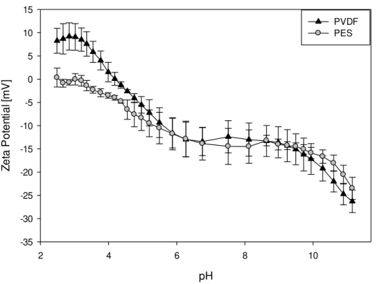
C. UFM Zeta Potential
membranes to attain a close value of -29.12 ± 1.36 mV and -26.29 ± 2.44 mV, respectively at pH 11.15.
pH
2 4 6 8 10
Z
e
ta
P
o
te
n
ti
a
l [
m
V
]
-35 -30 -25 -20 -15 -10 -5 0 5 10 15
PVDF PES
Figure 4.1: Evolution of Zeta potentials of PES and PVDF UFMs as a function of pH
However there are notable differences, for almost the entire range of pH 2.5 to 11, PES membrane showed a negative zeta potential except at very acidic pH (pH 2.5–3) were the membrane surface potential was close to zero. For the pH range 2.5 to 4 PVDF membrane showed positive zeta potential; for instance at pH 2.5, PVDF zeta potential was recorded at 8.26 ± 2.67 mV as opposed to 0.35 ± 0.35 mV recorded at the same pH for PES. The other notable difference was the point of zero charge; PVDF attained this point at pH 4.17 while PES attained it at pH 2.96. Interestingly though near the pH of EDUF operation, which is 7.8, PES and PVDF show close zeta potential measurements; -13.20 ± 1.14 mV and -12.37 ± 3.46 mV, respectively. Two iso-potential points, the pH where both membranes attain the same zeta potential, were identified from the pH titration of the two UFMs (figure 4.1), one at pH 5.87 and the second at pH 8.63.
Similar trend of zeta potential evolution with pH for PES and PVDF membranes have been reported before (Kim K., et al., 1996). The observation that the PVDF membranes showing a
dissociation of ions like H+, K+, Cl- and OH- from the solution onto the membrane surface (Kim
K., et al., 1996). Surface dissociable groups could explain the negative zeta potential observed
for PES membranes as the membrane has previously been reported to behave as a weakly acidic material (Lara R., and Benavente J., 2009). Due to the close zeta potentials exhibited by the PES and PVDF membranes at the working pH (7.8) (-13.20 ± 1.14 for and -12.37 ± 3.46, respectively) we expect no significant difference in terms of peptide migration with respect to the membranes on the basis of this little difference in zeta potential.
4.1.2. Electrodialysis with Ultrafiltration Membranes (EDUF) Parameters
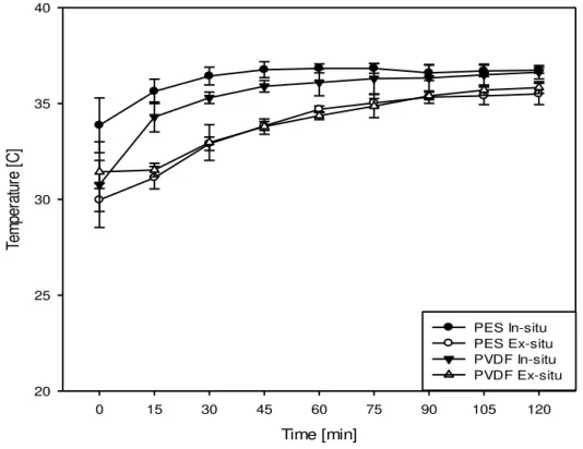
4.1.2.1. Evolution of System Temperature
Figure 4.2 depicts the variation of temperature in the feed compartment as a function of time. No statistically significant difference evolution of temperature with time was observed for the different membranes used but temperature significantly changed with time (P<0.001) during the first half of EDUF. Statistically significant difference in initial temperature was observed for PES ex-situ experiments (29.97 ± 0.60 0C) and PES in-situ experiments (at 32.87 ± 1.43 0C)
(P<0.001). System temperature generally increased from initial values to 35-36 0C within the
Time [min]
0 15 30 45 60 75 90 105 120
Te
m
pe
ra
tu
re
[
C
]
20 25 30 35 40
PES In-situ PES Ex-situ PVDF In-situ PVDF Ex-situ
Figure 4.2: Evolution of feed temperature as a function of time during 120 minutes of EDUF
The increase in temperature can be explained by joule heating, where the passage of current through a system heats the system (Firdaous et al ., 2009), and also the effect of pumping used
for re-circulating solutions in the four compartments of feed, A
-RC , C+RC and the electrolyte
solution (near the cathode and near the anode) compartments. Such phenomena are well documented in EDUF systems and previously reported elsewhere (Firdaous et al., 2009). The
slight variation in temperature between in-situ and ex-situ experiments could be due to minor variations in room temperatures that are less likely to affect EDUF parameters. Here it is important to note that the temperature in the recirculation system stabilizes near 360C and never
over passes 370C making the use of external heating system to maintain the feed temperature at
optimum for protein digestion by trypsin (370C) a non-necessity.
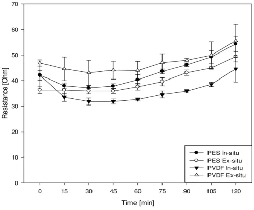
4.1.2.2. Evolution of System Resistance
significant difference in system resistance is also seen for the different UFMs within in-situ (P<0.001) and also within ex-situ (P<0.001) experiments.
In all the experiments a similar trend of evolution of system resistance is observed; resistance decreased during the first 45 minutes of EDUF and it slightly increased during the next 75 minutes. For all the EDUF experiments maximum system resistance was attained at t=120 minutes, this maximum resistance was the highest for PVDF ex-situ at a value of 55.49 ± 6.42
Ω and the lowest for PVDF in-situ at a value of 44.55 ± 5.14 Ω. As can be seen from figure 4.3,
experiments which started with higher resistance maintained a higher resistance throughout the EDUF experiment till the end while those experiments that started with a lower resistance maintained it till the end compared to the other experiments.
Time [min]
0 15 30 45 60 75 90 105 120
R
e
si
st
a
nce
[O
hm
]
0 10 20 30 40 50 60 70
PES In-situ PES Ex-situ PVDF In-situ PVDF Ex-situ
Figure 4.3: Evolution of system resistance as a function of time during 120 minutes EDUF
During initial times of EDUF, the rise in temperature reported in the above section (4.1.2.1) is accompanied by a corresponding decrease in system resistance. This suggests an expected inverse relationship between temperature and system resistance (Yang H., et al., 2008) at initial
times of EDUF. As we cross the initial 45-60 minutes of EDUF we observed a constant system temperature (figure 4.1) but a slight, and continuous, increase in resistance was also observed (figure 4.3). This could be attributed to the increasingly lower concentration of K+ and Cl- ions
in the A
compartments giving rise to a lower current for the same applied voltage, hence a higher resistance.
On a closer look the variation of the system resistance for the different experimental condition it is observed that PVDF ex-situ experiments exhibited a higher resistance than the rest of the other three experiments. This could be attributed to the significantly lower conductivity exhibited by the PVDF membranes used as UFM-I and UFM-II in the PVDF ex-situ experiment. In this experiment the UFM-I and UFM-II had conductivities of 2.7 ± 0.32 mS/cm and 3.13 mS/cm ± 0.12 mS/cm, respectively, compared to the other membranes which have conductivities close to 5 mS/cm. For instance UFM-I and UFM-II in PVDF in-situ experiments had conductivities of 5.23 ± 0.05 mS/cm and 4.67 ± 0.19 mS/cm, respectively and showed the lowest system resistance when used in EDUF. Experiments with PES membranes showed no significant difference in system resistance evolution for ex-situ/in-situ but when compared to their PVDF counterparts PES experiments exhibited a slightly higher system resistance compared to PVDF membranes with relatively close conductivity values (Example, PES in-situ experiments, UFM-I and UFM-II had conductivities of at 5.94 ± 0.19 mS/cm and 4.98 ± 0.31 mS/cm but had a system resistance slightly higher than PVDF in-situ experiment mentioned above). This observation suggests that, when the conductivities are close to each other, PVDF membranes impart lesser system resistance than PES.
4.1.2.3. Evolution of Conductivity in the Different Compartments
The evolution of conductivity as a function of time in the A
-RC for the in-situ and ex-situ
experiments with both UFMs is presented in figure 4.4. In all the experiments a linear decrease of conductivity is observed regardless of membrane type and digestion strategy used. ANOVA showed no significant difference in A
-RC conductivity for membrane type or strategy of
digestion but there was a significant effect of time (P<0.001) on A
-RC conductivity. Average
extent of demineralization in A
-RC for all the EDUF experiments was calculated to be 67.46 ±
2.00 % and there was no statistically significant difference in extent of demineralization in the A
Time [min]
0 15 30 45 60 75 90 105 120
C
o
nu
ct
iv
ity
[
m
S
/cm
]
0.5 1.0 1.5 2.0 2.5 3.0 3.5
PES In-situ PES Ex-situ PVDF In-situ PVDF Ex-situ
Figure 4.4: Evolution of conductivity in the anionic peptide recovery compartment (A
-RC) for
in-situ and ex-in-situ experiments with PES and PVDF membranes
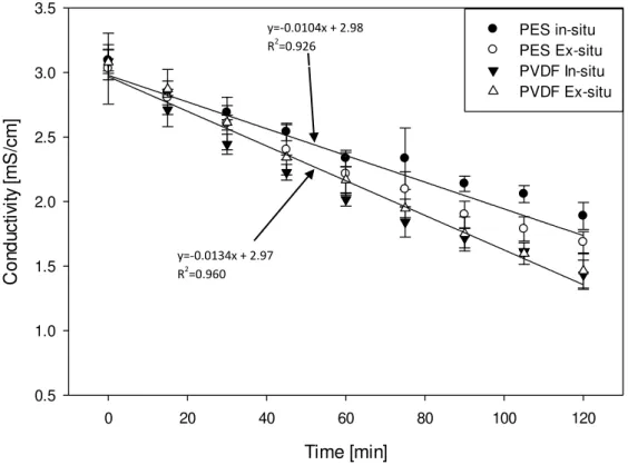
A similar trend of linearly decreasing conductivity with time is also observed for the C+ RC for
both UFMs and for both ways of digestion as indicated by the global regression lines in figure 4.5. Marginally significant difference in C+
RC conductivity evolution with time was observed for
PES vs PVDF membrane types (P<0.04) as tested by ANOVA which also indicated a significant effect of time (P<0.001) on C+
RC conductivity. For the extent of demineralization in
the C+
RC significant difference (P<0.001) was observed for PES and PVDF membranes with
mean values of (across in-situ and ex-situ experiments) 39.43 ± 6.22 % and 52.22 ± 3.43 %, respectively. Average extent of demineralization in C+
RC for all the EDUF experiments was
calculated to be 45.83 ± 8.16 %. This is confirmed by slope of the global regression line for the decrease of conductivity in C+
RC, with PES and PVDF membranes, which are -0.0104 mS cm-1
min-1 and -0.0134 mS cm-1 min-1, respectively. Hence, there was a slight difference on the rate at
which conductivities changed for the two membranes. This is also related to the system resistance evolution reported in the previous section where system resistance were generally lower for PVDF membranes than PES when the two types of membranes have closer values of conductivity.
Time [min]
0 20 40 60 80 100 120
C
o
n
d
u
c
ti
vi
ty
[
m
S
/c
m
]
0.5 1.0 1.5 2.0 2.5 3.0 3.5
PES in-situ PES Ex-situ PVDF In-situ PVDF Ex-situ
Figure 4.5: Evolution of conductivity in the cationic peptide recovery compartment (C+ RC) for
in-situ and ex-situ experiments with PES and PVDF membranes
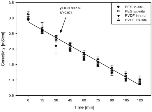
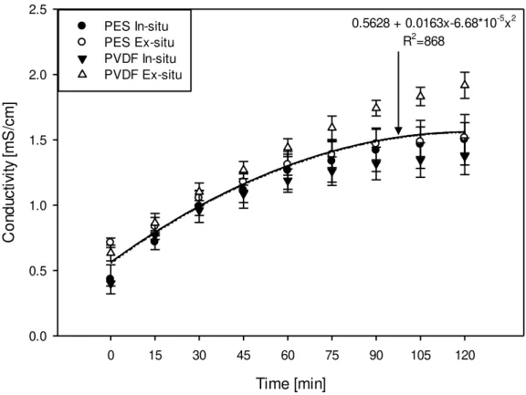
As the conductivity in the two peptide recirculation compartments were decreasing, the conductivity in the feed recirculation compartment was increasing as indicated in figure 4.6. ANOVA indicated no significant difference in feed compartment conductivity for PES/PVDF membranes used or for the digestion strategies employed but there was a significant effect of EDUF duration on the compartment conductivity (P<0.001). The average initial conductivity in the feed compartment for all the experiments was 0.656 ± 0.144 mS/cm and it reached 1.577 ± 0.244 mS/cm at the end of EDUF (2hrs).
y=-0.0104x + 2.98 R2=0.926
Time [min]
0 15 30 45 60 75 90 105 120
C
o
n
d
u
ct
iv
it
y
[m
S
/cm
]
0.0 0.5 1.0 1.5 2.0 2.5
PES In-situ PES Ex-situ PVDF In-situ PVDF Ex-situ
0.5628 + 0.0163x-6.68*10-5x2 R2=868
Figure 4.6: Evolution of conductivity in the feed recirculation compartment for PES in-situ, PES ex-situ, PVDF in-situ and PVDF ex-situ EDUF experiments
The average extent of mineralization in the feed recirculation compartment across all experiments was calculated to be 63.8 ± 8.20%. ANOVA showed that mineralization (starting from initial time to final time) in this compartment had a statistically significant difference for in-situ vs ex-situ experiments (P<0.001) at mean values of 68.83 ± 3.6 % and 58.9 ± 8.23 %, respectively.
The decrease in conductivity in the A
-RC is due to the migration of Cl- ions towards the anode
passing through the AEM to end up in the electrolyte recirculation compartment near anode. At the same time the K+ ions migrate towards the cathode passing through the feed recirculation on
their way to electrolyte solution near the cathode (Figure 2.1). The same phenomenon can explain the decrease of conductivity in the C+
RC, the K+ ions migrate to the electrolyte solution
near the cathode passing through the CEM while the Cl- ions migrate to the electrolyte
recirculation near the cathode passing through the feed compartment. The increase in conductivity in the hydrolysate compartment is due to the transfer of K+ ions migrating across
UFM-I from the A
-RC and the transfer Cl- ions migrate across UFM-II from C+RC (Doyen, et al.,
in A
-RC and C+RC and increasing conductivities in the feed recirculation compartment were
reported before (Roblet C., et al., 2013). It is important to note that conductivities are directly
affected by the NaOH and HCl added in all compartment to keep the pH of all the compartments at 7.8 to avoid the possibility of having peptides assume different charges in different compartments if the pH had changed from one compartment to the other.
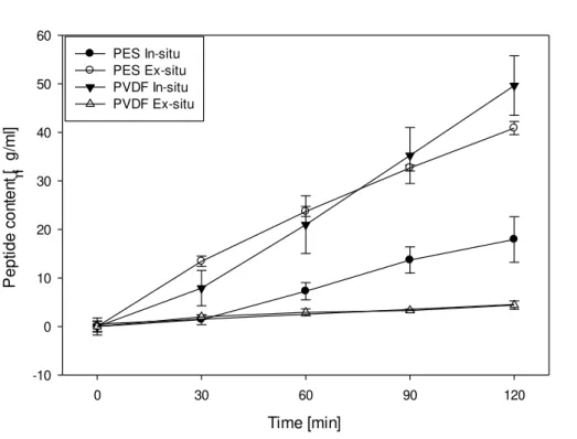
4.1.3. Total Peptide Migration
Figure 4.7 below shows the total peptide that migrated into the A
-RC with time for different
digestion strategy used with both PES and PVDF UFMs. ANOVA indicated total peptide migration in the A
-RC is significantly affected by membrane type (P<0.001), strategy of
digestion (P<0.001) and time (P<0.001). Double interactions were detected between membrane type and ex-situ/in-situ digestion (P<0.001), membrane and time (P<0.001), ex-situ/in-situ with time (P<0.001). Triple interactions were recorded between membrane type, ex-situ/in-situ digestion and time (P<0.003).
All experiments showed a similar trend of roughly linear increase, though at different rates, of total peptide migration to the A
-RC as a function of time. The rates of migration began to differ at
initial minutes except for PES in-situ and PVDF ex-situ experiments which indicated a similar, and very small, rate of total peptide migration till the end of the first 30 minutes of EDUF treatment.
Time [min]
0 30 60 90 120
P e p ti d e c o n te n t [ g /m l] -10 0 10 20 30 40 50 60 PES In-situ PES Ex-situ PVDF In-situ PVDF Ex-situ
Figure 4.7: Total peptide migration into the A
-RC as a function of time for PES and PVDF
![Table 1: Thickness (mm) and conductivity (mS/cm) of UFMs and IEMs used in EDUF experiments Thickness [mm] Conductivity [mS/cm] UFM Material Digestion Strategy Membrane Type](https://thumb-eu.123doks.com/thumbv2/123dok_br/16496062.733565/28.892.87.821.138.724/thickness-conductivity-experiments-thickness-conductivity-material-digestion-strategy.webp)