1. Serviço de Ortopedia do Centro Hospitalar Lisboa Norte; Serviço de Ortopedia Hospital de Santa Maria
Long head of biceps:
from anatomy to treatment
ACTA REUMATOL PORT. 2015;40:26-33
AbsTrAcT/resuMO
The long head of the biceps (LHB), the tendinous struc-ture of the proximal brachial biceps, has its well-known anatomy, which contrasts with its limited current func-tional characterization. Various forms of proximal an-chor and intra-articular route, important for the cor-rect interpre tation of its contribution to the pathology of the shoulder as well as the treatment methodology, are described. Knowledge of its biomechanics results mainly from cadaveric studies that contradict each other. Already the few studies in vivo indicate a depressant and stabilizing action, anterior, for the hume -ral head. Its patho logy is rarely isolated because it is al-most always corre lated with rotator cuff or labrum pathology. It can be divided into 3 major groups (in-flammatory, instability and traumatic) and subdivided according to its location. The anterior shoulder pain is the initial symptom of pathology of LHB Its perfect characterization is dependent on the associated in-juries. Clinical tests are multiple and only their combi-nation allows better sensitivity and specificity for LHB pathology. The arthro-MRI and dynamic ultrasound are able to increase pro per diagnostic of the pathology of LHB. Treatment ranges from conservative and surgical. The latter includes the repair, tenotomy and tenodesis of LHB, which can be performed by open or arthros -copic methodology. The author intends to review ex-isting lite rature on all aspects related to the long head of the biceps from anatomy to treatment, presenting the latest results.
Keywords: Long head of the biceps; Anatomical va
-riants; Rotator cuff; Labrum; Tenodesis; Tenotomy
Sarmento M1
ANATOMY
The long head of the biceps (LHB) originates from the supraglenoid tubercle of the scapula in continuity with the glenoid labrum1,2. The insertion is located medial
to the glenoid articular rim, creating a subsynovial re-cess, which may be posterior, predominantly pos-terior, anterior and pospos-terior, and also predominantly anterior2.
In the anterosuperior part of its insertion and in its continuity with the labrum, three anatomical variants have been described. In the study by Rao et al. normal insertion was found in 86 % of patients and in the re-maining 14 % there was an isolated sublabral foramen (3.3%), one sublabral foramen associated with a mid-dle cord-like glenohumeral ligament (8.6%) and complete absence of anterior and superior labrum associa -ted with a cord-like of middle glenohumeral ligament (1.5 %)3.
In its intraarticular portion of the LHB takes several variants (12), dependent on anatomic criteria and its mechanic behavior to arthroscopic mobilization4. These
are grouped into 4 large families and are dependent on the migration of LHB during embryonic growth, pass-ing for an extra-articular structure to an intrarticular one through the joint capsule5:
1. “meso” family: in this group LHB has a free move-ment beneath the rotator cuff;
2. “adherent” family: LHB is very adherent to the rota-tor cuff;
3. “split” family: the LHB is divided intra - articularly; 4. absence of LHB.
In the “meso” family there are 5 types. In the first there is a small cord, vascularized, between LHB and the rotator cuff. In another there is a synovial band from medial to lateral that is never in stress between the LHB and the rotator cuff. In a third type, the presence of a pulley based in the rotator cuff involves the LHB with-out trapping it and allowing it to slide freely. When this
pulley allows movement without letting it slide, we are in the presence of the fourth kind. Finally, there is the presence of a vascular synovial sheath and not just a pulley with freedom of movement without sliding of the LHB.
In the “adherent” family, and its first type, there is a strong and partial grip, from the medial part of the LHB extending down to the lateral part of the rotator cuff and that is in tension when placing the limb in abduc -tion, pulling the supraspinatus inferiorly. When this adherence is essentially lateral, leaving the me dial part of the LHB free, relaxing when the limb in abduc tion, we are in the presence of the second type. In the third type there is no mesotenon but a thick synovial cove -ring both the LHB in front and behind in continuity with the synovial capsule and not allowing any mo-bility. Finally, rare, the LHB is completely com pliant in the thickness of the supraspinatus without insertion in the medial glenoid tubercle.
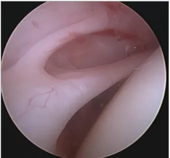
In the “split” family we have 2 types: in the first, the LHB originates partially on the surface of the supraspinatus and partially in the glenoid, joining be-fore emerging the groove and in the second the source is single but in emergency it is divided with a part out to the groove and another to join the most lateral part of the capsule, relaxing with adduction without limi-tation of the LHB slipping (Figure 1).
Finally we have a lack of embryological LHB, the fourth family4.
The vascularization of the most proximal part of the LHB is made from ascending vessels of the anterior humeral circumflex artery while the distal part is irri-gated by branches of the brachial and deep brachial ar-teries6. There is a hypovascular area from 1.2 to 3 cm
from the origin, which corresponds to the sliding part in its groove7.
The network of innervation, sensitive and sympa-thetic, it is also more exuberant in their anchor inser-tion than distally in the muscle-tendon juncinser-tion8.
Intra-articular tendon LHB has a diameter of 5--6mm and a length of 9 cm9and slides, on average, 18
mm inside the joint in anterior flexion and internal ro-tation movement as compared with the neutral posi-tion10.
Upon entering the bicipital groove, the LHB under-goes a twist of 30°-40°11and is stabilized by the
mor-phology of the groove that has a depth of 4 mm and an opening angle of the medial wall which can reach 56°12.
The remaining stability is conferred to it by the roof of the biceps pulley, which consists of fibbers of superior glenohumeral ligament, coracohumeral ligament, supraspinatus and subscapularis, insertion of the pec-toralis major tendon and falciform ligament13,14.
The short head of the biceps originates from the coracoid apophysis in the most lateral part of the con-joined tendon. It forms the medial part of the biceps mass and in its distal insertion, in the proximal bicipi-tal tuberosity of the radius bone, both suffer an exter-nal rotation of 90°. So the LHB has a more proximal insertion conferring supinator function while the short head, more distal, has essentially flexion function of the elbow15,16.
FuNcTION
Most biomechanical studies on the function of the LHB were performed on cadavers and focused on its effect on the stability of the glenohumeral joint, with con-troversial results17.
It is relatively consensual its stabilizing action of the glenohumeral joint when the limb is in abduction and external rotation18,19.
In vivo biomechanical studies shown a proximal mi-gration of the humeral head when the LHB was absent or when unstimulated, so it could be concluded that it acts as a humeral head depressor20,21. There has also
been an anterior translation of the shoulders when compared to the contralateral22.
FIGure 1.Split LHB, with a unique origin at the supraglenoid tubercle and divided at the emergence of the groove
PATHOLOGY
Diseases of the LHB can be classified into 3 broad groups: (1) inflammatory, (2) instability or (3) trau-matic. Meanwhile we can subdivide each group by anatomical location, the pathophysiological process and the state of the LHB23,24.
LHb TeNDINITIs
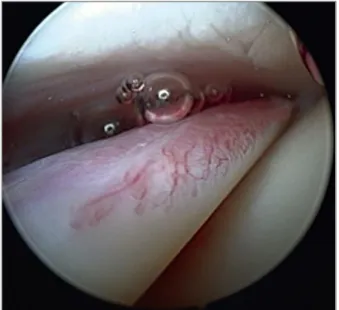
The inflammatory process of LHB is associated with pathology related to surrounding tissues like the rota-tor cuff (90%), the subacromial impingement or the glenohumeral arthritis. Its isolated primary inflamma-tory process is rare (5% of cases)25,26(Figure 2).
This tendinopathy is characterized by chronic in-flammatory process, fibrotic degeneration and de-creased tenoblastic capacity associated with increa sing release of neurotransmitters such as CGRP (calcitonin gene related peptide) and P substance27.
If this process becomes hypertrophic might cause a blocking of the tendon in its intra-articular sliding por-tion or in the groove by narrowing at this level - “ hour--glass biceps “ - which acts as trigger lesions in the fin-ger flexors of the hand28.
LHb ruPTure
The ongoing process of the inflamed tendon friction with the movements of the shoulder can lead to a process of macroscopic delamination of the tendon (Figu -re 3), with partial and subsequently complete ruptu-re. The ruptures are mainly located at the origin of the tendon or at the emergence of groove, and this corres -ponds to the hypovascular zone7,17. When they occur
they are generally associated with a symptomatic relief and a deformity of the distal biceps mass migration called the Popeye signal17,22. These ruptures are more
common over the 50’s years, and with a higher inci-dence (96%) than the short head portion or the distal biceps29.
INsTAbILITY
LHB becomes very unstable due to the non-integrity of its emergence in its groove. This instability can range from subluxation to complete dislocation both to the
medial or posterior lateral side. In the first case, it is associated with rupture of the subscapularis tendon (Fi -gure 4) and the second with previous rupture of the supraspinatus30. Habermeyer et al. divided these ins
-tabilities into 4 groups after arthroscopic visualization: type I - isolated lesions of the uppermost part of the su-perior glenohumeral ligament (SGHL), type II - SGHL injury associated with partial articular supraspinatus rupture, type III – SGHL injury associated with ruptu re of the articular part of the subscapularis and type -IV SGHL injury combined with partial rupture of the supraspinatus and subscapularis11.
sLAP LesION
This entity refers to the insertional lesions of the LHB
FIGure 2.Inflammatory signals in the intraarticular portion of the LHB
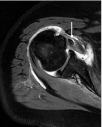
at labrum level, normally starting at a posterior level and subsequently extend to anteriorly (Superior Labrum Anterior and Posterior), beginning by peeling or by grin ding, with or without involvement of the LHB tendon itself (Figure 5).
The classic description of this injury made by Sny-der et al. in 1990 has 4 types and is still the most wide-ly used31. Later, more types were added (type V to X),
despite its limited clinical applicability and generating therapeutic guidelines32,33. In type I there is a fraying
of the labrum and integrity at the biceps anchor. In type II, the LHB anchor is detached from its bed. In type III there is a basket handle rupture of the anchor that falls into the glenohumeral joint and type IV this basket handle is associated with rupture of the tendon extending through and along the LHB31.
cLINIcAL PreseNTATION
The typical history of LHB is the appearance of pain in the anterior aspect of the shoulder, of insidious onset that worsens over time associated with signs/symp-toms of rotator cuff or subacromial impingement.
Spo-radically feeling/observation of instability of the tendon at the level of the groove and distal migration of the biceps mass when complete rupture may occur – Po -peye’s sign34.
The awakening or worsening of pain on palpation on the path of LHB on the groove and at the level of pectoralis major insertion is very suggestive and should be supplemented with oriented clinical test for LHB35,36.
In the broad spectrum of clinical test to assess the integrity of the LHB, the bear hug and upper cut were the most sensitive (0.79 and 0.73, respectively) while the belly press and Speed’s test the most specific ( 0.85 and 0.81 respectively). The association of upper cut and Speed’s test seems to be the best positive predic-tor group of tests we have to identify pathology of LHB36.
We should always evaluate for the possibility of pathology of the cervical spine, shoulder girdle and scapular- thoracic joint with which it makes differen-tial diagnosis34.
cOMPLeMeNTArY DIAGNOsTIc bY IMAGIOLOGY
Conventional radiographs and MRI may allow to
con-FIGure 4.LHB instability, medialized because a concomitant subscapularis tendon tear
FIGure 5.SLAP lesion with a well-defined plane between the supraglenoid tubercle and the LHB tendon origin
firm the diagnosis but mostly can determine associa -ted lesions of the rotator cuff34. Conventional
radiolo-gy at AP, lateral and axillary views, allows to exclude degenerative pathology of the glenohumeral and acromioclavicular joints, and the presence of bone di -sease causing impingement37. MRI nevertheless has li
-ttle diagnostic capacity for intra -articular lesions of LHP, LHP, and the sensitivity and specificity may slight-ly increase with arthro-MRI38. Ultrasonography (US)
is useful in confirming tears or dislocations but espe-cially in dynamic instabilities39. Regarding the latter,
comparing its diagnostic capacity prior to surgery and intraoperative findings, US identifies 90 % of normal LHB and 88 % of full-thickness tears of the LHB but its diagnostic abili ty of intermediate lesions, as partial tears or inflamma tory lesions drops to 27 and 22 %, re-spectively40-42. It has the advantage of being an
inex-pensive examination and easier to access, but very op-erator dependent41,42. Although no study with the same
characte ristics for MRI exist, this demonstrated a ca-pacity of overdiagnosis for partial tears of the LHB and underdiagnosis for inflammatory disease43.
TreATMeNT
Diseases of the LHB can be treated conservatively for technical reconstruction or tenodesis/tenotomy. We cannot ignore, as we have previously reported, that isolated lesions of LHB are rare and therefore its treat-ment methodology is dependent on the associated and concomitant lesions22.
cONserVATIVe
It includes changes in daily activities, anti - inflammatory and analgesic medications, cryotherapy and phy -siotherapy treatments for associated pathology17. A
corticosteroid injection into the sheath of the LHB, the subacromial space or intra-articular, with or without ultrasound support, can result in symptomatic relief, which is oriented to the associated lesions17. The
in-jection under ultrasound support is demonstrably more accurate: 86.7 % of localization in tendon sheath versus 26.7 % when the injection is made in a non--guided manner44.
As isolated pathology of the LHB is rare, also isola -ted injection to the sheath of the LHB is not very com-mon and it is usually associated with subacromial
bur-sa and/ or intra-articular treatments. Injection of the subacromial bursa or of the gleno-umeral joint, when effective, turns out to have a direct action on the pri-mary causal mechanism of the pathology and secondarily in LHB. The injection to the LHB sheath is usual -ly done with the patient in a sitting position after iden-tifying the bicipital groove by palpation and tender-ness45.
Local anesthesia is most commonly made with li-docaine or bupivacaine46. Regarding the choice of
cor-ticosteroids, it can be divided into: 1) acetates (methyl-prednisolone acetate, betamethasone acetate, hydro-cortisone acetate and dexamethasone acetate) versus phosphates (prednisolone sodium phosphate, be-tamethasone sodium phosphate), 2) fluorinated (be-tamethasone sodium phosphate, dexamethasone sodi-um phosphate, triamcinolone hexacetonide and triamcinolone acetonide) versus nonfluorinated (pre -dnisolone, methylpre-dnisolone, hydrocortisone). In the first group, acetates are less soluble and therefore of major indication for chronic conditions compared with phosphates, more soluble, so more prone to be used in acute situations. Fluorinated corticosteroids, in the extraarticular soft tissues, are associated with much higher rate of tendon ruptures and skin and subcuta-neous atrophy, so their use should be avoided in this location/pathology if the treatment is not guided by image46.
surGIcAL PrOceDures
The ideal type of surgery to deal with the LHB remains unclear and continues to be a source of controver-sy14,47,48. Many forms of tenotomy and tenodesis are
de-scribed either arthroscopically or in open surgery. More consensual criteria than what surgical treatment per-form in the LHB are when to act on it: partial rupture of the LHB, involvement of more than 25-50 % of its diameter, longitudinal delamination of the tendon that interferes with their ability to migrate in the groove, medial subluxation or pulley ruptures associated subs -capularis injuries14,49.
There have been many attempts to answer which of the treatment methodologies gets the best results. In a systematic review48in which were considered 5 stu
-dies of tenotomies, 8 stu-dies of tenodesis and 8 which compared the two techniques, no significant diffe -rences in clinical outcomes were observed, both in per-centage of success as in failure, except for the presence
of Popeye’s sign (more prevalent in patients undergo-ing tenotomies). However it was suggested a correct design of a prospective and randomized study to be a ble to achieve results with greater clinical evidence.
In the decision on tenotomy/tenodesis algorithm it must also be considered the fact that the tenotomy has a simpler, faster and shorter post-operative recovery procedure while tenodesis consumes more surgical time, may have an associated cost of implant and there is greater restriction in the initial stage of rehabilita-tion34.
While tenotomy is a consensual technical proce-dure, performed by arthroscopy at the LHB origin in the supraglenoid tubercle, and it should be verified its retraction into the groove and if it does not, remove the intra-articular stump50. The same consensus cannot be
said for tenodesis.
Remains relevant the discussion of open or arthros -copic fixation technique approach, location and an-chor point. The open approach is more appropriate at lower risk of recurrence in patients whose major con-cern is cosmetic, and also the most preferred in younger patients, athletes and heavier workers17. It is
difficult to determine the correct length-tension rela-tionship of the biceps mass and hence the numerous anchor points already attempted: lesser tuberosity51,
coracoid process51, 52, bicipital groove53, the transverse
humeral ligament, short head of the biceps6, the
pec-toralis major tendon6,9or subpectoral bone tunnel35,53.
Lately the controversy is centred on the location in the proximal half or the distal half of the bicipital groove/subpectoral. Lutton et al. in a retrospective case control study concluded that the most distal location favors the lower incidence of residual pain54.
Further-more, the revision rate is much lower (8 %) when the fixation is subpectoral, compared to arthroscopic proxi mal tenodesis (45%)55.
The method of attachment may be performed by the use of anchor sutures, soft tissues sutures, bone tunneling or interference screw56-58.
The technical reconstruction/repair is particularly useful at the treatment of SLAP lesions. The SLAP I, more frequent in elderly patients, are rarely an isola -ted source of pain, so the mechanical debridement is the most appropriate treatment32. The lesions of SLAP
II, typical in the active young people, benefits in its an-choring fixation, which may be achieved with a single double wire anchor or with two anchors with a single wire for stabilizing the anterior and posterior biceps insertion pillars32,59. Type III lesions are usually trea ted
with removal of the basket handle lesion and type IV depend on the degree of involvement of the tendinous portion of the LHB: greater involvement of the tendon requires tenotomy of the LHB with or without teno -desis; small involvements only debridement60.
cONcLusION
The long head of biceps remains a little-known structu re of a functional standpoint, which contrasts with its anatomy, where it is known a huge number of intra-articular variants and their relationship to the an-terior and superior labrum.
The clinic manifestations are the most consistent criteria in decision making since the amount of provocative tests alone do not combine good speci-ficity and sensitivity, allied by imagiological diagnos-tic procedures.
The determination of treatment is very dependent on the associated and concomitant diseases, since the pathology of isolated LHB is uncommon. The choice of treatment is not easier because of the lack of clini-cal studies that support accurate and clear guidelines. It is uniformly accepted the need for well-designed studies to firstly clarify the role of LHB in the kine-matics of the shoulder and in the other hand to help the choice of the best method of treatment for each disease process.
cOrresPONDeNce TO
Marco Sarmento
Serviço Ortopedia Hospital de Santa Maria Av. Prof. Egas Moniz
Lisboa, Portugal
E-mail: marco.sarmento@sapo.pt
reFereNces
1. Cooper DE et al. Anatomy, histology, and vascularity of the gle-noid labrum. An anatomical study. J Bone Joint Surg Am 1992;74(1): 46-52.
2. Tuoheti Y, et al. Attachment types of the long head of the biceps tendon to the glenoid labrum and their relationships with the glenohumeral ligaments. Arthroscopy 2005;21(10):1242--1249.
3. Rao AG et al. Anatomical variants in the anterosuperior aspect of the glenoid labrum: a statistical analysis of seventy-three ca-ses. J Bone Joint Surg Am 2003;85-A(4):653-659.
4. Dierickx C et al. Variations of the intra-articular portion of the long head of the biceps tendon: a classification of embryologi-cally explained variations. J Shoulder Elbow Surg, 2009;18(4):556-565.
5. Tena-Arregui JB, Puerta-Fonolla J, Murillo Gl. Arthroscopic stu-dy of the shoulder joint in fetuses J Shoulder Elbow Surg. 2005;1114-1119.
6. Becker DA, Cofield RH. Tenodesis of the long head of the bi-ceps brachii for chronic bicipital tendinitis. Long-term results. J Bone Joint Surg Am 1989;71(3): p. 376-381.
7. Cheng NM et al. The arterial supply of the long head of biceps tendon: Anatomical study with implications for tendon ruptu-re. Clin Anat 2010;23(6): p. 683-692.
8. Alpantaki K et al. Sympathetic and sensory neural elements in the tendon of the long head of the biceps. J Bone Joint Surg Am 2005;87(7): p. 1580-1503.
9. Ahrens PM and Boileau P. The long head of biceps and asso-ciated tendinopathy. J Bone Joint Surg Br 2007;89(8): p.1001--1009.
10. Braun S et al. Biomechanical evaluation of shear force vectors leading to injury of the biceps reflection pulley: a biplane fluo-roscopy study on cadaveric shoulders. Am J Sports Med 2010;38(5): p. 1015-1024.
11. Habermeyer P et al. Anterosuperior impingement of the shoul-der as a result of pulley lesions: a prospective arthroscopic stu-dy. J Shoulder Elbow Surg 2004;13(1): p. 5-12.
12. Cone RO et al. The bicipital groove: radiographic, anatomic, and pathologic study. AJR Am J Roentgenol 1983;141(4): p. 781-788.
13. Bicos J. Biomechanics and anatomy of the proximal biceps ten-don. Sports Med Arthrosc, 2008;16(3): p.111-117.
14. Nho SJ et al. Long head of the biceps tendinopathy: diagnosis and management. J Am Acad Orthop Surg 2010;18(11): p. 645-656.
15. Athwal GS, Steinmann SP, Rispoli DM. The distal biceps ten-don: footprint and relevant clinical anatomy. J Hand Surg Am 2007;32(8): p. 1225-1229.
16. Hutchinson HL, Gloystein M, Gillespie M. Distal biceps tendon insertion: an anatomic study. J Shoulder Elbow Surg 2008;17(2): p. 342-346.
17. Khazzam M et al. Disorders of the long head of biceps tendon. J Shoulder Elbow Surg 2012;21(1): p. 136-145.
18. Itoi E et al. Stabilising function of the biceps in stable and uns-table shoulders. J Bone Joint Surg Br 1993;75(4): p. 546-550. 19. Rodosky MW, Harner CD, Fu FH. The role of the long head of the biceps muscle and superior glenoid labrum in anterior sta-bility of the shoulder. Am J Sports Med 1994;22(1): p. 121--130.
20. Warner, J.J. and P.J. McMahon, The role of the long head of the biceps brachii in superior stability of the glenohumeral joint. J Bone Joint Surg Am, 1995. 77(3): p. 366-372.
21. Kido T et al. The depressor function of biceps on the head of the humerus in shoulders with tears of the rotator cuff. J Bone Joint Surg Br 2000;82(3): p. 416-419.
22. Elser F et al. Anatomy, function, injuries, and treatment of the long head of the biceps brachii tendon. Arthroscopy 2011;27(4): p. 581-592.
23. Walch G et al. Subluxations and dislocations of the tendon of the long head of the biceps. J Shoulder Elbow Surg 1998;7(2): p. 100-108.
24. Slätis P, Aalto K. Medial dislocation of the tendon of the long head of the biceps brachii. Acta Orthop Scand 1979;50(1): p. 73-77.
25. Favorito PJ, Harding WG, Heidt RS. Complete arthroscopic examination of the long head of the biceps tendon. Arthrosco-py 2001;17(4): p. 430-432.
26. Hsu SH, Miller SL, Curtis AS. Long head of biceps tendon pat-hology: management alternatives. Clin Sports Med 2008;27(4):
p. 747-762.
27. Murthi AM, Vosburgh CL, Neviaser TJ. The incidence of pa -thologic changes of the long head of the biceps tendon. J Shoul-der Elbow Surg 2000;9(5): p. 382-385.
28. Boileau P, Ahrens PM, Hatzidakis AM. Entrapment of the long head of the biceps tendon: the hourglass biceps-a cause of pain and locking of the shoulder. J Shoulder Elbow Surg 2004;13(3): p. 249-257.
29. Carter AN, Erickson SM. Proximal biceps tendon rupture: pri-marily an injury of middle age. Phys Sportsmed 1999;27(6): p. 95-101.
30. Lafosse L et al. Anterior and posterior instability of the long head of the biceps tendon in rotator cuff tears: a new classifi-cation based on arthroscopic observations. Arthroscopy 2007;23(1): p. 73-80.
31. Snyder SJ et al. SLAP lesions of the shoulder. Arthroscopy 1990;6(4): p. 274-279.
32. Bedi A, Allen A. Superior labral lesions anterior to posterior-evaluation and arthroscopic management. Clin Sports Med 2008;27(4): p. 607-630.
33. Maffet MW, Gartsman GM, Moseley B. Superior labrum-biceps tendon complex lesions of the shoulder. Am J Sports Med 1995;23(1): p. 93-98.
34. McDonald LS et al. Disorders of the proximal and distal aspects of the biceps muscle. J Bone Joint Surg Am 2013;95(13): p. 1235-1245.
35. Mazzocca AD et al. Clinical outcomes after subpectoral biceps tenodesis with an interference screw. Am J Sports Med 2008;36(10): p. 1922-1929.
36. Ben Kibler W et al. Clinical utility of traditional and new tests in the diagnosis of biceps tendon injuries and superior labrum anterior and posterior lesions in the shoulder. Am J Sports Med 2009;37(9): p. 1840-1847.
37. Ditsios K et al. Long head of the biceps pathology combined with rotator cuff tears. Adv Orthop 2012;2: p. 405-472. 38. Pfirrmann CW et al. Subscapularis tendon tears: detection and
grading at MR arthrography. Radiology 1999;213(3): p. 709--714.
39. Teefey SA et al. Ultrasonography of the rotator cuff. A compa-rison of ultrasonographic and arthroscopic findings in one hun-dred consecutive cases. J Bone Joint Surg Am 2000;82(4): p. 498-504.
40. Skendzel JG et al. Long head of biceps brachii tendon evalua-tion: accuracy of preoperative ultrasound. AJR Am J Roentge-nol 2011;197(4): p. 942-948.
41. Iannotti JP et al. Accuracy of office-based ultrasonography of the shoulder for the diagnosis of rotator cuff tears. J Bone Joint Surg Am 2005;87(6): p. 1305-1311.
42. Papatheodorou A et al. US of the shoulder: rotator cuff and non-rotator cuff disorders. Radiographics 2006; 26(1): p. e23. 43. Mohtadi NG et al. A prospective, double-blind comparison of magnetic resonance imaging and arthroscopy in the evaluation of patients presenting with shoulder pain. J Shoulder Elbow Surg 2004;13(3): p. 258-265.
44. Hashiuchi T et al. Accuracy of the biceps tendon sheath injec-tion: ultrasound-guided or unguided injection? A randomized controlled trial. J Shoulder Elbow Surg 2011;20(7): p. 1069--1073.
45. Tallia AF, Cardone DA. Diagnostic and therapeutic injection of the shoulder region. Am Fam Physician 2003;67(6): p. 1271--1278.
46. Skedros JG, Hunt KJ, Pitts TC. Variations in corticosteroid/anes-thetic injections for painful shoulder conditions: comparisons among orthopaedic surgeons, rheumatologists and physical medicine and primary-care physicians. BMC Musculoskelet Di-sord 2007;8: 63.
47. Hsu AR et al. Biceps tenotomy versus tenodesis: a review of cli-nical outcomes and biomechacli-nical results. J Shoulder Elbow Surg 2011;20(2): p. 326-332.
48. Frost A, Zafar MS, Maffulli N. Tenotomy versus tenodesis in the management of pathologic lesions of the tendon of the long head of the biceps brachii. Am J Sports Med 2009;37(4): p. 828-833.
49. Sethi N, Wright R, Yamaguchi K. Disorders of the long head of the biceps tendon. J Shoulder Elbow Surg 1999;8(6): p. 644--654.
50. Walch G et al. Arthroscopic tenotomy of the long head of the biceps in the treatment of rotator cuff tears: clinical and radio-graphic results of 307 cases. J Shoulder Elbow Surg 2005;14(3): p. 238-246.
51. Burkhead W et al. The biceps tendon. 4th ed. The shoulder. Vol. 2. 2009, Philadelphia: Saunders Elsevier.
52. Gumina S et al. Rupture of the long head biceps tendon trea-ted with tenodesis to the coracoid process. Results at more than 30 years. Int Orthop 2011;35(5): p. 713-716.
53. Hitchcock HH, Bchtol CO Painful shoulder; observations on the role of the tendon of the long head of the biceps brachii in its causation. J Bone Joint Surg Am 1948;30A(2): p. 263-273. 54. Lutton DM et al. Where to tenodese the biceps: proximal or
distal? Clin Orthop Relat Res 2011;469(4): p. 1050-1055. 55. Sanders B et al. Clinical success of biceps tenodesis with and
without release of the transverse humeral ligament. J Shoulder Elbow Surg 2012;21(1): p. 66-71.
56. Boileau P et al. Arthroscopic biceps tenodesis: a new technique using bioabsorbable interference screw fixation. Arthroscopy 2002;18(9): p. 1002-1012.
57. Romeo AA, Mazzocca AD, Tauro JC. Arthroscopic biceps te-nodesis. Arthroscopy 2004;20(2): p. 206-213.
58. Castagna A et al. Chondral print on humeral head: an indirect sign of long head biceps tendon instability. Knee Surg Sports Traumatol Arthrosc 2007;15(5): p. 645-648.
59. Burns JP, Bahk M, Snyder SJ. Superior labral tears: repair ver-sus biceps tenodesis. J Shoulder Elbow Surg 2011;20(2): p. S2--8.
60. Gregush RV, Snyder SJ. Superior labral repair. Sports Med Arth-rosc 2007;15(4): p. 222-229.