ALS pathogenesis: role of motor neuron-derived exosomes in microglia activation and dysfunction
112
0
0
Texto
(2)
(3) UNIVERSIDADE DE LISBOA Faculdade de Farmácia Research Institute for Medicines (iMed.ULisboa) Neuron Glia Biology in Health and Disease Group. ALS pathogenesis: role of motor neuron-derived exosomes in microglia activation and dysfunction. Sara Filipa Castro da Costa Pinto Dissertação de Mestrado. Orientador: Dora Maria Tuna de Oliveira Brites Investigadora Coordenadora e Professora Catedrática Convidada Faculdade de Farmácia da Universidade de Lisboa Co-orientador: Ana Rita Mendonça Vaz Botelho, PhD Bolseiro de Pós-Doutoramento da FCT Faculdade de Farmácia da Universidade de Lisboa. MESTRADO EM CIÊNCIAS BIOFARMACÊUTICAS 2016.
(4) The studies presented in this thesis were performed in the Neuron Glia Biology in Health and Disease group, at the Research Institute for Medicines (iMed.ULisboa), Faculty of Pharmacy, Universidade de Lisboa, under the supervision of Dora Brites, Ph.D. and Ana Rita Vaz, Ph.D.. ii.
(5) Part of the results discussed in this thesis was presented in the following occasions: Paper to be submitted in a special issue by invitation Cunha C., Gomes C., Pinto S., Barbosa M., Vaz A.R., Brites D. Pathological role of exosomes in neuroinflammation and in ALS-disease mechanisms. Frontiers in Neuroscience, section Neurodegeneration, for the Research Topic titled "Exosomes: Role in Cell Function, Neurodegeneration and Therapy”, hosted by Diana K Sarko, Cindy McKinney (accepted abstract) (Annex VI.1) Communications in National Scientific Meetings Brites D., Pinto S., Cunha C., Gomes C., Barbosa M., Cunha C., Vaz A.R. Exosomes as mediators of neuroinflammation and pathogenicity in ALS. 30ª Reunião do Grupo de Estudos de Envelhecimento Cerebral e Demência, June 3-4th, 2016, Hotel Açores, Lisboa (oral presentation) (Annex VI.2) Pinto S., Barbosa M., Cunha C., Vaz A.R., Brites D. Extracellular vesicles from ALS motorneurons trigger microglia activation. 8th Postgraduate iMed.ULisboa Students Meeting, July 14th, 2016 (poster presentation) (Annex VI.3). The work presented in this master thesis was supported by Santa Casa da Misericórdia de Lisboa (ELA Project 2016) and by Fundação para a Ciência e Tecnologia – FCT, Portugal (UID/DTP/04138/2013. to. iMed.ULisboa,. SFRH/BPD/76590/2011. to. SFRH/BD/91316/2012 to Cunha C. and SFRH/BD/102718/2014 to Gomes C.).. iii. Vaz. A.R.,.
(6) iv.
(7) Abstract Exosomes are nanosized (30-100 nm) extracellular vesicles that are formed by nearly all types of cells and derive from the endocytic pathway and intracellular multivesicular bodies. They are released when the multivesicular bodies fuse with the plasma membrane. Exosomes mediate intercellular communication and have an important role in the spreading of neurodegenerative diseases, probably also of Amyotrophic Lateral Sclerosis (ALS). Recently, release of exosomes derived from motor neuron (MN)-like NSC-34 cells overexpressing human superoxide dismutase 1 mutated in G93A (mSOD1), was suggested to be implicated in cell-to-cell transfer of mSOD1 toxicity. However, how the uptake of such exosomes by receptor cells, such as microglia, contributes to their activation or loss of function was never investigated. Here we evaluated a selected set of promising markers and mediators of inflammatory response to establish: (i) the pro- and anti-inflammatory microRNA profile in NSC-34 MN-like cell line and in their derived exosomes; (ii) alterations in microglia function and generated polarized microglia subtypes triggered by the mSOD1 MN-derived exosomes; and (iii) the cellular distribution of labelled exosomes in a MN-microglia co-culture system. For that, we used mouse NSC-34 cells expressing either wild-type SOD1 (wt) or the G93A mutation (mSOD1) and the mouse N9 microglial cell line. Exosomes were isolated from the cell culture medium by differential ultracentrifugation and incubated with microglia for 2, 4 and 24 hours, or with the MN-microglia co-cultures for 24 hours. We assessed microglia phagocytic ability and senescence, nitric oxide (NO) production, matrix metalloproteinase (MMP)-2 and MMP-9 activity in the extracellular media, nuclear factorkappa B (NF-κB) activation, and gene and microRNA expression by quantitative Real-Time PCR. We observed that the overexpression of microRNA (miR)-124 in mSOD1 MNs was reproduced in their derived exosomes. Such exosomes led to a loss of microglia phagocytic ability, acute release of NO, MMP-2 and MMP-9, and interleukin (IL)-1β and tumor necrosis factor (TNF)-α expression, together with lasting NF-κB activation and delayed increase of senescent-like cells. Interestingly, the early decrease in miR-124 and miR-146a expression induced by both types of exosomes was followed by their increase after 24 hours of incubation with the mSOD1 MN-derived exosomes, where enhanced miR-155 expression was similarly observed. Finally, we observed that the distribution of exosomes was preferentially towards microglia than to MNs, in the co-culture system. Preliminary data also suggest that mSOD1-associated exosomes increase the microglial expression of IL-1β and TNF-α, together with that of alarmin HMGB1. However, further studies are needed to confirm and assess the relevance of these pilot results. Overall, data highlight exosomes from mSOD1 MNs as inducers of microglia activation and dysfunction, different microglia subsets and inflammatory mediators’ production. Keywords: exosomes; amyotrophic lateral sclerosis; microglia activation/dysfunction; neuroinflammation-associated mediators; microRNA profiling. v.
(8) vi.
(9) Resumo Exossomas. são. nano-vesículas. extracelulares. (30-100. nm). formadas. por. praticamente todos os tipos de células e que derivam da via endocítica e corpos multivesiculares intracelulares. Estas vesículas são libertadas aquando da fusão do corpo multivesicular com a membrana plasmática das células. Os exossomas mediam a comunicação intercelular e têm um papel importante na disseminação de doenças neurodegenerativas,. como. possivelmente. a. Esclerose. Lateral. Amiotrófica. (ELA).. Recentemente foi proposto o envolvimento de exossomas provenientes das células motor neuron-like NSC-34, transfetadas com a superóxido dismutase 1 humana com mutação em G93A (mSOD1), na propagação da toxicidade desta proteína. Contudo, o efeito da incorporação destes exossomas pelas células recetoras, tal como as células de microglia, na sua ativação ou perda de função nunca foi investigado. No presente estudo, pretendeuse avaliar um conjunto específico de marcadores e mediadores da resposta inflamatória, de forma a estabelecer: (i) o perfil de microRNAs pro- e anti-inflamatórios na linha celular NSC34 e nos seus respetivos exossomas; (ii) alterações na funcionalidade das células de microglia, assim como os diferentes subtipos de polarização desencadeados pela exposição aos exossomas provenientes dos neurónios motores mSOD1; e (iii) a distribuição celular de exossomas marcados num sistema de co-cultura neurónios-microglia. Para isso, foram usadas células NSC-34 que expressam tanto a proteína SOD1 normal (wt) como mutada em G93A (mSOD1) e a linha celular de microglia N9. Os exossomas foram isolados do meio de cultura celular por ultracentrifugação diferencial e incubados com a microglia durante 2, 4 e 24 horas, ou nas co-culturas durante 24 horas. Os parâmetros avaliados foram a capacidade fagocítica da microglia e a sua senescência, produção de óxido nítrico (NO) e atividade das metaloproteinases (MMP)-2 e MMP-9 no meio extracelular, ativação do factor nuclear-kappa B (NF-κB), e expressão génica e de microRNAs por PCR quantitativo em tempo real. Nos neurónios motores mSOD1 foi observada a sobre-expressão do microRNA (miR)-124, que se reproduziu nos exossomas provenientes destas células. Estes exossomas induziram a perda da capacidade fagocítica da microglia, libertação aguda de NO, MMP-2 e MMP-9, e expressão da interleucina (IL)-1β e factor de necrose tumoral (TNF)-α, juntamente com a ativação prolongada do NF-κB e aumento tardio do número de células do tipo senescente. Curiosamente, a diminuição inicial na expressão do miR-124 e miR-146a induzida pelos dois tipos de exossomas foi seguida pelo seu aumento após 24 horas de incubação com os exossomas dos neurónios motores mSOD1, sendo igualmente observado o aumento da expressão do miR-155. Por fim, verificou-se que, quando em sistema de co-cultura, os exossomas são preferencialmente internalizados pelas células da microglia do que pelos neurónios motores. Dados preliminares sugerem ainda que os exossomas associados aos neurónios mSOD1 aumentam a expressão de IL-1β e TNF-α na microglia, juntamente com a alarmina HMGB1. No entanto, são necessários mais estudos que confirmem e atestem a vii.
(10) relevância destes resultados. Os resultados obtidos evidenciam os exossomas derivados dos neurónios motores mSOD1 como indutores de ativação e disfunção da microglia, assim como diferentes subconjuntos de microglia e produção de mediadores inflamatórios. Palavras-chave: exossomas; esclerose lateral amiotrófica; ativação/disfunção da microglia; mediadores associados à neuroinflamação; perfil de microRNAs.. viii.
(11) Agradecimentos Como não poderia deixar de ser, o meu primeiro agradecimento é dirigido à Professora Doutora Dora Brites, por me ter recebido tão bem no seu grupo. Durante este ano tive a oportunidade de conhecer melhor, e de perto, o mundo da investigação e foi sem dúvida uma experiência muito enriquecedora, a todos os níveis. Muito obrigada por toda a ajuda, pelo apoio e incentivo ao longo deste trabalho! De seguida, um muito obrigado a ti, Rita! Agradeço toda a orientação ao longo deste percurso e toda a disponibilidade que sempre demonstraste para me ajudar, mesmo quando eu não te queria incomodar com os meus “problemas”. Obrigada por tudo o que me ensinaste ao longo deste ano, por todas as correcções e discussões de resultados, e ainda pelas palavras de incentivo para concluir esta etapa! Por tudo isto, e talvez por mais, obrigada. Um agradecimento também aos Professores Rui, Adelaide, Sofia e Alexandra, e ainda à Cláudia, pela simpatia e boa disposição com que me receberam. Carolina, isto já não deve ser novidade para ti, mas como é óbvio não podia deixar de te agradecer! Desde o início que fui acompanhando o teu trabalho e foi contigo que aprendi grande parte daquilo que sei hoje. Estiveste sempre lá para me ajudar quando algo corria menos bem, para oferecer soluções ou para esclarecer as minhas dúvidas existenciais. “Só problemas”! Sem dúvida que és a alma do grupo, com o teu empenho e boa disposição! Obrigada por me chamares tantas vezes a atenção para não falar tanto... Espero poder continuar a acompanhar o teu trabalho, que com certeza irá ser brilhante. Ao resto das meninas que me acompanharam ao longo deste ano: Cátia, nunca me irei esquecer do dia em que me aplaudiste depois da minha cantoria, foste o meu melhor público até agora! Obrigada pela simpatia e por também tu estares sempre disponível para me ajudar. Martinha, vieste sem dúvida dar mais alegria à cave com a tua boa disposição! Obrigada pela tua ajuda ao longo deste trabalho, foste essencial para que conseguisse cumprir alguns dos objectivos. Foi um prazer trabalhar e discutir resultados contigo. Muito boa sorte para esta nova fase da tua vida! Gisela, vou ter saudades de te ver “implicar” com a Marta… Foi um prazer enorme conhecer-te, oxalá houvesse mais tempo! Ainda, um beijinho para todas as outras meninas (e menino!) com que me fui cruzando na cave, nem que por pouco tempo: Maria, Carla, Mafalda e Filipe. Agora, um agradecimento hiper mega especial à Margarida e à Tânia! Vocês partilharam comigo esta nova fase desde o início e foram quem mais ouviu os meus desabafos, frustrações, piadas secas e conversas sem sentido. Não dá para transmitir por palavras tudo aquilo que tenho para vos agradecer, mas fica só a ideia de que sem vocês isto não teria metade da piada!. ix.
(12) Por fim, obrigada à minha família e amigos! Ao Luís, mesmo não percebendo nada desta tese, sempre demonstraste interesse em saber aquilo que andava a fazer e a descobrir. Que me continues a aturar, a mim e às minhas células, por muito mais tempo! Às minhas “bestis”, um obrigado não chega, já são muitos anos a virar frangos! Aos meus “putxis”, Laura e Tiago, os melhores manos que podia ter! E por fim, o mais importante, aos meus pais, sem os quais nada disto seria possível. Obrigada por todos os dias me incentivarem a ser uma pessoa melhor, tanto a nível pessoal como profissional. Aprendo muito com vocês todos os dias e espero que também tenham aprendido alguma coisa aqui com a jovem! Muito obrigada, do fundo do coração!. x.
(13) Index ABBREVIATIONS.............................................................................................................. xvii I. INTRODUCTION ................................................................................................................ 1 1. Extracellular Vesicles and Exosomes – Role in Homeostasis and Cellular Dysfunction..... 1 1.1. Biogenesis of Exosomes ............................................................................................. 2 1.1.1. Mechanisms of Biogenesis ................................................................................... 2 1.2. Characteristics of Exosomes ....................................................................................... 3 1.2.1. Membrane Composition........................................................................................ 3 1.2.2. Cargo of Exosomes .............................................................................................. 4 1.2.3. Isolation Methods ................................................................................................. 7 1.3. Biological Functions of Exosomes ............................................................................... 8 1.3.1. Exosomes in Interneuronal Communication.......................................................... 9 1.3.2. Exosomes in Disease Spread ..............................................................................13 1.3.3. Exosomes as Disease Biomarkers ......................................................................14 2. Amyotrophic Lateral Sclerosis (ALS): Cellular and Vesicular Players that Contribute to Motor Neuron Injury..............................................................................................................15 2.1. Motor Neuron Degeneration – Involvement of SOD1 .................................................17 2.2. ALS as a Non Cell-Autonomous Disease ...................................................................18 2.2.1. Oligodendrocytes and Schwann Cells .................................................................18 2.2.2. Astrocytes ...........................................................................................................19 2.2.3. Microglia ..............................................................................................................20 2.2.4. Exosomes ...........................................................................................................20 3. Microglia as Main Players in ALS .....................................................................................22 3.1. Microglia Reactivity ....................................................................................................23 3.2. Microglial Phenotypes ................................................................................................25 4. Exosomes as a Promising Therapeutic Strategy ..............................................................29 4.1. Passage Across the Blood-Brain Barrier ....................................................................30 4.2. Small Interference RNA Delivery ................................................................................30 4.3. Exosomal MicroRNA Modulation ................................................................................31 5. Aims .................................................................................................................................33 II. MATERIALS AND METHODS .........................................................................................35 1. Materials...........................................................................................................................35 1.1. For cell culture ...........................................................................................................35 1.2. Supplements and chemicals ......................................................................................35 xi.
(14) 1.3. Antibodies ..................................................................................................................35 1.4. Equipment..................................................................................................................36 1.5. Cell lines ....................................................................................................................36 2. Methods ...........................................................................................................................37 2.1. Cell lines ....................................................................................................................37 2.1.1. NSC-34 cell line ...................................................................................................37 2.1.2. N9 cell line ...........................................................................................................37 2.2. Cell treatments ...........................................................................................................37 2.2.1. NSC-34 cell line ...................................................................................................37 2.2.2. N9 cell line and incubation with exosomes from NSC-34 cells .............................37 2.2.3. NSC-34-N9 co-cultures .......................................................................................38 2.3. Differential ultracentrifugation.....................................................................................39 2.4. Labelling with PKH67 fluorescent probe.....................................................................40 2.5. Determinations ...........................................................................................................41 2.5.1. Microglial phagocytosis........................................................................................41 2.5.2. Microglial senescence .........................................................................................41 2.5.3. NF-κB activation ..................................................................................................42 2.5.4. Extracellular nitric oxide .......................................................................................43 2.5.5. Gelatin zymography.............................................................................................43 2.5.6. Quantitative Real-Time PCR ...............................................................................43 2.5.7. Exosome distribution ...........................................................................................45 2.6. Statistical analysis......................................................................................................45 III. RESULTS .......................................................................................................................47 1. NSC-34 inflamma-miR profiling indicates that only miR-124 is upregulated in mSOD1 MNs, which is recapitulated in cell-derived exosomes ..........................................................47 2. Exosomes from mSOD1 MNs induce loss of microglia phagocytic ability and increase the number of senescent cells ....................................................................................................48 3. Evaluation of microglial inflammatory response to exosomes released by wt MNs and mSOD1 MNs ........................................................................................................................51 3.1. Exosomes from mSOD1 MNs lead to an early microglial production of inflammatory mediators ..........................................................................................................................51 3.2. Microglia increasingly release pro-inflammatory cytokines upon interaction with exosomes from mSOD1 MNs ...........................................................................................52 3.3. Exosomes from mSOD1 MNs trigger a delayed upregulation of inflammatory miRNAs .........................................................................................................................................55 4. Exosomes from MN-microglia donors preferentially distribute in microglia when both recipient cells are considered ...............................................................................................56 xii.
(15) IV. DISCUSSION..................................................................................................................59 Concluding remarks ..........................................................................................................67 New perspectives and approaches ...................................................................................67 V. REFERENCES ................................................................................................................69 VI. ANNEX ...........................................................................................................................87 Annex VI. 1 .......................................................................................................................87 Annex VI. 2 .......................................................................................................................89 Annex VI. 3 .......................................................................................................................91. xiii.
(16) Figure Index I. INTRODUCTION Figure I. 1 – Biological characteristics of exosomes: biogenesis, general composition and cargo ................................................................................................................................ 6 Figure I. 2 – Exosomal uptake by recipient cells occurs by different mechanisms ............. 9 Figure I. 3 – Involvement of exosomes in interneuronal communication ..........................12 Figure I. 4 – ALS is a neurodegenerative disease, resulting from the degeneration of UMNs and/or LMNs .........................................................................................................16 Figure I. 5 – Role of glial cells and exosomes in the progression and dissemination of ALS ........................................................................................................................................22 Figure I. 6 – Inflammatory response of microglia cells in the presence of mutant SOD1 ..25 Figure I. 7 – Microglia assume different phenotypes during ALS disease progression .....27 Figure I. 8 – Therapeutic uses of exosomes in neurological diseases ..............................32 Figure I. 9 – Schematic representation of the specific aims of the present thesis .............34 II. MATERIALS AND METHODS Figure II. 1 – Schematic representation of the experimental model for the isolation of exosomes and incubation in the recipient cells ................................................................39 Figure II. 2 – Schematic procedure for the isolation of exosomes from the extracellular media ...............................................................................................................................40 III. RESULTS Figure III. 1 – Exosomes from mSOD1 MNs contain an increased expression of microRNA (miR)-124, reflecting their cells of origin ...........................................................................47 Figure III. 2 – Exosomes from mSOD1 MNs produce a lasting decrease in the microglia phagocytic ability .............................................................................................................49 Figure III. 3 – Microglia exposed to exosomes from mSOD1 MNs display signs of dysfunction, with increased SA-β-gal activity and dystrophic features ..............................50 Figure III. 4 – Microglia exposure to exosomes from mSOD1 MNs lead to an immediate microglial release of NO, MMP-9 and MMP-2 ..................................................................52 Figure III. 5 – NF-κB is activated in the microglia exposed to exosomes from mSOD1 MNs, leading to increased expression of TNF-α and IL-1β...............................................54 Figure III. 6 – Microglial HMGB1 expression is maintained unchanged upon incubation with exosomes from wt and mSOD1 MNs ........................................................................54 Figure III. 7 – Expression of microRNA (miR)-124, miR-146a and miR-155 in microglia cells after 24 hours of incubation indicate the acquisition of several microglia phenotypes ........................................................................................................................................56 Figure III. 8 – Exosomes released from motor neurons (MNs) and microglia are preferentially sorted in microglia that in MNs ....................................................................57 IV. DISCUSSION Figure IV. 1 – Schematic representation of the major findings in this thesis .....................66 xiv.
(17) Table Index II. MATERIALS AND METHODS Table II. 1 – List of primer sequences used in qRT-PCR. .................................................44 III. RESULT Table III. 1 – Expression of TNF-α, IL-1β and HMGB1 in microglia co-cultured with wt and mSOD1 motor neurons (MNs), in the presence and absence of added exosomes isolated from microglia-MN co-cultures (either wt or mSOD1) .......................................................58. xv.
(18) xvi.
(19) Abbreviations ALS. Amyotrophic Lateral Sclerosis. AMPA. α-amino-3-hydroxy-5-methyl-4-isoxazolepropionic acid. BBB. Blood-brain barrier. BSA. Bovine serum albumin. 2+. Ca. Calcium. CD14. Cluster of differentiation 14. CNS. Central nervous system. CSF. Cerebrospinal fluid. CX3CL1. Fractalkine. DIV. Days in vitro. DLS. Dynamic light scattering. DMEM. Dulbecco’s modified Eagle’s medium. EAAT. Excitatory amino acid transporter. ER. Endoplasmic reticulum. ESCRT. Endosomal Sorting Complexes Required for Transport. EVs. Extracellular vesicles. fALS. Familial Amyotrophic Lateral Sclerosis. FBS. Fetal bovine serum. G418. Geneticin 418 sulfate. GluR2. Glutamate receptor 2. HMGB1. High-mobility group box 1. Hsp70. Heat shock protein 70. Iba1. Ionized calcium-binding adaptor molecule 1. IFN. Interferon. IL. Interleukin. ILV. Intraluminal vesicle. Inflamma-miRs. Inflammatory microRNAs. iNOS. Inducible nitric oxide synthase. LPS. Lipopolysaccharide. MFG-E8. Milk-fat globule EGF factor-8. MHC. Major histocompatibility complex. miRNAs. MicroRNAs. MMP. Matrix metalloproteinase. MN. Motor neuron. mRNA. Messenger RNAs. mSOD1 MNs. Mutant SOD1 motor neurons (NSC-34/hSOD1G93A) xvii.
(20) MVB. Multivesicular body. NEAA. Nonessential amino acids. NF-κB. Nuclear factor-kappa B. NMDA. N-methyl-D-aspartate. NO. Nitric oxide. PBS. Phosphate-buffer saline. PDL. Poly-D-Lysine. Pen/Strep. Penicillin/Streptomycin. PNS. Peripheral nervous system. qRT-PCR. Quantitative Real-Time PCR. Rab. Ras-related in brain. ROS. Reactive oxygen species. RPMI. Roswell Park Memorial Institute. RT. Room temperature. sALS. Sporadic Amyotrophic Lateral Sclerosis. SA-β-gal. Senescence-associated β-galactosidase. siRNA. Small interfering RNA. SOD1. Superoxide dismutase 1. TEM. Transmission electron microscopy. TLR. Toll-like receptor. TNF-α. Tumor necrosis factor-α. wt MNs. Wild-type motor neurons (NSC-34/hSOD1wt). xviii.
(21) I. INTRODUCTION 1. Extracellular Vesicles and Exosomes Homeostasis and Cellular Dysfunction. –. Role. in. Almost all living cells release extracellular vesicles (EVs) that facilitate and modulate intercellular communication by carrying proteins, lipids and nucleic acids. Increasing evidences suggest that soluble factors and EVs within the secretome provide a major contribution to paracrine activity, generating a tissue microenvironment that may be either beneficial or neurotoxic. The release of membrane-enclosed vesicles from cells was described more than 40 years ago. Originally, these EVs were thought to bud directly from the plasma membrane (Crawford, 1971). However, in 1985, a research using transmission electron microscopy (TEM) showed that, during maturation of reticulocytes to erythrocytes, small vesicles were formed by inward budding inside an intracellular endosome, leading to the formation of a multivesicular body and to vesicle sorting (Pan et al., 1985). In 1987, the word “exosome” was proposed for these EVs and they were first isolated from immature sheep reticulocytes (Johnstone et al., 1987). Later, in 1989, it was established that such vesicles had biological value, helping maturing erythrocytes get rid of transferrin receptors and other unnecessary proteins (Johnstone et al., 1989). Nowadays, EVs are categorized into exosomes and ectosomes (also called microvesicles or shed vesicles), based on their mechanisms of biogenesis and biophysical properties, such as size and surface protein markers. Exosomes are homogenous small particles, with 30-100 nm in size and a cup-shaped appearance by TEM, and are derived from the endocytic recycling pathway. Consequently, exosomes contain specific endosomal proteins that are often used as exosomal markers. Ectosomes constitute a more heterogeneous population of EVs, with 100-1000 nm in size. They are produced directly through the outward budding and fission of membrane vesicles from the plasma membrane, so their surface markers are largely dependent on the composition of the membrane of origin (Lee et al., 2012b; Kastelowitz and Yin, 2014). Despite their differences, the functions of ectosomes are largely analogous to those of exosomes (Cocucci and Meldolesi, 2015). Along the years, accumulating evidence shows that several cell types have the capacity to secrete EVs, including neuronal and glial cells. From now on, we will be focused on exosomes in this Introduction Section, since isolation method and experimental work indicated in the present work only considered exosomes. 1.
(22) Chapter I. Introduction. 1.1. Biogenesis of Exosomes Through exocytosis, cells deliver newly synthetized proteins and/or lipids to either the plasma membrane or the extracellular space. By the opposite process of endocytosis, cells remove plasma membrane components and deliver them to internal compartments called endosomes, from where they can be recycled to the plasma membrane or delivered to lysosomes for degradation (Alberts et al., 2008). These processes complement each other, maintaining homeostasis in the cell membrane. The endosomal system consists of primary endocytic vesicles, early endosomes and late endosomes, also called multivesicular bodies (MVBs). Early endosomes are located near the plasma membrane where they contact with the primary endocytosed vesicles, resulting in the transfer of cargo and addition of membrane. Early endosomes then mature into late endosomes (or MVBs) and during this process they accumulate intraluminal vesicles (ILVs) in their lumen. This is possible by invagination of the MVB membrane, which creates a membrane enclosed compartment in which the lumen is equivalent to the cytoplasm of the cell. The ILVs that are formed sequester proteins, lipids and nucleic acids that are specifically sorted. Interestingly, the fact that two membrane inversions occur during the formation of an exosome, one during the endocytic internalization and other during the formation of the ILVs, allows exosomes to contain transmembrane cargo in the same orientation relative to the plasma membrane (Klumperman and Raposo, 2014). When the MVB is formed, it can have several fates: (i) degradation; (ii) recycling; or (iii) exocytosis. Degradation occurs via fusion of the MVB with lysosomes, allowing the cell to remove excessive membrane and unnecessary proteins. MVBs can also traffic to the Golgi for redistribution and recycling of their cargo, or fuse with the plasma membrane, leading to release of ILVs into the extracellular environment as exosomes (Figure I.1). Consequently, when released, exosomes can communicate with the surrounding cells, having a role in several physiological processes. However, in pathological conditions, exosomes can carry toxic forms of aggregated proteins that would be targeted for degradation, under normal conditions, contributing to the spreading and progression of neurodegenerative diseases, such as Amyotrophic Lateral Sclerosis (ALS).. 1.1.1. Mechanisms of Biogenesis At the limiting membrane of MVBs, some mechanisms act jointly to allow vesicular formation and cargo sorting. The main mechanism of biogenesis requires the Endosomal Sorting Complexes Required for Transport (ESCRT) machinery (Kowal et al., 2014). These protein complexes are transiently recruited from the cytoplasm to the endosomal membrane, where they function sequentially in the sorting of transmembrane proteins and in the. 2.
(23) Chapter I. Introduction. formation of ILVs. Many studies use ESCRT inhibition as a tool to inhibit secretion of exosomes. However, MVBs and ILVs can also form in the absence of ESCRT machinery. This was demonstrated by the inactivation of four proteins of the four different ESCRT complexes, in mammalian cells, which still allowed the MVB formation (Stuffers et al., 2009). Ceramide and phosphatidic acid, two lipids present in the limiting membrane of MVBs, induce inward curvature of MVBs and thus formation of ILVs (Trajkovic et al., 2008; Ghossoub et al., 2014). After sorting and budding of ILVs, MVBs are transported to the plasma membrane, followed by docking and fusion of the lipid bilayers, so that exosomes can be released into the extracellular space. Several components of the endocytic machinery are involved in secretion of exosomes, such as Rab GTPases (Ras-related in brain), cytoskeleton regulatory proteins and SNAREs (soluble NSF-attachment protein receptor) (Théry et al., 2001; Fader et al., 2009; Colombo et al., 2014).. 1.2. Characteristics of Exosomes The composition and content of exosomes depends on the cell of origin, but proteins, lipids and nucleic acids are the main components, with some of them considered as hallmarks of the ILVs due to their endosomal origin. In the past few years, numerous studies have reported changes in protein and lipid composition, as well as in RNA content, of secreted vesicles induced by different extracellular environments or different physiological states of the secreting cells. Hypoxia, inflammation and cancer were some of the conditions studied (de Jong et al., 2012; Tauro et al., 2013). These variations between normal and diseased cells, depending on cell context, have garnered much interest in exosomes as disease biomarkers and may prove vital to the understanding of the involvement of exosomes in pathological disorders.. 1.2.1. Membrane Composition The exosomal membrane is mainly composed by transmembrane proteins and lipids (Figure I.1). Proteomic studies revealed that exosomes contain a specific subset of proteins, some of which depend on the donor cell type, whereas others are from endosomes, the plasma membrane and the cytosol. Proteins from other intracellular organelles, like nucleus, mitochondria, endoplasmic reticulum (ER) and Golgi complex, are minimal (Théry et al., 2001). Exosomes expose at its surface the extracellular domain of transmembrane proteins. Surface transmembrane proteins serve as exosomal markers and include major histocompatibility complex (MHC) class I and II molecules, adhesion molecules (such as. 3.
(24) Chapter I. Introduction. integrins), several members of tetraspanins (CD9, CD63, CD81 and CD82) and lipid raftassociated proteins (including flotillin-1) (Théry et al., 2001; Wubbolts et al., 2003; Subra et al., 2010; Baietti et al., 2012). Exosomes also contain proteins that are involved in specific cell functions, probably addressing exosomes to target cells, through receptor-ligand interactions (Segura et al., 2005; Purushothaman et al., 2016). Moreover, the presence of proteins involved in all major neuropathological hallmarks of neurodegenerative diseases, such as amyloid-β for Alzheimer’s disease, α-synuclein for Parkinson’s disease, and Cu/Zn superoxide dismutase 1 (SOD1) for ALS, suggests the involvement of exosomes in dissemination of these diseases, as discussed in Section 1.3.2. Most interesting, if exposed at the surface, these proteins may be recognized by therapeutic agents. Besides proteins, exosomes contain abundant phospholipids composing their limiting membrane. Although the published works do not provide a completely unified view of this composition, in a general way exosomes contain lipid rafts enriched in cholesterol, glycosphingolipids, sphingomyelin and ceramide, together with phosphatidylserine, a lipid usually present at the cytosolic side of the plasma membrane (Wubbolts et al., 2003; Laulagnier et al., 2004; Trajkovic et al., 2008; Llorente et al., 2013). It has been reported that different lipid compositions are directly related to rigidity and delivery efficiency of exosomes to other cells, hence contributing to the diffusion of certain diseases (Parolini et al., 2009). Exosomes can also have saccharide groups on their outer surface. It was observed that exosomes are enriched in mannose, polylactosamine, α-2,6 sialic acid and complex N-linked glycans, which indicates that glycosylation has a role in protein sorting (Batista et al., 2011).. 1.2.2. Cargo of Exosomes Another interesting feature of exosomes is their cargo and how it can modulate the recipient cells. Exosomes are known to contain within the cytosolic lumen various types of cytosolic proteins and nucleic acids (Figure I.1). It is believed that proteins are target for entry into ILVs by ubiquitination of their cytosolic domains and are recognized by the ESCRT machinery (Katzmann et al., 2001). Cytosolic proteins include cytoskeletal proteins, like tubulin and actin, annexins and Rab proteins, all participating in intracellular transport and membrane fusion, as well as molecules involved in signal transduction, such as syntenin, and heat shock proteins, such as Hsc70. Exosomes may likewise contain specific proteins involved in the MVB biogenesis, including Alix and Tsg101 (Théry et al., 2001; Wubbolts et al., 2003; Subra et al., 2010). Regarding nucleic acids, exosomes have been reported to contain significant amounts of messenger RNAs (mRNA) and small RNAs, including microRNAs (miRNAs). The RNA species were termed “exosomal shuttle RNA” for their properties of being delivered to. 4.
(25) Chapter I. Introduction. another cells and altering their gene expression and protein profiles (Valadi et al., 2007; Iguchi et al., 2010; Montecalvo et al., 2012). Interestingly, many of the mRNA found was not present in the cytoplasm of the donor cell, suggesting a preferential sorting of certain mRNAs to exosomes (Valadi et al., 2007). Concerning small RNA species, the nature of miRNAs present in exosomes is not entirely clear. However, since exosomes fuse and release their miRNA content in recipient cells and repress target mRNAs, inhibiting their expression, it is believed that these miRNAs play a functional role in intercellular communication. For instance, in normal conditions, exosomal miR-124a is transferred from neurons to astrocytes and regulates the levels of the excitatory amino acid transporter 2 (EAAT2), involved in synaptic modulation. In the spinal cord of end-stage SOD1G93A mice, the mouse model of ALS, miR-124a is selectively reduced and, probably because it will be reduced in the released exosomes, it may determine the pathological loss of EAAT2 and neuronal excitotoxicity demonstrated to be associated (Morel et al., 2013). The mechanism of miRNA sorting is not yet clear, but it also seems to be preferential for some of them. This sorting may be controlled by specific RNA-binding proteins that deliver miRNAs into the MVB. Recently, it was described the involvement of one particular protein, the protein heterogeneous nuclear riboprotein A2B1 (hnRNPA2B1) (Villarroya-Beltri et al., 2013). This protein recognizes and binds specific RNA motifs in miRNAs, called EXOmotifs, and controls their sorting into exosomes when sumoylated, pointing to sumoylation as controlling the binding of hnRNPA2B1 to miRNAs. Other non-coding RNAs were also found enriched in exosomes, relative to its cellular content, including vault-RNA, γ-RNA and specific transfer RNAs (Nolte’T Hoen et al., 2012). Moreover, it was also reported that exosomes released from astrocytes and glioblastoma cells contained mitochondrial DNA (Guescini et al., 2010).. 5.
(26) Chapter I. Introduction. Figure I. 1 – Biological characteristics of exosomes: biogenesis, general composition and cargo. Extracellular vesicles are composed by ectosomes and exosomes. Ectosomes are produced directly through the outward budding and fission of the plasma membrane. Exosomes are derived from the endocytic recycling pathway and are formed by the inward budding of the multivesicular body (MVB) membrane, originating the intraluminal vesicles (ILVs) in their luminal space. After being formed, MVBs can be (i) degraded via lysosomes, (ii) recycled to the Golgi network or (iii) released into the extracellular space as exosomes. Exosomal membrane is mainly composed by transmembrane proteins and lipid rafts, in the same orientation as the cell membrane, and the exosomal lumen is equivalent to cytoplasm. Surface transmembrane proteins serve as exosomal markers and include major histocompatibility complex (MHC) class I and II molecules, adhesion molecules, lipid raft-associated proteins and several members of tetraspanins. In a general way, lipid rafts are enriched in cholesterol, glycosphingolipids, sphingomyelin and ceramide. Furthermore, exosomes contain inside various types of cytosolic proteins and nucleic acids. Cytosolic proteins include cytoskeletal proteins, proteins involved in intracellular transport and membrane fusion, molecules that participate in signal transduction, heat shock proteins, as well as proteins implicated in MVB biogenesis. Nucleic acids already identified in exosomes are messenger RNAs (mRNA) and microRNAs (miRNAs). Other small non-coding RNAs found in exosomes are vault-RNA, γ-RNA and specific transfer RNAs.. 6.
(27) Chapter I. Introduction. 1.2.3. Isolation Methods One major ongoing challenge in the isolation of exosomes is the lack of methods that allow its isolation with high purity and the precise characterization of exosome populations. Accurate discrimination between exosomes and microvesicles is also a challenge. Exosomes have been successfully purified from cell culture conditioned medium or body fluids. The original and most commonly used protocol for exosome purification involves differential ultracentrifugation (Raposo et al., 1996), which sequentially pellets cells, microvesicles and exosomes. Several variants of this method are used nowadays, in which the first centrifugation steps are replaced by a single filtration step (Théry et al., 2006). Other slightly modified versions of this protocol were designed for purifying exosomes from body fluids. Because of the viscosity of some fluids, it is necessary to dilute them and to increase the speed and length of centrifugation (Caby et al., 2005; Théry et al., 2006). A different purification. procedure. has. also. been. described,. using. ultrafiltration. instead. of. ultracentrifugation that is especially useful for purifying exosomes from large volumes of conditioned medium (Lamparski et al., 2002). When the goal is to isolate only a subpopulation of marker-positive exosomes, beads coated with antibodies specific for exosomal surface molecules can be used (Clayton et al., 2001; Caby et al., 2005). Furthermore, an extra purification step, after obtaining the pellet of exosomes, may be through a sucrose cushion. This step eliminates most of contaminants, such as proteins nonspecifically associated with exosomes or large protein aggregates, which are sedimented by centrifugation but do not float on a sucrose gradient (Théry et al., 2006). In addition to these traditional isolation techniques, in the last few years several kits have been commercialized, such as ExoQuick™ and Total Exosome Isolation™. Recently, Van Deun and colleagues (2014) have compared different types of exosome isolation protocols and showed that both differential ultracentrifugation and density gradient centrifugation originates the purest exosome preparations, which are enriched in exosomal marker proteins, when compared with commercial kits. Most importantly, it was shown that differential ultracentrifugation results in a high yield of protein and RNA, thus making it a suitable method for exosome isolation. However, it must be taken into account that, after obtaining the isolated EVs, they have to be characterized to confirm the specific presence of exosomes (Arroyo et al., 2011; Van Deun et al., 2014). Further characterization requires at least two different techniques, including complementary biochemical, mass spectrometry and imaging techniques. Exosomes can be examined using dynamic light scattering analysis (DLS) or TEM (Raposo et al., 1996; Jella et al., 2014). To visualize specific markers, exosomes can be previously immunolabelled with antibodies against proteins known to be exposed on exosomal membranes (van Niel et al., 2001). Also,. 7.
(28) Chapter I. Introduction. quantification of exosomes can be made by nanoparticle tracking analysis (NTA), which allows determining the mean size of the population of vesicles analyzed and the particle concentration estimation in the sample (Soo et al., 2012). Furthermore, to characterize the purified vesicles as exosomes, it is important to show that most of the common exosomal proteins (as well as more specific proteins) are present, which can be accomplish by immunoblotting.. 1.3. Biological Functions of Exosomes Nowadays, it is known that exosomes are involved in normal homeostasis, as well as in pathological conditions, serving as mediators of cell-to-cell communication. Cells utilize exosomes to dispose of unwanted proteins or to exchange signals with neighboring cells. Once released from a cell, exosomes are internalized into recipient cells, transferring exosomal molecules from one cell to another. Exosomes transfer both proteins and functional RNA species, resulting in alterations in the recipient cell (Valadi et al., 2007; Skog et al., 2008). For instance, exosome-mediated small interfering RNA (siRNA) delivery has been shown to knockdown target gene expression (Alvarez-Erviti et al., 2011a), and administration of exosomes loaded with luciferin substrate to luciferase-expressing cells results in production of bioluminescence (Montecalvo et al., 2012). Furthermore, exosomal uptake has been visualized directly, by using fluorescent lipid membrane dyes to stain EV membranes, such as PKH67 (Fitzner et al., 2011). Various mechanisms for exosomal uptake have been proposed, many involving exosomal protein interactions with the plasma membrane of target cells that facilitate subsequent endocytosis (Christianson et al., 2013). Exosomes that bind to cell surface receptors lead to the endocytic uptake of the vesicle, but direct fusion of exosomes with the plasma membrane is also possible (Parolini et al., 2009) (Figure I.2). Several studies propose endocytosis as the primary method of exosome uptake and visualization of exosomes inside cells occur within 15 minutes after initial introduction (Feng et al., 2010). Endocytosis can occur in many ways, including phagocytosis (Feng et al., 2010), macropinocytosis (Fitzner et al., 2011; Tian et al., 2014), clathrin-, caveolin- and lipid raftmediated endocytosis (Escrevente et al., 2011; Nanbo et al., 2013; Svensson et al., 2013; Tian et al., 2014), however there seems to be little agreement in the literature as to which mechanisms are most important. It is possible that, due to their heterogeneity, a population of exosomes can simultaneously trigger different gateways into a cell, depending on the cell type and its constituents. Recently, it has also been proposed that exosomes do not need to be internalized to lead to a phenotypic response. Receptor–ligand interactions between. 8.
(29) Chapter I. Introduction. exosomes and the cell surface may be sufficient to permit signal transduction and subsequent downstream signaling effects in the recipient cell (Raimondo et al., 2015). 2. Clathrin-mediated endocytosis. 2. Lipid raft-mediated endocytosis. 2. Phagocytosis. 2. Caveolin-mediated endocytosis. Endosome. 1. Membrane fusion. 3. Ligand-receptor interaction 2. Macropinocytosis Recipient cell. Figure I. 2 – Exosomal uptake by recipient cells occurs by different mechanisms. When released from cells, exosomes can bind to neighboring cells, transferring exosomal molecules from one cell to another. They transfer both proteins and functional RNA species, producing changes in the recipient cell. The adhesion of exosomes to the recipient cell utilizes the interaction of various exosomal surface proteins and cellular receptors. Once bound, the exosome may fuse with the cellular membrane and transfer its content into the cytoplasm of the recipient cell (1), be endocytosed by phagocytosis, macropinocytosis, and by clathrin-, caveolin- and lipid raft-facilitation (2), or may elicit signal transduction via intracellular signaling cascades through ligand-receptor interaction (3). After being endocytosed, exosomes can fuse with the membrane of endosomes and release their content in the recipient cell, or remain in the endosome to be recycled/degraded.. 1.3.1. Exosomes in Interneuronal Communication Initially, exosomes were thought to be a mechanism for cells to remove unwanted proteins however, they also appear to have a function in intercellular communication. Many studies have reported the release of EVs (ectosomes and exosomes) by all cells that comprise the central nervous system (CNS): neurons (Fauré et al., 2006), oligodendrocytes (Krämer-Albers et al., 2007), astrocytes (Fauré et al., 2006; Taylor et al., 2007) and microglia (Potolicchio et al., 2005). It is proposed that exosomes are linked to a number of different biological processes in the CNS, such as synaptic plasticity, neuroprotection, metabolic support and regulation of myelin membrane biogenesis (Figure I.3). Exosomes secreted by neurons have been implicated in synaptic plasticity. These can be incorporated into neurons or into surrounding glial cells however, the machinery for exosome targeting remains unclear. In neurons, MVBs are more abundant in soma and dendrites, being noted an association of MVBs with postsynaptic sites (Cooney et al., 2002), 9.
(30) Chapter I. Introduction. and the secretion of exosomes is regulated by glutamatergic synaptic activity. In the cortex and hippocampus, calcium (Ca2+) entry through ionotropic glutamate receptors, such as Nmethyl-D-aspartate (NMDA) and α-amino-3-hydroxy-5-methyl-4-isoxazolepropionic acid (AMPA) receptors, activates the MVB fusion to the plasma membrane and, thereby, exosome secretion. Recently, Chivet and colleagues (2014) demonstrated that exosomes released upon synaptic activation bind specifically to other neurons, and not to glial cells. The secreted exosomes can carry AMPA glutamate receptor 2 (GluR2) subunits as a way of regulating the amount of these postsynaptic receptors and, consequently, adjusting the strength of neuron's excitatory synapses, maintaining homeostasis (Lachenal et al., 2011). Synaptic plasticity can also be achieved by the delivery of specific proteins and miRNAs via exosomes. This was observed for synaptotagmin 4, a membrane-trafficking protein, that when released by presynaptic terminals and transmitted to postsynaptic cells, activates retrograde signaling and synaptic growth (Korkut et al., 2010), and also for miR-124a that, as previously mentioned, is released by neurons and can modulate the expression of EAAT2 in neighboring astrocytes, consequently controlling the glutamate uptake levels (Morel et al., 2013). Furthermore, neuron-derived exosomes can regulate neuritic elimination through microglial activation. It was found that neurite removal is accelerated when microglial cells are pre-incubated with exosomes, by upregulating complement factors in microglia (Bahrini et al., 2015). Besides the transfer of exosomes from neuron to neuron (or to other cells), glial cells can also release exosomes that are internalized by neurons. It is proposed a bidirectional communication between neurons and oligodendrocytes. Release of the neurotransmitter glutamate by neurons triggers oligodendroglial exosome secretion into the periaxonal space, through NMDA and AMPA receptors on the surface of oligodendrocytes (Frühbeis et al., 2013), whereas these exosomes, containing myelin and stress-protective proteins, help maintaining axonal integrity and improve cellular viability under conditions of cell stress, such as oxygen/nutrient deprivation (Krämer-Albers et al., 2007; Fröhlich et al., 2014). Neurons internalize oligodendroglial exosomes by endocytosis and utilize their cargo. Furthermore, oligondendrocyte-derived exosomes can communicate with other cells in the brain. It has been shown that oligodendroglial exosomes inhibit differentiation of oligodendrocytes and control myelin formation, in an autocrine way (Bakhti et al., 2011), and are taken-up by microglia, by macropinocytosis, which confirms the role of these cells in removing debris (Fitzner et al., 2011). Interestingly, such exosomes do not provoke an inflammatory response by microglia, since they are preferentially taken-up by microglia that do not have antigenpresenting capacity. In the peripheral nervous system (PNS), Schwann cells also communicate with neurons, contributing to axonal regeneration. After nerve damage,. 10.
(31) Chapter I. Introduction. Schwann cells dedifferentiate and proliferate, remove myelin and axonal debris, and support axonal regeneration (Chen et al., 2005). On the other hand, regenerating axons express adhesion molecules that promote Schwann cells alignment and migration (Yamauchi et al., 2008). A recent study demonstrates that Schwann cells secrete exosomes that can be selectively internalized by axons, increasing axonal regeneration. These exosomes carry on their surface the p75-neurotrophin receptor, a protein expressed by dedifferentiated Schwann cells (Lopez-Verrilli et al., 2013). Astrocytes can also mediate neuroprotection through exosomes. In response to oxidative and heat stress, astrocytes secrete elevated levels of heat shock protein 70 (Hsp70) and synapsin I, a synaptic vesicle-associated protein implicated in neural development, in association with exosomes. This is associated with an increase in the survivability of neighboring neurons during injury (Taylor et al., 2007; Wang et al., 2011). Interestingly, astrocyte-derived exosomes have been reported to contain mitochondrial DNA, which can have some relevance in diseases involving mitochondrial alterations (Guescini et al., 2010). Furthermore, an important role of astrocytes in the CNS is the scavenging of extracellular glutamate through EAATs. These transporters have been reported in EVs, such as exosomes, suggesting a possible role in reducing excitotoxicity at an extracellular level (Gosselin et al., 2013). Concerning microglia-derived exosomes, they have been proposed to provide metabolic support to neurons. Early studies demonstrated that microglia secrete exosomes containing the expected exosomal proteins, as well as a set of proteins previously reported for B cell- and dendritic cell-derived exosomes. They also carry the surface-bound aminopeptidase N (CD13) and the monocarboxylate transporter 1 (MCT1), as well as metabolically active enzymes and chaperones. Exosomal CD13 degrades enkephalins, enabling the bind of the ligands to the opioid receptor and thus influencing neuronal cyclic adenosine monophosphate (cAMP) levels. The presence of the lactate transporter MCT1, together with glycolytic enzymes, may attest their role in delivering energy substrates to neurons (Potolicchio et al., 2005). Recently, it has been suggested that release of serotonin from neurons induces secretion of microglia-derived exosomes. This suggests a neurotransmitter-dependent signaling pathway in microglial cells that regulates exosome release (Glebov et al., 2015). Moreover, recent evidences suggest an exosome-mediated inflammasome signaling. When there is a trauma in the CNS, inflammasome is activated with secretion of exosomes containing inflammasome protein cargo into the cerebrospinal fluid (CSF). These exosomes then fuse with target cells to activate the innate immune response in peripheral tissues (De Rivero Vaccari et al., 2016). It is believed that exosomes trigger toll-like receptors (TLRs) in. 11.
(32) Chapter I. Introduction. monocytic cells, but possibly also in other immune cells, and induce the nuclear factor-kappa B (NF-κB) activation and release of cytokines (Bretz et al., 2013; Chow et al., 2014). Furthermore, dendritic cell-derived exosomes increase inflammation through the NF-κB pathway in a similar way to that of lipopolysaccharide (LPS) (Gao et al., 2016). Exosomes containing Hsp70, for instance astrocyte-derived exosomes, can also lead to NF-κB activation and tumor necrosis factor (TNF)-α release in macrophages (Anand et al., 2010).. Figure I. 3 – Involvement of exosomes in interneuronal communication. Many studies have reported the release of exosomes by neurons, oligodendrocytes, astrocytes and microglia. It is proposed that these exosomes are linked to a number of different biological processes in the central nervous system, such as synaptic plasticity, neuroprotection and metabolic support. Neuron-derived exosomes can carry AMPA glutamate receptor 2 (GluR2) subunits as a way of adjusting the strength of neuron's excitatory synapses, synaptotagmin 4 (Syt4), a membrane-trafficking protein that, when released by presynaptic terminals and transmitted to postsynaptic cells, activates retrograde signaling and synaptic growth, and the microRNA(miR)-124a that modulates the expression of excitatory amino acid transporters (EAATs) in neighboring astrocytes. Glial cells can also release exosomes that are internalized by neurons. Oligodendrocyte-derived exosomes contain myelin and stress-protective proteins that help maintaining axonal integrity and improve cellular viability under conditions of cell stress. These exosomes can also be taken-up by microglia, which indicates the role of these cells in removing debris. Astrocytes mediate neuroprotection through exosomes containing elevated levels of the heat shock protein 70 (Hsp70) and synapsin I, a synaptic vesicle-associated protein implicated in neural development. Interestingly, astrocyte-derived exosomes have been also reported to contain mitochondrial DNA (mtDNA) and EAATs. Concerning microglia-derived exosomes, they have been proposed to provide metabolic support to neurons. Microglia secrete exosomes carrying the surfacebound aminopeptidase N (CD13) and the monocarboxylate transporter 1 (MCT1), as well as metabolically active enzymes and chaperones. In the peripheral nervous system, Schwann cells also communicate with neurons through exosomes, contributing to axonal regeneration. Schwann cells secrete exosomes that carry on their surface the p75-neurotrophin receptor (p75NTR), a protein expressed by dedifferentiated Schwann cells. 12.
(33) Chapter I. Introduction. 1.3.2. Exosomes in Disease Spread It has been shown that part of the physiological role of exosomes is their ability to influence other cells through their protein and RNA based cargo, though this also happens in pathological conditions. In fact, many studies have noted an increased production of EVs in diseased states when compared with non-diseased ones, such as in cancer and senescence (Parolini et al., 2009; Lehmann et al., 2012), as well as alterations in their composition and cargo (de Jong et al., 2012; Tauro et al., 2013). These findings suggest that these vesicles can contribute to the spreading of the disease in pathological conditions. One mechanism associated to disease spreading involves the transfer of miRNAs and pathogenic proteins by exosomes, an issue highly explored in oncology. For example, glioblastoma-derived exosomes contain elevated levels of miR-21 and oncogenic receptors, such as EGFRvIII, that are taken-up by normal cells in the tumor environment, leading to further tumor growth and metastasis (Skog et al., 2008). Furthermore, exosomes may modulate the tumor environment into a more favorable niche for tumor growth and metastasis (Park et al., 2010). Besides cancer, exosomes are also implicated in the spread of neurodegenerative diseases. Indeed, since exosomes can be found in almost all body fluids, there might be no distance restriction in its propagation. Thus, exosomes could play an important role in transmitting the pathogenic proteins from the CNS to the PNS. Since the discovery of prion diseases, the concept has emerged that a protein could be a transmissible pathogen and exosomes have been suggested to contribute to their release from host cells to other cells. Alzheimer’s disease is characterized by neurofibrillary tangles composed of hyperphosphorylated tau, and by extracellular deposits of the amyloid-β peptide, called amyloid plaques (Masters et al., 1985; Grundke-Iqbal et al., 1986). In neurons, it is proposed that amyloid-β is sorted to the MVBs and released in association with exosomes, being a component of the exosomal membrane. Consequently, this peptide can spread to the surrounding cells, forming aggregates in such cells. This is supported by the finding of exosomal proteins in the plaques of diseased brains (Rajendran et al., 2006). Furthermore, exosome-associated tau was found in human CSF, suggesting that tau may also be secreted via this mechanism (Saman et al., 2012). Similarly, in Parkinson’s disease, α-synuclein, a presynaptic neuronal protein that forms toxic inclusions, has similarly been detected in exosomes. Once again, exosomes can transfer and propagate the toxic inclusions to other cells (Emmanouilidou et al., 2010; Alvarez-Erviti et al., 2011b) and, more recently, it was proposed that exosomes accelerate the conversion of monomeric to fibrillar aggregates, since they provide catalytic environments for α-synuclein aggregation (Grey et al.,. 2014).. Exosome-mediated. transfer. has. also. been. described. in. ALS.. This. neurodegenerative disease, characterized by the degeneration of motor neurons, has been. 13.
(34) Chapter I. Introduction. recently associated with exosomes through the propagation of mutant and misfolded SOD1 and its cell-to-cell transmission (Grad et al., 2014b). This topic will be further detailed in Section 2, as it is the main focus of the present thesis.. 1.3.3. Exosomes as Disease Biomarkers As previously mentioned, exosomes can be found in most circulating body fluids, such as plasma (Caby et al., 2005), saliva (Ogawa et al., 2008), urine (Pisitkun et al., 2004) and CSF (Street et al., 2012), suggesting their role in intercellular communication and disease dissemination. Thus, exosomes can be used as potential biomarkers for many pathological conditions, while providing an easy and noninvasive way to evaluate the “donor conditions”. Biomarkers are defined as indicators of a biological or pathological condition and include DNA, RNA, proteins and metabolites. The most advantageous feature of exosomes as biomarkers is the use of their cell- and condition-specific cargo. Besides the regular exosomal proteins associated to their endosomal origin, exosomes contain disease-specific proteins or RNAs that can be used to determine the state or progress of the disease. For instance, misfolded SOD1, a protein involved in the motor neuron degeneration in ALS, is found exposed at the outer surface of the exosomal membrane, unlike native SOD1 that normally resides in the exosomal lumen (Kim et al., 2013). This may allow its recognition and deactivation by potential pharmacological and immunological therapeutics (Grad et al., 2014b). In the context of RNA-based biomarkers, they can be easily determined by standard methods, like quantitative Real-Time PCR (qRT-PCR) and microarrays. Exosomes have been shown to contain miRNAs implicated in many diseases that seem to be promising for diagnosis (Cheng et al., 2014). Skog and colleagues (2008) were the first to demonstrate that vesicle-secreted miRNAs can be used for diagnosis of glioblastoma and to determine the stage of disease. These exosomes do not simply replicate the miRNA profile of the cell of origin but also contain a specific set of these genes, suggesting a selective packaging of miRNAs into exosomes. More interestingly, miRNAs are known to be dysregulated in neurodegenerative diseases. For instance, miR-9, miR-125b, miR-146a and miR-155 are increased in the CSF and extracellular fluid of Alzheimer’s disease patients (Alexandrov et al., 2012). In Parkinson’s disease, miR-7 expression is found decreased (Junn et al., 2009). Moreover, significant upregulation of miR-146a and miR-155 is found in ALS patients (Koval et al., 2013). Lately, it was found that miR-146a and miR-155 are present in exosomes from dendritic cells, reason why we hypothesize that they may serve as biomarkers (Alexander et al., 2015).. 14.
(35) Chapter I. Introduction. An interesting discovery for brain diseases was the observation that exosomes are able to cross the blood-brain barrier (BBB). The BBB is a physiological barrier between the brain and circulating blood, formed by brain capillary endothelial cells. These cells are linked by tight junctions between them and are surrounded by the basement membrane, as well as by pericytes and astrocytes, forming the neurovascular unit. The BBB confers protection to the CNS, since it restricts the influx and efflux of molecules (Chow and Gu, 2015). Whether exosomes are internalized by endothelial cells and undergo transcytosis, or affect the permeability/integrity of the BBB is still unknown. However, not only exosomes in the circulating fluids can provide information regarding the cells in the CNS, but also can target a specific cell type and deliver protein or nucleic acid content into such cell. Exosomes have been successfully studied for therapy by taking advantage of their low immunogenicity and unique delivering capability (Sun et al., 2010; Alvarez-Erviti et al., 2011a; Zhuang et al., 2011), as will be discussed in Section 4.. 2. Amyotrophic Lateral Sclerosis (ALS): Cellular and Vesicular Players that Contribute to Motor Neuron Injury Amyotrophic Lateral Sclerosis (ALS), also known as Lou Gehrig’s disease, is a fatal adult-onset neurodegenerative disorder, involving the degeneration of the upper and lower motor neurons (MNs), from the motor cortex, brainstem and ventral horn of the spinal cord (Rowland and Shneider, 2001) (Figure I.4). It affects the muscles of mobility, speech, swallowing and respiration, but usually does not affect the cognitive function, since only 5% of patients develop frontotemporal dementia (Phukan et al., 2007). Typically, the age of onset is around 48 years and patients die 2 to 5 years after due to respiratory failure. The diagnosis is clinical, based on the physical examination, and is usually supported by electrophysiological or neuropathological studies and neuroimaging and laboratory tests to exclude other diseases (Hardiman et al., 2011). Etiologically, ALS can be divided into two categories, sporadic (sALS) and familial (fALS), with 90-95% of cases with no known cause. Both genetic and non-genetic forms of ALS have been described with a remarkable similarity in terms of disease progression and clinical manifestation (Lilo et al., 2013). In fALS, some of the causative genes identified were in Cu/Zn superoxide dismutase 1 (SOD1), transactive response DNA-binding protein (TARDBP), fused in sarcoma (FUS) and chromosome 9 open reading frame 72 (C9ORF72) (Ajroud-Driss and Siddique, 2015). Recently, new gene variants were found to be responsible for 3% of all ALS patients. The new gene, called NEK1, was the result of a big data analysis in ALS research, in more than 1,000 fALS and 6,000 sALS individuals. The study was conducted in 11 countries and involved 80 researchers. NEK1 has multiple roles, 15.
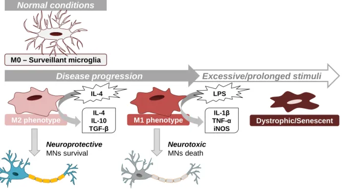
(36) Chapter I. Introduction. including maintenance of neuronal cytoskeleton, mitochondrial membrane regulation and DNA repair. Understanding the role of NEK1 may provide novel drug driven therapies (Kenna et al., 2016). Since the first SOD1 missense mutation was discovered in 1993 (Rosen et al., 1993), more than 180 different mutations have been identified. Mutations in SOD1 account for approximately 20% of the fALS cases. It is believed that these mutations lead to a gain of function where the misfolding and accumulation/aggregation of this protein in neurons and glial cells causes toxicity (Hayashi et al., 2016). Although the main hallmark is MN injury, glial cells were shown to be actively implicated in this disorder and its progression, through the release of soluble factors (Lasiene and Yamanaka, 2011). More recently, it has also been described the involvement of exosomes in the cell-to-cell transmission of the disease (Basso et al., 2013; Grad et al., 2014b).. Figure I. 4 – ALS is a neurodegenerative disease, resulting from the degeneration of UMNs and/or LMNs. Upper motor neurons (UMNs) prolong their axons from the motor cortex through brainstem to spinal cord, connecting with the lower motor neurons (LMNs). In Amyotrophic Lateral Sclerosis (ALS), MN degeneration begins focally in the central nervous system and then spreads contiguously, ultimately leading to an impairment of signal transmission from MNs to muscle. This leads to weakness and muscle atrophy and, ultimately, to muscle paralysis. Nowadays it is know that, besides MN degeneration, neuroinflammation has an important role in the progression of the disease and, eventually, on the onset as well.. 16.
(37) Chapter I. Introduction. 2.1. Motor Neuron Degeneration – Involvement of SOD1 SOD1, a small 153-amino acid protein, is a cytosolic and mitochondrial antioxidant enzyme, converting superoxide to molecular oxygen and hydrogen peroxide (Klug et al., 1972). Wild-type SOD1 is a highly stable dimer, retaining significant conformation and enzymatic activity in the presence of strong protein denaturants. However, it is prone to misfolding and subsequent aggregation in the MNs, when aberrantly oxidized or mutated (Bosco et al., 2010). Indeed, new evidences suggest that the transitory formation of highly toxic trimeric SOD1 species is involved in the neurotoxic phenomena in ALS (Proctor et al., 2016). Although to date the majority of cases are unrelated to mutations in the SOD1 gene, it is suggested the existence of a common pathogenic pathway involving this gene, since SOD1-containing inclusions are seen in cases of sALS and fALS. This observation indicates that aggregation of this protein is a common event in ALS disease (Gruzman et al., 2007; Grad et al., 2014b). However, other common mechanisms in sALS and fALS were identified, such as astrocyte-mediated toxicity (Haidet-Phillips et al., 2011; Meyer et al., 2014; Re et al., 2014). Recently, astrocytes in ALS have been shown to reduce the expression of MHC class I molecules on MNs, increasing their susceptibility to astrocyte-induced cell death (Song et al., 2016). However, exactly how these astrocytes become toxic remains unclear. It is believed that SOD1 displays “prion-like” properties. In vitro assays showed that wild-type and mutant SOD1 can seed aggregation of themselves (self-seeding) and of each other (cross-seeding) and can be transferred to other cells (Chia et al., 2010). Furthermore, misfolding of SOD1 can persist even in the absence of the original misfolded source in cell culture and, when exogenously applied, mutant SOD1 can be efficiently taken-up into living cells and trigger the aggregation of endogenously expressed SOD1 (Grad et al., 2011; Münch et al., 2011; Furukawa et al., 2013). Recently, it was discovered that SOD1 can enter the vesicle-mediated secretory pathway, becoming selectively incorporated into exosomes and exit the cell, which can explain the cell-to-cell transfer of toxicity (Gomes et al., 2007; Basso et al., 2013; Grad et al., 2014a). However, it is still unknown how SOD1 is packaged on exosomes. One explanation can be that the presence of misfolded and aggregated proteins begins to overwhelm protein clearance pathways, leading the cell to release exosomes containing these proteins. Another possibility is that mutant and misfolded SOD1 is released from dying cells and becomes bound to vesicles in the extracellular space that were secreted by other cells. The mechanism by which aggregation of SOD1 causes MN death and degeneration is not completely known, but several hypothesis have been proposed: disruption of axonal transport (Bilsland et al., 2010), glutamate excitotoxicity (Rothstein et al., 1995; Howland et al., 2002), mitochondrial dysfunction (Cozzolino et al., 2009), ER stress (Kikuchi et al., 2006),. 17.
Imagem


+7
Documentos relacionados