Novembro, 2014
Maria Inês Fazendeiro Cunha
Licenciada em Biologia
Exploring deregulated signals involved in Motor
neuron-Microglia cross-talk in ALS
Dissertação para obtenção do Grau de Mestre em Genética Molecular e Biomedicina
Orientador: Dora Maria Tuna de Oliveira Brites,
Investigadora Coordenadora e Professora Catedrática Convidada, Faculdade de Farmácia da Universidade de Lisboa
Co-orientador: Ana Rita Mendonça Vaz, PhD,
Faculdade de Farmácia da Universidade de Lisboa
Júri:
Presidente: Prof.ª Doutora Margarida Casal Ribeiro Castro Caldas Braga
Arguente: Doutora Cláudia Guimas de Almeida Gomes
v
Exploring deregulated signals involved in Motor neuron-Microglia cross-talk in ALS
Copyright © Maria Inês Fazendeiro Cunha, Faculdade de Ciências e Tecnologia, Universidade Nova de Lisboa.
vii Part of the results discussed in this thesis was presented in the following meetings:
Cunha MI, Cunha C, Vaz AR, Brites D. Studying microglial-motoneuron cross-talk in ALS pathology. 6th iMed.UL Postgraduate Students Meeting, Lisbon, July 2, 2014. [Abstract and Poster]
Vaz AR. Motoneuron degeneration and glial reactivity in ALS: insights from cellular to animal models. Neuroscience Seminars at IMM 2012, Instituto de Medicina Molecular, Universidade de Lisboa, Lisbon, Portugal, June 9, 2014. [Oral Communication (by invitation)]
This work was supported by grants PTDC/SAU-FAR/118787/2010 (DB) and
xi
AGRADECIMENTOS
Obrigada Professora Dora. Obrigada por me ter feito parte integrante do seu Grande grupo, pela confiança e pela disponibilidade sempre demonstrada em ajudar e ensinar. Mas acima de tudo, obrigada pela inspiração que é a sua energia a discutir resultados! Houvesse mais tempo. Obrigada, Professora!
Rita, agradeço-te o tempo que disponibilizaste a ajudar-me a construir este projecto tão
importante para mim e a tentar elucidar as minhas ‘bigger pictures’.. Obrigada!
Professor Rui, Professora Alexandra, Adelaide e Sofia, agradeço-vos a disponibilidade com que sempre se mostraram em ajudar no que fosse preciso e as sugestões dadas que contribuíram sem dúvida para melhorar o meu trabalho. Obrigada.
“Ó Carolina eu preciso de você”, já dizia o Seu Jorge! E é bem verdade..foste essencial em
qualquer altura deste meu percurso e é até ‘assustador’ como tu sempre, sempre, sempre, literalmente
apareceste quando eu precisava da tua ajuda. Fosse segunda, terça, quarta..fosse sábado! Se sou uma pessoa de sorte, o meu factor sorte nesta etapa passou por ter tido o teu apoio e aconselhamento. A tua força de espírito é contagiante. Obrigada!!
Cátia, não teria sido o mesmo se não tivesse tido alguém para quem cantar..! Muito obrigada pela tua ajuda sempre, por esclareceres as minhas duvidas existenciais, e claro, o teu rato foi indispensável!! Obrigada!
Filipa, há pelo menos duas coisas que vou certamente guardar comigo: uma conversa de “2 minutos”, que mudaram completamente a minha forma de encarar este ano de tese, e o teu abraço
quando soube que tinha entrado no programa. Desejo-te a melhor sorte em concretizares também tu a tua tese, sei que vai correr tudo bem. Obrigada!
Cláudia, obrigada pelo teu apoio sempre, mas principalmente nas minhas maratonas de Western..certamente caricatas! Obrigada!
Catarina, Cátia e Rui..e isto é que é um ano de tese de mestrado.. Obrigada por todos os momentos partilhados, desejo que concretizem com sucesso esta etapa! Rui, obrigada pelo teu apoio desde o ínicio, foi (e é!) muito importante. Obrigada pela cumplicidade, pela amizade..Desejo-te a melhor sorte! Obrigada!
Inês F., Marta, Andreia e Gonçalo..Obrigada!
xii
semana só para ter os géis..que desespero!! Haja sentido de humor e uma companhia como a tua, para hoje me lembrar desse momento com vontade de rir. Obrigada!
Idálio, ainda ontem falávamos sobre objectivos. Hoje falamos sobre conclusões..Obrigada pela tua presença durante este ano, a ajuda, os desabafos, as preocupações, a troca de ideias, as saídas para desanuviar..! Obrigada!
Rita.. a aventura em que nos pusemos..ou melhor, as aventuras em que nos pomos! Estiveste sempre, estás sempre, e como tal este foi mais um ano em que a tua amizade fez a diferença no meu percurso.. Obrigada SS!
Gisela, e este ano, esta tese.. sem ti? Que desagradável!! Acho que ‘só’ te posso agradecer por tornares Tudo mais fácil. A ‘pressão’ com os prazos, acredita, fez toda a diferença! Tornaste-te um apoio indispensável. Obrigada pela cumplicidade..pela tua presença! É importante. Obrigada!
Obrigada Rita e Ulisses, pela ajuda dada em concretizar todo este ano, este trabalho, esta vivência, em papel. Muito obrigada!
Obrigada irmãos, especialmente a ti João (e Joana)..Palavras são poucas. Obrigada!
E, claro..Obrigada à pessoa que tornou possível estar neste momento a escrever os
agradecimentos de uma tese de mestrado e para a qual um ‘Obrigada’ não deixa de ser muito, muito pouco.. Obrigada Mãe!
xiii
ABSTRACT
Amyotrophic Lateral Sclerosis (ALS) is the most common neurodegenerative disease affecting motor neurons (MNs). Neuroinflammation has shown to be a prominent pathological feature, highlighted by the presence of activated microglia, which may exert either beneficial or detrimental effects. Mutated MNs may release factors able to induce different microglial responses. However, how cells differently modulate each other remains elusive. Therefore, a better understanding of the MN-microglia signaling pathways compromised in ALS is warranted. Here, we aim (i) to uncover signaling pathways underlying MN injury and (ii) to dissect how MNs are modulating microglial response as well as the contribution of healthy microglia to rescue MN dysfunction. We focused on fractalkine-CX3XR1 axis, MFG-E8-mediated phagocytosis and HMGB1-TLR4 signaling.
For this we used a MN-like cell line (NSC-34) stably transfected with human SOD1, either wild-type (wtMNs) or with G93A mutation (mMNs), alone or in mixed cultures with N9 microglial cell line. We observed a compromised viability of microglia in the presence of mMNs, yet they were more activated, as suggested by the increase of CD11b mRNA expression. The dysfunctional mechanisms associated with increased NO and decreasedglutamate production by mMNs were not recovered by the presence of healthy microglia. However, the increased activity of matrix metalloproteinase -9 observed in mMNs was decreased in the presence of microglia. In addition, mMNs presented accumulation of membrane-fractalkine and, in mixed cultures, CX3CR1 mRNA expression was up-regulated in their presence. Furthermore, we showed that mMNs expressed higher levels of MFG-E8, which were further increased in the presence of microglia. Finally, both HMGB1 and TLR4 levels were also increased in mMNs, mainly in the presence of microglia.
Together, these results highlight an impairment of microglial function caused by MN dysfunction and support the development of immunomodulatory strategies restoring both healthy state of microglia and MNs.
xv
RESUMO
A Esclerose Lateral Amiotrófica (ELA) é uma das doenças neurodegenerativas mais frequentes, que afeta os neurónios motores (NMs). A neuroinflamação tem vindo a ser apontada como caracteristica da ELA, sobretudo pela presença de microglia activada que pode ter efeitos benéficos ou detrimentais. Os NMs mutados podem libertar fatores capazes de induzir diferentes respostas microgliais. Contudo, as vias de sinalização intercelular não estão ainda bem esclarecidas, tornando-se necessário um melhor entendimento das vias de sinalização comprometidas entre os NMs e a microglia na ELA. Pretendeu-se: (i) descobrir as vias pelas quais os NMs sinalizam o seu comprometimento e (ii) explorar de que forma estes modulam a resposta da microglia, assim como a contribuição da microglia saudável em recuperar a função dos NMs. Focámo-nos nas vias fractalquina-CX3CR1, fagocitose mediada pelo MFG-E8 e a sinalização HMGB1-TLR4.
Utilizou-se uma linha celular de NMs (NSC-34) transfetados com a proteina humana SOD1, normal ou com mutação G93A, em mono-cultura ou em cultura-mista com uma linha celular de microglia (N9). Verificámos uma microglia comprometida na presença dos NMs mutados, mas mais ativada, com base no aumento de expressão do mRNA do CD11b. O aumento de produção de NO e diminuição de glutamato não foram recuperados na presença de microglia, no entanto a atividade aumentada da metaloproteinase-9 foi reduzida. Os NMs mutados demonstraram acumulação de fractalquina membranar e, nas culturas-mistas, a expressão do mRNA do CX3CR1 estava aumentada na sua presença. Verificou-se ainda que os NMs mutados expressam níveis superiores de MFG-E8, aumentados na presença da microglia. Mais ainda os níveis de HMGB1 e TLR4 encontravam-se igualmente aumentados nos NMs mutados, e principalmente na presença da microglia.
Os resultados obtidos evidenciam um comprometimento da função microglial causada pela disfunção dos NMs e apoiam o desenvolvimento de estratégias imunomoduladoras que recuperem a funcionalidade quer da microglia quer dos NMs.
xvii
TABLE OF CONTENTS
ABBREVIATIONS ... XXIII
I. INTRODUCTION ... 1
1. AMYOTROPHIC LATERAL SCLEROSIS ... 1
1.1. GENETICS ... 2
1.1.1. Mutations in SOD1 ... 3
1.1.2. Other mutations ... 3
1.2. SPORADIC ALS ... 5
1.3. COMPROMISED HOMEOSTASIS OF MNS ... 6
1.3.1. Oxidative Stress – accumulation of free radicals ... 7
1.3.2. Excitotoxicity – glutamate activity ... 7
1.3.3. Protein misfolding and aggregation ... 8
1.3.4. ER stress ... 8
1.3.5. Mitochondrial dysfunction and impairment of axonal transport ... 9
1.3.6. Cell death pathways ... 10
1.4. THE ROLE OF GLIAL CELLS IN ALS ... 11
1.4.1. Schawn cells and oligodendrocytes ... 11
1.4.2. Astrocytes ... 11
1.4.3. Microglia ... 12
2. MICROGLIA: A KEY PLAYER IN THE PATHOPROGRESSION OF ALS ... 15
2.1. VARIABLE ROLE: FROM SURVEILLANT TO NEUROPROTECTIVE OR NEUROTOXIC ... 15
2.2. MOTOR NEURON-MICROGLIA CROSS-TALK IN ALS ... 17
2.2.1. Ligand-receptor interaction ... 17
2.2.1.1. Fractalkine ... 17
2.2.1.2 High mobility group box 1 ... 18
2.2.2. Phagocytosis ... 18
3. ALS RESEARCH MICE MODELS... 20
3.1. IN VITRO MODELS ... 20
3.1.1. Primary cultures and cell lines ... 20
3.1.2. Mixed cultures and co-cultures ... 21
3.1.3. Organotypic cultures ... 21
3.2. IN VIVO MODELS ... 21
3.2.1. Transgenic mSOD1 mice ... 21
xviii
5. AIMS... 25
II. MATERIALS AND METHODS ... 27
1. MATERIALS ... 27
1.1. CHEMICALS ... 27
1.2. ANTIBODIES ... 27
1.3. EQUIPMENT ... 28
2. METHODS ... 28
2.1. CELL LINES ... 28
2.1.1. NSC-34 cell line ... 28
2.1.2. N9 cell line ... 28
2.2. CELL CULTURES ... 28
2.2.1. NSC-34 mono-culture ... 28
2.2.2. NSC-34 and N9 mixed-cultures ... 29
2.3. IMMUNOCYTOCHEMISTRY ... 30
2.4. WESTERN BLOT ASSAY ... 30
2.5. GELATIN ZYMOGRAPHY ... 30
2.6. QUANTIFICATION OF NITRITE LEVELS ... 31
2.7. MEASUREMENT OF EXTRACELLULAR GLUTAMATE ... 31
2.8. FLOW CYTOMETRY ... 31
2.9. QRT-PCR ... 32
2.10. STATISTICAL ANALYSIS ... 32
III. RESULTS ... 33
1. CHARACTERIZATION OF MMN-MICROGLIA MIXED CULTURES ... 33
1.1. MICROGLIAL CELLS ARE MORE VULNERABLE, YET MORE ACTIVATED, IN THE PRESENCE OF MMNS THAN WTMNS . 33 1.2. MARKERS OF NEUROINFLAMMATION ARE DIFFERENTLY MODULATED BY THE PRESENCE OF MMNS ... 35
2. COMMUNICATION BETWEEN MICROGLIA AND MNS IS MODIFIED WHEN CELLS EXPRESS MUTATED HSOD1 (MMNS) ... 37
2.1. DEREGULATION OF FKN/CX3CR1 SIGNALING IS OBSERVED WHEN MNS EXPRESS MUTATED HSOD1(MMNS) .. 37
2.2. NEURONAL MFG-E8 INCREASES IN MMNS IN THE PRESENCE OF HEALTHY MICROGLIA ... 39
2.3. INTRACELLULAR HMGB1 LEVELS ARE HIGHER IN MMNS THAN IN WTMNS, MAINLY IN THE PRESENCE OF MICROGLIA……. ... 41
2.4. TLR4 EXPRESSION IS UP-REGULATED IN MMNS, MAINLY IN THE PRESENCE OF HEALTHY MICROGLIA ... 42
IV. DISCUSSION... 45
xix
xxi
TABLE OF FIGURES
FIGURE I.1. ALS SYMPTOMS ARE A RESULT OF UMNS AND/OR LMNS DEGENERATION SIGNS ... 2
FIGURE I.2. MOLECULAR MECHANISMS LEADING TO COMPROMISED HOMEOSTASIS OF MNS IN ALS ... 6
FIGURE I.3.GENERAL OVERVIEW OF MICROGLIA PHENOTYPES ... 13
FIGURE I.4.THE ROLE OF GLIAL CELLS IN ALS ... 14
FIGURE I.5.MICROGLIA’S SHIFT FROM SURVEILLANT TO NEUROPROTECTIVE OR NEUROTOXIC IN ALS IS THOUGHT TO BE THROUGH INTERACTION WITH MNS ... 16
FIGURE I.6.MOLECULAR PATHWAYS INVOLVED IN MN-MICROGLIA CROSS-TALK FOCUSING ON FKN/CX3CR1 AXIS, MFG-E8-MEDIATED PHAGOCYTIC PATHWAY AND HMGB1 SIGNALING ... 19
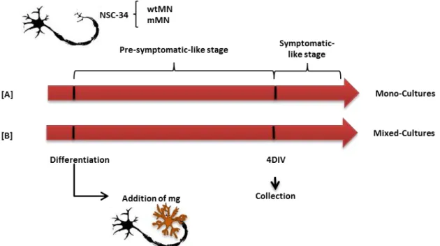
FIGURE II.1.SCHEMATIC REPRESENTATION OF THE EXPERIMENTAL MODEL OF MONO- AND MIXED CULTURES ... 29
FIGURE III.1.MIXED CULTURES WITH MUTATED MOTOR NEURONS (MMNS) AND MMNS ALONE, EVIDENCE DECREASED VIABILITY AND INCREASED APOPTOSIS, AS COMPARED WITH WILD TYPE (WT)MNS. ... 34
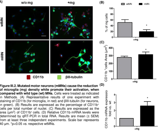
FIGURE III.2.MUTATED MOTOR NEURONS (MMNS) CAUSE THE REDUCTION OF MICROGLIA (MG) DENSITY WHILE PROMOTE THEIR ACTIVATION, WHEN COMPARED WITH WILD TYPE (WT)MNS ... 35
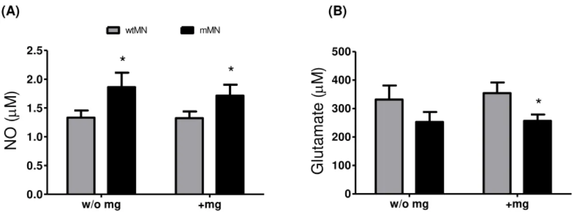
FIGURE III.3.MUTATED MOTOR NEURONS (MMNS), WHEN COMPARED WITH WILD TYPE (WT)MNS, INCREASINGLY PRODUCE NO, INDEPENDENTLY OF THE PRESENCE OF MICROGLIA (MG), AND RELEASE LESS GLUTAMATE, AN EFFECT AGGRAVATED BY MG... 36
FIGURE III.4.ONLY MMP-9 IS INCREASINGLY RELEASED BY MUTATED MOTOR NEURONS (MMNS) VS. WILD TYPE (WT)MNS AND BOTH MMP-2 AND MMP-9 LEVELS SHOW TO DECREASE BY THE PRESENCE OF MICROGLIA (MG) ... 36
FIGURE III.5.MUTATED MOTOR NEURONS (MMNS), WHEN COMPARED WITH WILD TYPE (WT)MNS, INCREASINGLY EXPRESS FRACTALKINE (FKN) WHICH ACCUMULATES IN THE MEMBRANE, INSTEAD OF BEING RELEASED ... 38
FIGURE III.6.EXPRESSION OF CX3CR1 IN MICROGLIA (MG) INCREASES IN THE PRESENCE OF MUTATED MOTOR NEURONS (MMNS), WHEN COMPARED WITH WILD TYPE (WT)MNS ... 39
FIGURE III.7.INCREASED LEVELS OF LACTADERIN/MFG-E8 IN MUTATED MOTOR NEURONS (MMNS) AS COMPARED WITH WILD TYPE (WT) CELLS AND MAINLY IN THE PRESENCE OF HEALTHY MICROGLIA, MAY COMPROMISE PHAGOCYTOSIS ... 40
FIGURE III.8.INTRACELLULAR HMGB1 IS INCREASED IN MOTOR NEURONS EXPRESSING MUTATED HSOD1(MMNS), EITHER ALONE OR IN MIXED CULTURE WITH MICROGLIA ... 42
FIGURE III.9.TLR4 LEVELS INCREASED IN MUTATED MOTOR NEURONS (MMNS), AS COMPARED WITH WILD TYPE (WT) CELLS AND MAINLY IN THE PRESENCE OF HEALTHY MICROGLIA (MG), MAY DERIVE FROM THE UP-REGULATION OF HMGB1... 43
xxiii
ABBREVIATIONS
ALS Amyotrophic Lateral Sclerosis
AMPA α-amino-3-hydroxy-5-methyl-4-isoxazole propionic acid
ATF6 Activating transcription factor 6
Bcl2 B-cell lymphoma 2
BiP Binding immunoglobulin protein
BMAA β-N-Methylamino-L-alanine
CNS Central nervous system
CX3CL1 Chemokine (C-X3-C motif) ligand 1 CX3CR1 Chemokine (C-X3-C motif) receptor 1
Cyt c Cytochrome c
DMEM Dulbecco’s modified Eagle’s medium
Drp1 Dynamin related protein 1
EAAT Excitatory amino acid transporters
ER Endoplasmic reticulum
fALS Familial Amyotrophic Lateral Sclerosis FBS Fetal bovine serum
FKN Fractalkine
FUS Fused in Sarcoma
FWR Forward
GFAP Glial fibrillary acidic protein
GLAST Glutamate aspartate transporter
GLT1 Glial glutamate transporter 1
GUDCA Glycoursodeoxycholic acid HMGB1 High mobility group box 1 Hsp70 Heat shock protein 70 IGF-1 Insulin-like growth factor 1
IL Interleukin
iNOS Inducible nitric oxide synthase
IRE1 Inositol requiring enzyme 1
LC3 Microtubule-associated protein light chain 3
LMN Lower motor neuron
MFG-E8 Milk fat globule-EGF factor 8 Mfn1 Mitofusin 1
Mg Microglia
MMP Matrix metalloproteinase
MN Motor neuron
mSOD1 Mutated Superoxide dismutase 1
xxiv
NF-kB Nuclear factor-KB
NO Nitric oxide
OH Hydroxyl radical
PDI Protein disulphide isomerase
PDL Poly-D-lysine
PERK PKR-like ER kinase
PKR Double-stranded RNA-activated protein kinase
PS Phosphatidylserine
RAGE Receptor for advanced glycation endproducts
REV Reverse
ROS Reactive oxygen species
sALS Sporadic Amyotrophic Lateral Sclerosis
SOD1 Superoxide dismutase 1
TARDBP Transactive response DNA-binding protein TDP-43 TAR-DNA binding protein
TLR Toll-like receptor
TLS Translocated in liposarcoma
TNF-α Tumor necrosis factor α
UBC Umbilical cord stem cell
UDCA Ursodeoxycholic acid
UMN Upper motor neuron
UPR Unfolded protein response
1
I. INTRODUCTION
1. Amyotrophic Lateral Sclerosis
Amyotrophic Lateral Sclerosis (ALS) is the most common adult-onset neurodegenerative disease affecting motor neurons (MNs). It is characterized by the selective loss of MNs in the motor cortex (upper motor neurons - UMNs) as well as in the brainstem and in the spinal cord (lower motor neurons – LMNs). The disease begins focally, in the central nervous system (CNS), and spreads contiguously leading to progressive degeneration (Figure I.1). In 1869 the french neurobiologist and clinician Jean-Martin Charcot first described and diagnosed the first cases of ALS and found that the symptoms vary depending on the location of the lesion (Kumar et al., 2011) resulting in a clinical presentation of muscle weakness, spasticity, atrophy and ultimately, paralysis, together with a slurred speech, dysphasia and dysarthria, culminating in death, in the majority of cases by respiratory failure. Typically the cognitive functions of the brain remain undamaged so the patient is aware of the disease progression. This relentless disease usually has a fast rate of progression being the typical age of onset around 48 years and patients usually dye within 3-5 years from symptoms onset. ALS has a worldwide incidence of 1 to 2 new cases per 100,000 individuals each year and a prevalence of 4 to 6 cases per 100,000 individuals (Chen et al., 2013).
Exploring deregulated signals involved in Motor neuron-Microglia cross-talk in ALS
2
Figure I.1. ALS symptoms are a result of UMNs and/or LMNs degeneration signs. Upper motor neurons
(UMNs) localize in the motor cortex and connect with lower motor neurons (LMNs) present in brainstem and spinal cord. In ALS, motor neuron (MN) degeneration begins focally in CNS and then spreads contiguously, ultimately leading to an impairment of signal transmission from MNs to muscle. Clinically this is represented by progressive muscle weakness, spasticity, atrophy, paralysis and death, mostly by respiratory failure, within 3 to 5 years from symptoms onset.
Concerning etiology, approximately 90% of ALS cases have unknown cause and are non-genetic, commonly classified as sporadic ALS (sALS), and only 5-10% of cases are genetic forms, generally known as familial ALS (fALS). Interestingly, both the non-genetic and the genetic forms of ALS are suggested to have common pathogenic mechanisms and share the clinical features (Lilo et al., 2013).
1.1. Genetics
I. Introduction
3
1.1.1.
Mutations in SOD1
In 1993, Rosen and colleagues found the first causative mutations in fALS within the gene encoding the enzyme SOD1 (Rosen et al., 1993). The gene maps to chromosome 21q22.1 and is composed of five exons encoding a 153 amino acid metalloenzyme. SOD1 is a very stable dimer ubiquitously expressed with a crucial function in cellular homeostasis. The protein binds copper and zinc ions and forms a homodimer that acts as dismutase removing toxic superoxide radicals by converting them to molecular oxygen and hydrogen peroxide, thus preventing ROS toxicity (Keller et al., 1991), and was found to localize in the cytoplasm, nucleus, lysosomes and intermembrane space of mitochondria.
So far, 177 SOD1 mutations have been described counting for 20% of genetic forms and 2-7% of non-genetic forms (Al-Chalabi, 2014) and with few exceptions all SOD1 mutations are dominant. The majority of the mutations in SOD1 are missense within all five exons affecting the functional domains of the protein, like the glycine-to-alanine substitution at position 93 (SOD1G93A) which seems to be particularly vulnerable since it is point mutated to all 6 possible residues in fALS (Turner and Talbot, 2008). Nevertheless, nonsense mutations, insertions and deletions were also reported (Shaw and Valentine, 2007).
SOD1-ALS is thought to originate as a distal axonopathy, being neuromuscular junctions the first regions of the MNs to degenerate in a process known as ‘dying back’ (Fischer et al., 2004). Clinically, patients with mutated SOD1 (mSOD1) present mostly with lower limb onset, with predominance of LMN features, and young age-of-onset (Gordon, 2013) but the time of onset and duration of disease may be different according to the type of mutations (Boillée et al., 2006a).
It is believed that a gain of toxic function mechanism is the main pathway for SOD1 neurotoxicity (Pasinelli and Brown, 2006). It was reported from analysis of mSOD1 transgenic mouse models that mice lacking SOD1 did not develop the disease (Shefner et al., 1999) and transgenic mice overexpressing human SOD1G93A develop ALS clinical features (Gurney et al., 1994). The mechanism underlying this toxicity may be, for instance, through SOD1 aggregation that is likely to be an early event in the disease as it appears at disease onset and its abundance increases along ALS progression (Wang et al., 2002).
1.1.2.
Other mutations
In addition to SOD1 mutations in the genes coding for TARDBP, FUS and C9ORF72 are closely associated with typical clinical phenotype (Chen et al., 2013).
TDP-Exploring deregulated signals involved in Motor neuron-Microglia cross-talk in ALS
4
43 is usually localized in the nucleus of neurons and some glial cells (Gendron et al., 2010). Although its physiological function in the nervous system is currently unknown, as it is a DNA-RNA binding protein, it is predicted to be involved in mRNA splicing and nucleo-cytosolic RNA transport (Mackenzie and Rademakers, 2008). Hence, abnormal modified (phosphorylated and truncated) TDP-43 promoting its aggregation together with cellular mislocalization of TDP-43 to the cytoplasm of affected neurons leads to the hypothesis that TDP-43 mediated neurotoxicity is likely to result from the loss of normal TDP-43 function, as well as from a toxic gain of function conferred by TDP-43 inclusions (Gendron et al., 2010). To date, several studies have report in total nearly 50 mutations in fALS and sALS (about 3% and 1.5%, respectively), mostly involving the C-terminal glycine-rich region of the protein (Lattante et al., 2013). So currently, TARDBP mutations seem to have comparable incidence of SOD1 mutations in sALS. In both lower MNs and glia TDP-43 positive inclusions are consistent feature of all sporadic cases and fALS without SOD1 mutations (Tan et al., 2007).
Mutations in the gene encoding FUS / translocated in liposarcoma (TLS) have been identified in a subset of patients with familial ALS and, less common, with sporadic ALS (Liscic and Breljak, 2011). FUS is a DNA/RNA binding protein predominantly expressed in cell nuclei in physiological conditions that is involved in transcriptional regulation, RNA and miRNA processing, and mRNA transport. Similar to TDP-43, most mutations are clustered in the C-terminal region rich in arginine and glycine, which encodes also for nuclear localization signal (Lagier-Tourenne and Cleveland, 2009). Studies in postmortem tissue (brain and spinal cord) harboring FUS/TLS mutations revealed an increased cytoplasmic FUS-staining and cytoplasmic inclusions in MNs (Kwiatkowski et al., 2009; Vance et al., 2009), leading to the hypothesis that FUS mutations may contribute to ALS pathogenesis through the formation of cytoplasmic inclusions and/or the loss of the physiological nuclear functions of the protein, analogous to TDP-43. More than 30 mutations have been identified and all except one have an autosomal dominant pattern (Kabashi et al., 2011). Clinically, mutations in this gene are usually associated with age of onset younger than 40 years, survival of less than three years and onset in the upper limbs (Millecamps et al., 2010).
The striking functional and structural similarities of TDP-43 and FUS, both DNA/RNA binding proteins, suggest that abnormal RNA processing is a crucial event in ALS pathogenesis although the mechanisms underlying protein aggregation and the consequent neurodegeneration remain currently unknown.
I. Introduction
5 other features. Byrne et al. (2012) suggest that the C9ORF72 repeat expansion may be inherited in an autosomal dominant way with an expanded phenotype of neurodegeneration and variable penetrance.
1.2. Sporadic ALS
Despite all the progress achieved so far, the causes of large majority of sALS remain unknown but it is thought to be probably associated with a combination of genetic and environmental factors. Environmental factors associated with ALS have been studied for many years and recent advances
have shed light to the cyanotoxin β-N-Methylamino- L-alanine (BMAA). Interestingly, food containing
BMAA has been found on Guam, a well-known focus of ALS/parkinsonism-dementia complex (AL-SPDC) and high concentrations of BMAA were found in the postmortem brain tissue (Cox et al., 2003). This non-protein neurotoxic amino acid can be misintegrated into the protein structure, in the place of L
-serine (Dunlop et al., 2013) which may induce protein misfolding and cell death.
Although to date most cases are unrelated to SOD1 gene mutations, the chance that sporadic and familial ALS may converge on a common pathogenic pathway involving abnormal SOD1 species must be taken into account. For instance, SOD1 immunoreactive protein aggregates were found in spinal cord of both human fALS and sALS cases (Gruzman et al., 2007) and wild-type SOD1 (wtSOD1) was shown to acquire binding and toxic properties of mSOD1 through oxidative damage (Ezzi et al., 2007; Rakhit et al., 2004). Also, wtSOD1 can interact with Heat shock protein 70 (Hsp70) as well as chromogranin B, inducing tumor necrosis factor α (TNF-α) and inducible nitric oxide synthase (iNOS),
leading to a dose dependent cell death (Ezzi et al., 2007). Moreover, oxidized wtSOD1 and mSOD1 were reported to share a conformational epitope that is not present in normal wtSOD1, and both recombinant oxidized wtSOD1 and wtSOD1 immunopurified from human sALS tissues have been shown to inhibit kinesin-based fast axonal transport, similar to SOD1-fALS (Bosco et al., 2010).
Exploring deregulated signals involved in Motor neuron-Microglia cross-talk in ALS
6
1.3. Compromised Homeostasis of MNs
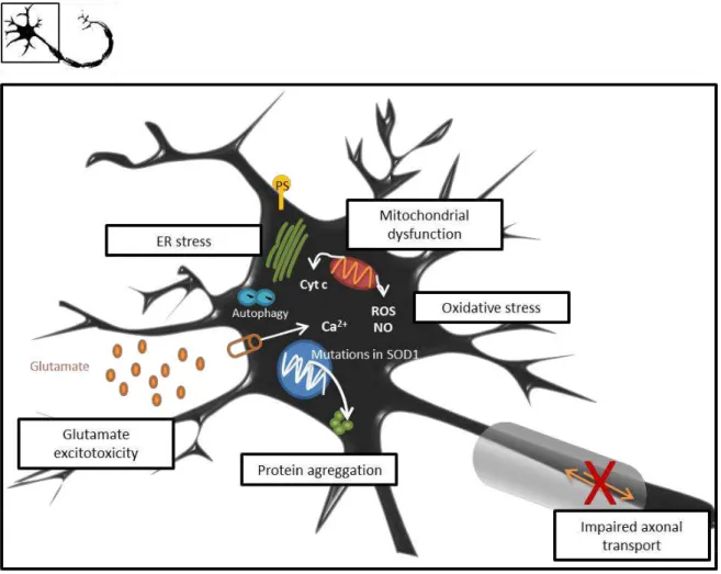
A main hallmark of ALS is MN degeneration and identification of SOD1 mutations has enabled development of animal and cell culture models from which it was possible to uncover some of the mechanisms triggering MN injury, schematically represented in Figure I.2. Although the precise mechanism by which SOD1-mediated toxicity triggers ALS pathogenesis remains unknown, several hypotheses have been proposed such as oxidative stress, glutamate excitotoxicity, protein aggregation, endoplasmic reticulum stress, mitochondrial dysfunction, axonal transport abnormalities, as well as microglial activation and neuroinflammation. Given the variety of mechanisms already reported to be involved in the disease, it seems, most likely that ALS results from cumulative upstream abnormalities which will lead to a common pathogenic cascade.
Figure I.2. Molecular mechanisms leading to compromised homeostasis of MNs in ALS. Motor neuron (MN)
injury is believed to be a result of multiple dysfunctional processes. In SOD1-linked ALS, mutations in the gene will result in a protein with major propensity to aggregate with itself as well as with other proteins. These protein aggregates will cause endoplasmic reticulum (ER) stress and activate the unfolded protein response (UPR). Prolonged UPR triggers apoptosis, characterized by exposure of phosphatidylserine (PS), through ER-stress specific caspases and pro-apoptotic proteins, and cross-talk with mitochondrial intrinsic apoptotic pathway. Also mitochondrial dysfunction results in release of cytochrome c (cyt c) and production of higher levels of free radicals, as reactive oxygen species (ROS) and nitric oxide (NO), inducing apoptosis and increased oxidative stress. Increased number of autophagic vacuoles is also observed within MNs. Abnormalities in the axonal transport are reported in MNs, further resulting in accumulation of protein and organelles that cause axonal damage and, consequently, impaired signal transmission. Extracellular glutamate accumulation promoting excitotoxicity is reported in ALS. For instance, overstimulation of AMPA/kainate receptors leads to major influx of calcium (Ca2+)
I. Introduction
7
1.3.1.
Oxidative Stress
–
accumulation of free radicals
Oxidative stress, associated with the formation of reactive oxygen species (ROS), has been identified as one of the major causes of cellular injury in several neurodegenerative diseases. Oxidative stress is commonly defined as a disturbance in the balance between the production of ROS and
antioxidant defenses, where biological system’s ability to readily detoxify free radicals or easily repair
the resulting damage is compromised. Intracellular ROS may be instigated by leakage of electrons from the mitochondrial respiratory chain, leading to incomplete reduction of molecular oxygen during oxidative phosphorylation, generating superoxide radical anion and hydrogen peroxide. Free radicals, as superoxide and nitric oxide (NO), generated by nitric oxide synthase (NOS) are also produced by immune cells and modulate the expression of the immune response (Wink et al., 2011). Increased levels of superoxide reacting with NO lead to production of peroxinitrite, a potent oxidant, and slowly decomposition of hydrogen peroxide lead to the highly reactive hydroxyl radical (OH). Both peroxynitrite and hydroxyl radical are highly reactive oxidizing agents being able to damage proteins, lipids and DNA. This injury may include changing of protein conformation, altering cellular membrane dynamics by oxidation of unsaturated fatty acids and alterations in DNA and RNA species, as reviewed in Barber and Shaw (2010).
In ALS, the role of oxidative stress has been established in several studies. In fact, Shaw et al. (1995) reported that the levels of protein carbonyl, a biomarker of oxidative stress, were increased in the spinal cord of patients with sALS and Ferrante et al. (1997) reported that those levels were also increased in the motor cortex. In addition, Beal et al. (1997) showed increased concentrations of 3-nitrotyrosine, a marker of peroxynitrite-induced oxidation, in MNs of both familial and sporadic ALS patients. Moreover, using a transgenic mouse model of ALS (SOD1G93A), Andrus et al. (1998) proposed that the increased hydroxyl radical production associated with the SOD1G93A mutation may cause extensive protein oxidative injury, for instance, in SOD1 protein itself.
1.3.2.
Excitotoxicity
–
glutamate activity
An important pathophysiological process in both forms of ALS seems to be glutamate-mediated excitotoxicity. Glutamate is the main excitatory neurotransmitter in the mammalian CNS. Whereas it is important for normal nerve cell function, it becomes toxic in elevated concentrations. This phenomenon is known as excitotoxicity and arises when there is a rupture in the equilibrium between the release and re-uptake of glutamate that leads to increased synaptic glutamate concentrations and consequent excessive stimulation of glutamate receptors, resulting in augmented intracellular calcium levels, generation of ROS, consequent neuron damage and death. This may happen either as a consequence of excessive production or by failure of glutamate transporters, as the excitatory amino acid transporters (EAAT), mostly present in glial cells. MNs are known to have a high level of glutamatergic input and to be particularly vulnerable to excitotoxic cell death (Heath and Shaw, 2002).
Exploring deregulated signals involved in Motor neuron-Microglia cross-talk in ALS
8
transporter EAAT2, the major glutamate transporter responsible to remove glutamate from the synaptic cleft, is reduced by 90% in the ventral horn of paralyzed transgenic SOD1G93A rats (Howland et al., 2002). Moreover, by using primary cultures of MNs from murine spinal cord, Roy et al. (1998) have shown that normally non-toxic glutamatergic input, particularly via calcium-permeable AMPA/kainate receptors, is a major factor in the vulnerability of MNs to the toxicity of SOD1 mutants.
1.3.3.
Protein misfolding and aggregation
Protein aggregation is a hallmark of many neurodegenerative diseases, including ALS. The clumps of protein misfolded, either because of injury from ongoing cellular processes or through the heritage of an abnormal structure in genetic disorders, may affect normal MN functions and induce cell death. SOD1 inclusions constitute a primary feature of SOD1-fALS in both human ALS patients and animal models. These inclusions were reported to form before symptoms onset and to accumulate during disease progression and were found to occur in the cytoplasm, mitochondria and vacuoles of MNs, as well as axons and dendrites (Chattopadhyay and Valentine, 2009).
In addition, Wang and co-workers proposed that toxicity of aggregates is mediated by their size and that mSOD1 has an increased aggregation propensity, i.e., an increased likelihood of an unfolded protein to aggregate, toxic for ALS patients (Wang et al., 2008). In fact, aggregation is thought to be a consequence of the formation of misfolded SOD1 that escape from cellular quality-control and housekeeping mechanisms, and different ALS-associated mutations may increase SOD1 aggregation for several reasons, such as: a decrease in the negative charge of SOD1 without affecting stability or metal binding, a destabilization of protein native state, or an impairment of Cu or Zn binding by SOD1 (Shaw and Valentine, 2007). Interestingly, intracytoplasmic protein deposits strongly stained for SOD1 in the spinal cord of both sALS and fALS and, in ALS, are thought to consist of granule-coated fibrils that contain SOD1 as well as other proteins as reviewed by Chattopadhyay and Valentine (2009).
1.3.4.
ER stress
I. Introduction
9 compensated for, prolonged UPR triggers apoptosis through ER-stress specific caspases and pro-apoptotic proteins, and cross-talk with mitochondrial intrinsic pro-apoptotic pathway (Walker and Atkin, 2011). Regarding ALS, two studies reported that UPR was present in mice model SOD1G93A (Atkin et al., 2006; Kikuchi et al., 2006) and that mSOD1 was present inside the ER. Atkin et al. (2008) pointed out an up-regulation of the full spectrum of UPR markers in lumbar spinal cord tissue from human sALS, therefore suggesting ER stress as common phenomenon to all ALS cases. Interestingly, Saxena et al. (2009), by using SOD1G93A mice model, have observed that selectively vulnerable MNs were prone to ER stress and that the activation of UPR was correlated with microglial activation in lumbar spinal cord. Altogether, these findings highlight an important role for ER in the pathogenicity of ALS, although the underlying mechanisms remain elusive.
1.3.5.
Mitochondrial dysfunction and impairment of axonal transport
Mitochondria are involved in generation of intracellular ATP and in free radicals, buffering of intracellular calcium and initiation of programmed cell death. Free radicals, as ROS and NO, are produced in mitochondria and both mitochondria and its genetic material are sensitive to oxidative stress (Duchen, 2004). Hence, impairment of mitochondrial function, for instance through an imbalance between fission and fusion processes, results in lower energy production by neurons, diminished activity and lack of healthy mitochondria at synaptic terminals.
Studies in human ALS postmortem tissue (spinal cord) have shown both biochemical and morphological mitochondrial abnormalities (Sasaki and Iwata, 1996) as well as in mSOD1 transgenic mice (Jaarsma et al., 2000). mSOD1 was shown to be toxic to mitochondria affecting bioenergetics (Mattiazzi et al., 2002), protein import (Li et al., 2010) and the conformation of apoptotic proteins (Pedrini et al., 2010). Recent studies in our lab with NSC-34/hSOD1G93A cells, an in vitro model of MN degeneration in ALS, reported a significant impairment of mitochondria dynamic properties (Vaz et al., 2014). By analyzing dynamin related protein 1 (Drp1) and mitofusin 1 (Mfn1), two main proteins involved in fission and fusion processes, respectively, these cells demonstrated mitochondrial dysfunction essentially trough fission processes (Ferreira, 2013).
Exploring deregulated signals involved in Motor neuron-Microglia cross-talk in ALS
10
both anterograde and retrograde axonal transport are impaired due to dysfunction of kinesin and dynein, respectively (Ferreira, 2013).
1.3.6.
Cell death pathways
Evidence has accumulated pointing towards a programmed cell death of MNs in ALS resembling apoptosis. Apoptosis is characterized biochemically by exposure of phosphatidylserine (PS) on the outer leaflet of the plasma membrane, alterations in mitochondrial membrane permeability, which is thought to be regulated by proteins of the B-cell lymphoma 2 (Bcl2) family, release of intermembrane space mitochondrial proteins, as cytochrome c, and caspase-dependent activation (Elmore, 2007). Morphologically it is characterized by plasma membrane blebbing, nuclear fragmentation, formation of apoptotic bodies and chromatin condensation in the nuclear membrane (Elmore, 2007). In human ALS postmortem tissue, changes in the balance of pro- and anti-apoptotic members of the Bcl2 family were reported, either for expression and intracellular localization, usually resulting in predisposition for apoptosis in mMNs when compared to controls, and increased activities of caspases 1 and 3 were reported in the spinal cord, as reviewed by Sathasivam et al. (2001). Nuclear and cytoplasmic condensation and formation of apoptotic bodies were described in morphological studies. Furthermore, in cellular models of SOD1 ALS was observed that mSOD1 cells express higher amounts of PS on cell surface and increased cleavage/activation of caspase-9 (Sathasivam et al., 2001; Shaw, 2005; Vaz et al., 2014).
I. Introduction
11
1.4. The role of glial cells in ALS
Increased evidence indicates that MN death in ALS is a non-cell autonomous process. Although it was initially considered a secondary phenomenon, an increasing number of studies support the contributory role of non-neuronal cells in the selective MN degeneration.
1.4.1.
Schawn cells and oligodendrocytes
Myelination of axons allows more speed and energy efficiency of nerve conduction through the process of saltatory conduction in which there is neuronal action potential propagated between nodes of Ranvier (Waxman, 2006). This process is performed by oligodendrocytes in the CNS and Schwan cells in the peripheral nervous system.
Recently, oligodendrocytes were reported to play a relevant role in ALS since widespread degeneration was observed in the gray matter oligodendrocytes in the spinal cord of mSOD1 mice prior to disease onset (Kang et al., 2013). Even new oligodendrocytes were still formed they weren’t able to
maturate and therefore they were unable to mediate remyelination. Also it was suggested that oligodencrocytes injury together with demyelination and lack of metabolic support to neurons may contribute to accelerate disease progression in ALS (Philips et al., 2013).
Schwan cells, which are associated with the full length of peripheral axons that represent 90% of the volume of MNs, are also involved in non-conductive functions of MNs such as axonal development and regeneration and maintenance of neuromuscular synapses. These are processes disrupted in early stages of ALS models and thus injury in these cells may be somehow involved in ALS. For instance, Lobsiger et al. (2009) reported that decreased levels of mSOD1 within Schwann cells in a ALS mouse model promotes a significant acceleration of disease progression as well as a reduction of insulin-like growth factor 1 (IGF-1) in nerves, hypothesizing a protective role of mSOD1 within Schwann cells. Interestingly, Turner et al. (2010) stated no pathological effects of mSOD1 accumulation within Schwann cells to spinal MNs nor injurious to disease course in ALS model mice. In the other hand, Chen et al. (2010) suggested that Schwann cells are involved in the process of distal axonopathy in mouse ALS through their expression of iNOS inducing MN axonal damage at the nodes of Ranvier.
Taken together, these findings suggest an important role for myelinating glia in ALS pathogenesis.
1.4.2.
Astrocytes
Astrocytes constitute the major fraction of non-neuronal cells in the CNS and are emerging as major players in MN degeneration. In fact, Nagai et al. (2007), by using an in vitro model of either MNs derived from mouse embryonic spinal cord or MNs from mouse embryonic stem cells co-cultured with astrocytes expressing mSOD1, have reported that astrocytes promote death of wild-type MNs mediated by the release of soluble factors specific to astrocytes. Later, it was shown by Ferraiuolo et al. (2011a) that pro-NGF (nerve growth factor) is one toxic factor compromising MN survival in those conditions.
Exploring deregulated signals involved in Motor neuron-Microglia cross-talk in ALS
12
1999). Additionally, dysfunction of the glutamate transporters GLAST (the rodent homologue of human EAAT1) and GLT-1 (EAAT2), the transporters responsible for most of the glutamate uptake and mainly expressed by astrocytes, has been linked with ALS (Dumont et al., 2014). Interestingly, it was recently reported a population of astrocytes with an aberrant phenotype, highly proliferative and undifferentiated in the spinal cord of symptomatic transgenic SOD1G93A rats. This astroglial population was also capable of inducing MN death through the secretion of soluble factors, which still remain to uncover but are suggested to be cytokines, excitotoxins or trophic factors (Diaz-Amarilla et al., 2011).
1.4.3.
Microglia
Microglia are the brain immune cells that provide the first line of defense against invading microbes and can be the first to detect critical changes in neuronal activity via interactions with neurons. As the resident macrophages of the CNS, microglia are part of the innate immune response and play many significant functions in the normal brain and during neurodevelopment, by providing neurotrophic support (Benarroch, 2013). Microglia are from hematopoietic origin and are essential regulators for cell number and synapse formation during development, revealing changes within maturation process. As proposed in the literature, microglial regulation by the CNS microenvironment can change as a function of age and demands of CNS (Benarroch, 2013). In 2005, two studies showed that resting microglia are not inactive but rather in a state of surveillance. This state is represented by high motility of cells that sample their environment with continuously moving processes with a fast response to brain injury, being able to sense subtle changes through a variety of surface receptors such as purine-receptors or fractalkine (FKN or CX3CL1) receptor (Davalos et al., 2005; Nimmerjahn et al., 2005). When microglia become activated, they migrate towards the damaged cells and clear the debris and may undergo rapid morphological and functional activation, such as change from highly ramified cells towards a more amoeboid-like shape (Xiang et al., 2006), increased phagocytosis and antigen presentation. Also, microglial activation is associated with expression of a broad spectrum of factors that can modulate the functions of surrounding cells, such as chemokines, cytokines, ROS or neurotrophic factors (Nakamura, 2002) and surface markers, such as CD11b (Roy et al., 2006). The role of microglial cells has demonstrated to be very complex and the final result of their activation is likely to depend on the stimulus by which it occurs, the type of neuronal damage, the release of cytokines and the interplay with surrounding cells (Minghetti and Levi, 1998).
I. Introduction
13 later stages. More recently, it was observed that microglia is highly reactive in preclinical stages of ALS in the transgenic rat model with mSOD1G93A (Graber et al., 2010) however its ablation in spinal cord close to clinical onset has not shown to protect MNs (Gowing et al., 2008). Moreover, replacement of microglial cells expressing mSOD1 using clodronate liposomes significantly slowed disease progression and prolonged survival of the transgenic ALS mice (Lee et al., 2012).
In this way, a strong possibility concerning microglia role in ALS is that microglia may assume different phenotypes along the disease course, becoming activated in the early stages in anti-inflammatory-like phenotype and then, in the later stages, become pro-inflammatory and further dystrophic, with no ability to have an active immune response and further contributing to disease progression (Appel et al., 2011; Brites and Vaz, 2014).
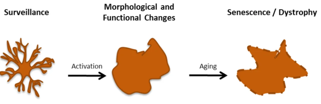
A general overview of the diversity of phenotypes that microglia may assume is schematically represented in Figure I.3, and will be further detailed in section I.2.1.
Figure I.3. General overview of microglia phenotypes. Under normal conditions, microglia remains highly
ramified in a surveillant state, sampling the environment. Activation may occur through multiple stimuli causing them to change from highly ramified cells to amoeboid-like shape, with an active immune response through increased phagocytosis and antigen presentation, release of cytokines and soluble factors capable of modulate the inflammatory process. However, microglia may become senescent or dystrophic with aging, showing fragmented cytoplasmic processes and low ability to respond to external stimuli.
Exploring deregulated signals involved in Motor neuron-Microglia cross-talk in ALS
14
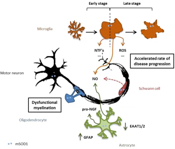
In summary, glial cells play an important role for ALS onset/progression. A schematic interaction between those cells with MNs, and the main molecules involved in such dialogue that can be altered in ALS, are summarized in Figure I.4.
Figure I.4. The role of glial cells in ALS. ALS is a non cell-autononous disease with glial cells exerting an
imperative role in its course. Injury in oligodendrocytes is translated in myelination abnormalities. The role of Schwann cells is somehow controversial; mutated SOD1 (mSOD1) in these cells is suggested to have a protective role although, at some point, they may be contributing to axonal damage of motor neurons (MNs) through increased levels of nitric oxide (NO). Astrogliosis is a feature of ALS with increased expression of glial fibrillary acidic protein (GFAP), and astrocytes are reported to release toxic soluble factors, as pro-nerve growth factor (pro-NGF), promoting injury in MNs. Also, decreased expression of the glutamate transporters, excitatory amino acid transporters 1 and 2 (EAAT1 and EAAT2, respectively) were observed, expected to be a cause of glutamate excitotoxicity. Microglia is thought to have a beneficial action in the early stages of the disease, through the release
of neurotrophic factors (NTFs), whereas it’s thought to contribute to an accelerated rate of disease progression in
I. Introduction
15
2. Microglia: a key player in the pathoprogression of ALS
2.1. Variable role: from surveillant to neuroprotective or neurotoxic
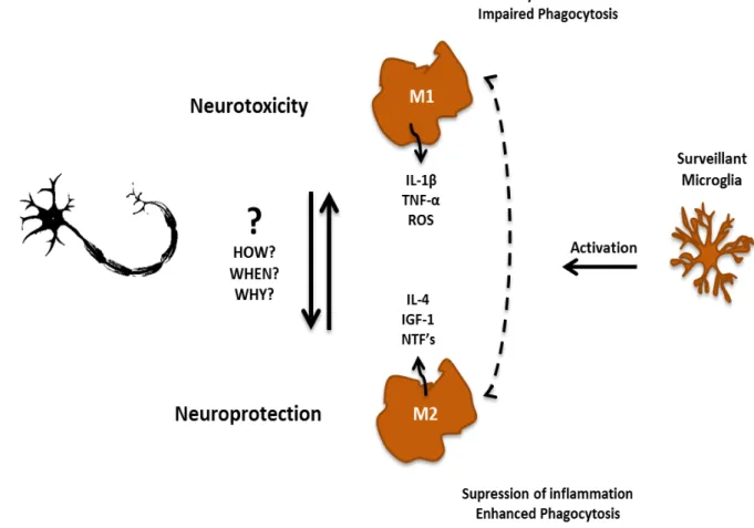
As seen previously, under normal conditions microglial cells have a surveillance function essential to keep CNS homeostasis. It is widely accepted that these cells make brief, repetitive contacts with synapses and rapidly retract (Benarroch, 2013). Activated microglia are very plastic cells that may exert various phenotypes and shift their activation state in agreement with microenvironmental changes. Their main role is to initiate appropriate responses, such as inflammation. Although neuroinflammation is crucial in order to protect CNS, uncontrolled and consequent chronic neuroinflammation is harmful and leads to cellular injury (Cherry et al., 2014).
There is increasing evidence highlighting neuroinflammation as a prominent pathological hallmark in ALS, not only in animal models but also in patients. One of the most noticeable features of neuroinflammation is the presence of activated microglia at sites of MN injury. So far, studies have shown that activated microglia may have distinct phenotypic states, being able to exert either a toxic or protective effect on neurons depending on the stimulus by which they are activated. Classically activated microglia (M1) are cytotoxic due to the secretion of ROS and pro-inflammatory cytokines such as interleukin (IL)-6, IL-1β, TNF-α, interferon-γ. Alternatively activated microglia (M2), in a general way,
Exploring deregulated signals involved in Motor neuron-Microglia cross-talk in ALS
16
Figure I.5. Microglia’s shift from surveillant to neuroprotective or neurotoxic in ALS is thought to be through
interaction with MNs. Depending on the type and duration of the stimulus by which microglia are activated, these
cells may turn into an anti-inflammatory phenotype, so called M2 phenotype, or into a cytotoxic phenotype (M1). M2 microglia, in general, have enhanced phagocytosis and act by the release of neurotrophic factors and anti-inflammatory cytokines such as insulin-like growth factor 1 (IGF-1) and interleukin(IL)-4, respectively, in a way to supress inflammation and help injured motor neurons (MNs). M1 microglia have impaired phagocytic ability and are cytotoxic due to secretion of pro-inflammatory cytokines, as IL-1β and tumor necrosis factor α (TNF-α), and
reactive oxygen species (ROS) and promote cells’ injury. It remains elusive the cause of this change of phenotypes, though in ALS MNs are believed to have a crucial role.
In addition to the activation of microglial cells, infiltrating lymphocytes at sites of MN injury may also constitute a feature of neuroinflammation in ALS pathogenesis (Henkel et al., 2009). CD4+ T cells are believed to cross-talk with microglia and promote their neuroprotective or neurotoxic phenotype being a possible intervenient in the MN-microglial dialogue in ALS. Indeed, Appel et al. (2010) reported that in mSOD1 mouse model, the lack of CD4+ T cells leads to faster progression of MN disease, implying a neuroprotective role of these cells. It also eliminates the early slow phase of disease together with increased pro-inflammatory and cytotoxic factors, diminished anti-inflammatory and neurotrophic factors as well as survival.
I. Introduction
17
2.2. Motor neuron-microglia cross-talk in ALS
2.2.1.
Ligand-receptor interaction
As seen previously, it is believed that there is an immunological shift from neuroprotection to neurotoxicity with major contributes to ALS disease progression (Appel et al., 2011). Once established an imperative role of microglial cells and their mutable action, the questions now are how, when and why this shift occurs and a strong possibility is that the answer lies in the cross-talk between MNs and microglia.
Neurons may modulate microglia phenotype through the release of molecules such as the CD200 neuronal glycoprotein, suggested to suppress the pro-inflammatory-like state of microglia, and the chemokine FKN, likely to maintain the resting/ramified microglial state or promote antioxidant effects (Suzumura, 2013). CD200 has been reported to promote a down-regulation of the activated state of microglia through interaction with its receptor CD200R, predominantly expressed by myeloid cells (Deckert et al., 2006). Regarding ALS studies, by analyzing transgenic SOD1G93A mice during pre-symptomatic and pre-symptomatic stages, Chen and co-workers observed a significant up-regulation of CD200R in the pre-symptomatic stage, though no alterations on CD200 were reported (Chen et al., 2004).
On the other hand, under pathological conditions, damaged neurons will release signals such as high mobility group box 1 (HMGB1) that will lead to microglial activation (Suzumura, 2013). Interestingly, in ALS context, extracellular mSOD1 was found to be able to induce activation of wild-type microglia to a pro-inflammatory-like state, in the same way the lipopolysaccharide (LPS) does (Zhao et al., 2010), by interacting with CD14, a receptor for misfolded proteins, which ligates TLR-2 and TLR-4, co-receptors, and activate a pro-inflammatory cascade, as suggested by Appel et al. (2011) (Figure I.6).
The aforementioned chemokine FKN and its receptor CX3CR1, as well as the alarmin HMGB1 are two promising intervenient that are likely to play an essential role in MN-microglia cross talk in ALS, and that will be further detailed below.
2.2.1.1.
Fractalkine
Exploring deregulated signals involved in Motor neuron-Microglia cross-talk in ALS
18
FKN was shown to have a beneficial action in an in vitro model, decreasing the secretion of pro-inflammatory cytokines and ROS by microglia activated with LPS, a previous mentioned strong exogenous activator of microglial cells (Mizuno et al., 2003). Nevertheless, in human Parkinson’s
disease patients, the plasma levels of soluble FKN were reported to be positively correlated with disease severity and progression (Shi et al., 2011). Regarding its receptor, CX3CR1 deficiency in transgenic mouse model of ALS was reported to trigger microglial neurotoxicity and it was proposed by the authors that enhancing CX3CR1 signaling could protect neurons against toxicity (Cardona et al., 2006).
2.2.1.2
High mobility group box 1
HMGB1 is a ubiquitous nuclear protein with multiple functions able to act as a transcription factor, promoting expression of pro-inflammatory cytokines, and to bind receptors as receptor for advanced glycation endproducts (RAGE) and TLR-2 or TLR-4, triggering a pro-inflammatory cascade (Figure I.6). It is known to be released from necrotic cells, retained by apoptotic cells or actively secreted by activated immune cells, such as monocytes, macrophages and microglia, in response, for example, to LPS stimulus (Zurolo et al., 2011). Lo Coco et al. (2007) described a progressive reduction of HMGB1 within MNs of SOD1G93A mice and a prominent immunoreactivity in the nucleus of glial cells and hypothesized that HMGB1 could be released from damaged MNs and act as inflammatory signal while contributing to proliferation and hypertrophy of glial cells. Casula et al. (2011) observed a cytoplasmic translocation of HMGB1 in activated microglia and astrocytes from spinal cord tissue of sALS patients, suggesting that extracellular HMGB1 is derived mostly from glial cells and arising the hypothesis that may exist a positive feedback loop leading to an amplified inflammatory response.
2.2.2. Phagocytosis
I. Introduction
19
Figure I.6. Molecular pathways involved in MN-microglia cross-talk focusing on FKN/CX3CR1 axis,
MFG-E8-mediated phagocytic pathway and HMGB1 signaling. Motor neurons (MNs) modulate microglia response
Exploring deregulated signals involved in Motor neuron-Microglia cross-talk in ALS
20
3. ALS research mice models
Due to the similarities previously mentioned for non-genetic and genetic ALS, mSOD1 animal and cellular models constitute the most common tool to study the disease and they are also important to perform parallel experiments in more than one model system to better understand different aspects of a process and in order to have more accurate results.
3.1. In vitro models
In vitro models, as cell or organotypic cultures, have an increased yield and homogeneity in comparison with in vivo models that allows more data output in a shorter time.
3.1.1.
Primary cultures and cell lines
Primary cultures refer to cells that are obtained from the tissue and maintained in culture. These may be composed of mixed cell types that can be further sorted in order to have the desired cell type to study. Primary spinal cord cultures have been used as a good model to study morphological, biochemical and electrophysiological characteristics of MNs (Tovar et al., 2009). They are obtained from 12-14 days-old rodent embryos, where the spinal cord is dissociated by mechanical and enzymatic procedures and plated on matrix-coated dishes. Although this culture system is very limited in terms of simulating in vivo conditions, it offers the possibility of studying, for instance, intracellular mechanisms such as whose triggering MN injury in SOD1-ALS. MNs can be induced to express various copies of the gene of interest in order to reproduce an ALS phenotype, as mSOD1G93A, through microinjection of the vector into the cells identified by their specific morphology (Tradewell et al., 2011). However, cultures of MNs without the trophic support of glial cells are known to be difficult to maintain for more than 2 weeks. Moreover, the use of primary cells from embryos may fail to reproduce some features of the adult phenotype.
I. Introduction
21 demonstrated the efficacy of the anti-inflammatory and anti-oxidant bile acid, glycoursodeoxycholic acid (GUDCA) (Vaz et al., 2014).
In vitro cultures of microglia allow the study of the activation state, released factors, motility, among others. The N9 murine microglial cell line was developed by immortalization of E13 mouse embryonic cultures with the v-myc or v-mil oncogenes of the avian retrovirus MH2. It has been largely used as illustrative of mouse microglial cells since it is derived from mouse brain and has many phenotypical characteristics of primary mouse microglia in producing substantial amounts of NO and various cytokines after stimulation (Stansley et al., 2012).
3.1.2.
Mixed cultures and co-cultures
Mixed cultures and/or co-cultures constitute a valuable tool for the study of specific interactions among cells being studied, discarding any external interference (Zhang and Fedoroff, 1996). Also, cells with a target mutation can be combined with healthy cells in order to better understand the role of which cell type in the disease. For instance, combining cultures of NSC-34, either transfected with wtSOD1 or mSOD1, and N9 cell line, is likely to be a valuable model to study the interaction between MNs and healthy microglia.
3.1.3.
Organotypic cultures
Organotypic cultures constitute a three-dimensional culture system which has biochemical and physiological properties more similar to in vivo tissue (Ravikumar et al., 2012). Organotypic slice cultures can be achieved from embryos and postnatal animals and the slices are obtained from 200-400 μm -thick transversal sections that are transferred into membrane inserts to a 6-well or 12-well culture plates and can be used for more than two months (Delfs et al., 1989). The slices may be used to perform multiple analyses like immunohistochemical staining or electrophysiological recordings. This culture system constitute a good model to preserve important processes that occur in ALS, as cellular interactions between MNs and neighboring cells, once the whole slice is cultivated in an organotypic
culture. Although this culture system allows dynamic studies with various drugs, it doesn’t completely
recapitulate what happens in vivo (Tovar et al., 2009). An important feature of these cultures is that they may constitute a first approach before using in vivo models, in order to reduce the number of animals sacrificed in experiments.
3.2.
In vivo
models
3.2.1.
Transgenic mSOD1 mice
Vertebrate models allow the establishment of landmarks of disease progression and the understanding of the functional consequences of gene mutations. The laboratory mouse Mus musculus is widely used as a study model because of its close genetic and physiological similarities to humans, the possibility of genome manipulation and its relatively high reproductive rate.
Exploring deregulated signals involved in Motor neuron-Microglia cross-talk in ALS
22
93 (G93A) and was shown to recapitulate the paralytic phenotype of ALS (Gurney et al., 1994). Therefore, this model is useful for examining the pathophysiology of MN degeneration in ALS, as briefly reviewed above, and has provided a tool for developing preclinical data on drugs that may be used to slow the course of the disease. Though G93A is currently the main study model, also G37R, G85R, G86R, among other mutations in SOD1, are used as study models of this disease, since they were shown to also recapitulate some classical features of MN degeneration (Turner and Talbot, 2008).
I. Introduction
23
4. Therapeutic approaches
–
recent findings
A century after it has been first described, there is still no effective treatment for ALS. The only FDA approved drug Riluzole possess anti-glutamatergic properties but may only increase survival by two or three months (Bensimon et al., 1994).
Given the evidence of non-cell autonomy in ALS, therapeutic shifted its focus from neuron to their interaction with non-neuronal cells. Stem cell research aims to replace damaged cells in injurious regions. For instance, regarding the determinant role of microglia in ALS course, replacing mutant microglia for healthy microglia may serve as a source of neurotrophic factors and provide protection delaying MN degeneration and disease progression. In fact, Lee et al. (2012) reported that replacing microglial cells by bone marrow transplantation in SOD1G93A mouse model slowed disease progression and prolonged survival elucidating the therapeutic potential of these cells.
In addition, treatments that may down-regulate toxic responses of innate and adaptive immune cells and up-regulate the beneficial responses could also slow ALS progression. For that, modulation of microglial phenotype is becoming an appealing neurotherapeutic strategy. Interestingly, bone marrow transplantation with stem cell factor, in transgenic SOD1G93A mice, was reported to change microglia phenotype towards a neuroprotective state, resulting in increased MN function and survival (Terashima et al., 2014).
Herewith, mesenchymal stem cells (MSCs) are arising as a promising therapeutic strategy given the relative availability and their potential for autologous cellular therapy (Kassem et al., 2004). Delivery of MSCs in SOD1G93A mice promotes multiple beneficial effects on disease course as improved motor function, decrease in MN loss and prolonged survival. Besides, intraspinal MSC transplants lead to improvement on neuroinflammation effects, astrogliosis and microglial activation. Moreover, MSCs are being considered to serve as vehicle to deliver neuroprotective factors to CNS. Intracerebroventricular injection of umbilical cord stem cells (UBCs), capable of differentiate into mesenchymal cells, in SOD1G93A mice diminished disease progression and enhanced survival up to 10% and those effects are thought to be mediated by production and release of anti-inflammatory cytokines and chemokines. Likewise, retro-orbital delivery of UBC’s improved neuromuscular transmission and intravenous
administration delayed disease progression by 15%, promoted anti-inflammatory responses, decreased microglial activation and enhanced survival by 20-25% (Lunn et al., 2014).
Exploring deregulated signals involved in Motor neuron-Microglia cross-talk in ALS
24
I. Introduction
25
5. Aims
With this thesis we aim to dissect the MN-microglia signaling pathways that are compromised in MN-microglia homeostasis, by using an in vitro model of ALS. We will focus on the production and release of mediators that are involved in cellular demise and neuroinflammation, as well as specific molecules involved in MN-microglia cross-talk that may be compromised.
Therefore, the specific aims are:
To uncover the pathways by which MNs are signaling their own injury. For this we will use mono-cultures of a MN-like cell line (NSC-34) stably transfected with human SOD1, either wild-type (wtMNs) or with G93A (mMNs) mutation.
27
II. MATERIALS AND METHODS
1. Materials
1.1. Chemicals
Dulbecco’s modified eagle’s medium-Ham’s F12 medium (DMEM-Ham’s F-12), DMEM high glucose w/o pyruvate, fetal bovine serum (FBS), penicilin/streptomycin, L-glutamine and nonessential
aminoacids (NEAA) were purchased from Biochrom AG (Berlim, Germany). RPMI-1640 medium,
poly-D-lysine (PDL), trypsin-EDTA solution (1X), trypsin-EDTA solution (10X), ATP, Hoechst 33258 dye,
bovine serum albumin (BSA) were purchased from Sigma-Aldrich (St.Louis, MO, USA). Geneticin 418 sulfate (G418) was obtained from Calbiochem (Darmstadt, Germany). L-glutamic acid kit and Triton
X-100 were obtained from Roche Diagnostics (Mannhein, Germany). Nitrocellulose membrane was obtained from Amersham Biosciences (Piscataway, NJ, USA). Cell lysis buffer® and LumiGLO® were purchased from Cell Signaling (Beverly, MA, USA). Luminaris HiGreen High ROX qPCR Master Mix was obtained from Thermo Scientific (Waltham, MA, USA). Trizol was obtained from Invitrogen
Corporation™ (Carlsbad, CA, USA). All the other chemicals were purchased either from Sigma-Aldrich or Merck.
1.2. Antibodies
Primary antibodies used in this work: goat anti-FKN [FKN (1:100)] and rabbit polyclonal anti-TLR4 (1:200) were purchased from Santa Cruz Biotechnology® (Santa Cruz, CA, USA), mouse anti-HMGB1 (1:200) from BioLegend® (San Diego, CA, USA), rabbit polyclonal anti-MFG-E8 (1:100), rat anti-CD11b (1:100), mouse anti-βIII-tubulin (1:250), mouse anti-β-actin (1:2000) was obtained from Sigma-Aldrich (St. Louis, MO, USA)
Secondary antibodies used in this work: FITC anti-mouse (1:227), Alexa Fluor® 488 anti-rabbit (1:1000) and Alexa Fluor® 594 anti-rat (1:1000) were obtained from Invitrogen Corporation™ (Carlsbad, CA,