F
ACULDADE DEE
NGENHARIA DAU
NIVERSIDADE DOP
ORTOBiomechanical analysis of the influence
of a cholesteatoma in human hearing
Maria Leonor Illa Mendonça
D
ISSERTATIONIntegrated Master in Bioengeneering
Supervisor: Carla Bibiana Monteiro França Santos | PhD Co-supervisors: Professor Maria Fernanda Gentil Costa
Professor Marco Paulo Lages Parente
c
Biomechanical analysis of the influence of a
cholesteatoma in human hearing
Maria Leonor Illa Mendonça
Integrated Master in Bioengeneering
Resumo
O sistema auditivo humano é um sistema complexo, com estruturas particulares que possibilitam a audição. O ouvido médio, estudado em detalhe nesta tese, é formado por uma cavidade na qual existe uma cadeia ossicular articulada de modo a transferir mecanicamente as informações sono-ras desde a membrana timpânica até ao ouvido interno, onde a informação é codificada e enviada para o cérebro, para interpretação. Além dos ossículos, músculos e ligamentos a eles conectados, há outra estrutura importante que atravessa o ouvido médio, o nervo da corda timpânica. Este nervo é uma ramificação do nervo facial, estando associado ao paladar e à produção de saliva. Es-ticar ou pressionar este nervo pode causar lesões permanentes nesta estrutura, levando à paralisia facial. Apesar de a sociedade ser inclusiva, as pessoas com paralisia facial podem sofrer alguns constrangimentos. As doenças do ouvido médio podem comprometer o funcionamento da corda do tímpano, ao esmagá-la, e a capacidade auditiva, reduzindo as vibrações da cadeia ossicular, o que resultará em menos informação a chegar ao ouvido interno. Uma dessas doenças é otite média. Se não for bem tratada, e caso dure mais de 3 meses, esta doença pode evoluir para um estado crónico, havendo maior probabilidade de desenvolvimento de um colesteatoma, uma massa benigna feita de detritos de pele. Nesta dissertação, estudou-se a influência do desenvolvimento do colesteatoma próximo da corda timpânica, de forma a avaliar as consequências dessa massa na audição e possível paralisia facial. Assim, foi utilizado um modelo do ouvido previamente desen-volvido, baseado no método dos elementos finitos. O modelo foi aprimorado através da modelação e adição da corda timpânica. Três colesteatomas de tamanhos diferentes foram criados na articu-lação entre o martelo e a bigorna e adicionados ao modelo. Um nível de pressão sonora de 60 e 80 dB SPL foi aplicado na membrana timpânica e realizou-se uma análise em estado estacionário para frequências de 100 Hz a 10 kHz. Para avaliar o impacto na função auditiva, foram analisados os deslocamentos da membrana timpânica e da base do estribo. Os resultados foram comparados com um caso saudável, sem tumor no ouvido médio. Foi demonstrado que o desenvolvimento de um colesteatoma leva a uma diminuição dos deslocamentos tanto no início como no fim da cadeia ossicular. Foram também realizados alguns testes atribuindo propriedades semelhantes aos ossícu-los ao tumor maior, já que os tumores se tornam em estruturas rígida e densas ao longo do tempo. Este caso levou a uma diferença ainda maior em termos de deslocamento. Além disso, como os colesteatomas são capazes de recrutar osteoclastos que degradam osso, a degradação dos ossículos também foi simulada. Os deslocamentos observados mostram uma diminuição considerável para o tumor maior. A fase da resposta do deslocamento foi também comparada para todas as situações estudadas. As diferenças de deslocamento observadas estão diretamente relacionadas com a perda auditiva, sendo possível concluir que o crescimento de um colesteatoma no ouvido médio levará a problemas auditivos. A influência do crescimento tumoral na corda timpânica foi também anal-isada. As tensões no nervo foram avaliadas em dois momentos: quando o tumor interage com ele pela primeira vez, empurrando-o para baixo, e quando o tumor o esmaga contra a bigorna. As tensões obtidas permitiram inferir sobre as consequências a nível do paladar e da paralisia facial, embora alguns estudos relatem que, quando a pressão diminui, é possível recuperar totalmente.
Abstract
The human auditory system is a complex system, with particular structures that play impor-tant roles, turning hearing possible. The middle ear, further explored in this dissertation, consists of a cavity filled with an ossicular chain articulated to mechanically transfer the sound information from the tympanic membrane to the inner ear, where the information is coded, being afterwards sent to the brain for interpretation. Besides the ossicles, muscles and ligaments attached, there is another important structure crossing the middle ear, the chorda tympani nerve. This nerve is one of the facial nerve branches, being associated to taste sensation and saliva production. Stretching or pressuring this nerve may cause injuries in this structure, that may result in permanent damage leading to facial paralysis. Despite the society being inclusive, people with facial paralysis may suffer some constraints. Diseases in the middle ear may compromise chorda tympani functioning, due to its pinching, as well as hearing ability, by reducing the vibrations in a certain point of the ossicular chain, which will result in a poor information reaching the inner ear. One of this con-ditions is otitis media. If this disease is not properly treated and lasts for more than 3 months, it may develop to a chronic otitis media. In this case, appearance and growth of a cholesteatoma, a benign mass made of skin debris, have higher probability. In this dissertation, the influence of cholesteatoma development near chorda tympani nerve was studied, in order to assess the conse-quences of this mass in both hearing and possible facial paralysis. To do so, a previously developed ear model based on the finite elements method was used. The model was improved by modeling and adding the chorda tympani nerve to it. Three different sized cholesteatomas were created in the connection between the malleus and the incus and assembled to the model. A sound pressure level of 60 and 80 dB SPL was applied in the tympanic membrane and a steady state analysis was performed for frequencies from 100 Hz to 10 kHz. To assess the impact on hearing function, the displacements of the tympanic membrane and of the stapes footplate were analysed. The results were compared with a healthy case, where no tumour existed in the middle ear. It was shown that the cholesteatoma development leads to a decrease in the displacements both at the beginning as well as at the end of the ossicular chain. Some tests were also performed assigning the larger tumour properties similar so the ossicles, as tumours become harder and denser structures along time. This case led to an even greater displacement difference. Furthermore, as cholesteatomas are able to recruit osteoclasts that degrade bone, the ossicles degradation was also simulated, by assigning tumour properties to part of the ossicles. The observed displacements show a consider-able decrease for the larger tumour in these conditions. The phase angle of the displacement was also compared for all the studied situations. The observed displacement differences are directly connected to hearing loss, being possible to conclude that cholesteatoma growth in the middle ear will lead to hearing problems. The influence of tumour growth on chorda tympani nerve was also analysed. The tensions felt in this nerve were assessed in two moments: when the tumour first in-teracts with it, pushing it down, and when the tumours squeezes it against the incus. The obtained tensions allowed to infer on the consequences regarding taste disturbance and facial paralysis, although some studies report that when pressure fades away, it is possible to fully recover.
Agradecimentos
Em primeiro lugar, quero deixar o meu sincero agradecimento à minha orientadora, Dra. Carla Santos, por toda a ajuda, por ser incansável e por ter sempre uma palavra motivadora. Muito obrigada pela sua disponibilidade. Foi um gosto trabalhar consigo.
Aos meus co-orientadores, Prof. Dr. Marco Parente, por todo o apoio e ajuda na resolução de problemas durante o desenvolvimento deste trabalho. A sua ajuda foi essencial para a realização desta tese, muito obrigada! E à Prof. Dra. Fernanda Gentil, por me ajudar e orientar em toda a parte clínica associada a este trabalho. Muito obrigada por todas as sugestões e conselhos! E agradecer também ao Prof. Dr. Renato Natal, que se manteve sempre por perto, contribuindo com importantes dicas para o sucesso deste projeto.
A toda a minha família, um grande obrigada por me manterem motivada ao longo de todo o meu percurso académico. Por manterem a vossa expectativa alta, levando-me sempre a querer fazer mais e melhor. Um agradecimento especial aos meus pais e às minhas irmãs, por nunca se cansarem de todas as minhas queixas e desabafos (ou cansarem, mas continuarem a aturá-los :) ). Aos professores que me acompanharam ao longo dos vários anos escolares, com especial destaque para a professora Adélia, o professor Luís, a professora Susana e o professor Jorge. Obrigada por me inspirarem e motivarem! Se esse é o papel de um professor, no que me diz respeito, a missão foi cumprida.
A quem verdadeiramente me acompanhou nestes 5 anos. Raquel, obrigada por estares sempre lá e seres a minha fiel companheira (e gémea, como muitos dizem) nesta jornada. Bruno, por todas as vezes em que reclamamos sobre tudo e por teres sempre as ideias mais fora da caixa. Pedro, Pedrão e Migas, por serem muito bons a criticar quando é preciso, mas serem também os melhores compinchas que podia ter ao longo destes anos. Um obrigada ao Luís e à Leonor, génios incompreendidos em Bio, mas que não conseguem cortar as raízes a este curso maravilhoso. E claro, aos preferidos ainda não mencionados (sem nenhuma ordem específica, para não ferir os mais sensíveis), Meneses, por todos os áudios e pela incrível energia que transmites ao grupo, Álvaro, por permitires que o nível de português e o nível de piada do grupo sejam inversamente proporcionais, Joana e Natália, por serem as melhores e mais adoradas biotecas com espírito biomédico, Gu, por seres, juntamente com o 3D, um dos grandes responsáveis por biomédica ser um grupo de amigos e não de colegas e Ana, por seres também um elemento essencial nesta difícil tarefa, sempre com ideias de eventos e atividades incríveis. A todos muito obrigada pela forcinha, videochamadas e mensagens diárias. Sem vocês não teria sido a mesma coisa.
A amizades que já têm uns aninhos. Raquel, por seres a minha confidente e me apoiares sempre! Catarina, por saber que posso sempre contar contigo! Kika, por nos termos aventurado juntas nesta loucura que é a Bioengenharia e por todas as boas histórias. Ao Picão, Luisinho, Bea, Matilde, Marta, Rodrigo, Bárbara e Gonçalo. Obrigada a todos por se manterem presentes.
Uma menção especial a quem, nos últimos 3 anos, me fez descobrir uma grande paixão. Sofia e toda a equipa Barquinho, obrigada por serem o espaço em que sempre me senti realizada! Quem diria que o que eu pensava que seria "apenas um part-time" me poderia trazer tanto. Não encarei
vi
como trabalho nem um dia que fosse. Obrigada por acreditarem em mim e por me apoiarem sempre! (E um grande agradecimento aos meus alunos, que são os melhores do mundo!)
And of course, Mireia, Charlotte D, Charlotte B, Katelynn, Belisa, Monica and Eva, thanks for all the good memories in Groningen! Erasmus would not have been the same without you girls!
Por fim, agradecer a todas as pessoas relacionadas com a dança que tiveram impacto na minha vida, por partilharmos juntos esta paixão. E uma menção especial à Raquel, por ser a minha boleia oficial para todos os eventos e por todas as memórias partilhadas.
A todos... muito obrigada!
“Não tenhamos pressa, mas não percamos tempo.”
José Saramago
Contents
Resumo i Abstract iii Agradecimentos v 1 Introduction 1 1.1 Motivation . . . 1 1.2 Context . . . 1 1.3 Goals . . . 2 1.4 Expected Contribution . . . 3 1.5 Structure . . . 3 2 The ear 5 2.1 Anatomy and Physiology . . . 52.1.1 External Ear . . . 5
2.1.2 Middle Ear . . . 6
2.1.3 Inner Ear . . . 11
2.2 Acoustics . . . 12
2.2.1 Frequency and Period . . . 12
2.2.2 Intensity and Pressure . . . 13
2.2.3 Timbre . . . 13
2.2.4 Impedance . . . 14
2.3 Middle Ear Diseases . . . 14
2.3.1 Otitis Media . . . 15
2.4 Biomechanical models of the ear . . . 21
3 Methodology 27 3.1 Ear model . . . 27
3.2 Cholesteatomas . . . 35
3.3 Chorda Tympani Nerve . . . 38
4 Results 43 4.1 Effects of cholesteatoma development in the middle ear on hearing . . . 43
4.2 Effects of cholesteatoma development in chorda tympani nerve . . . 52
5 Conclusion and future work 57
A CTN reported values 59
x CONTENTS
B Middle ear before and after the addiction of CTN 61
List of Figures
2.1 Human Ear . . . 6
2.2 Ossicular Chain . . . 7
2.3 Middle Ear Muscles and Chorda Tympani Nerve . . . 8
2.4 Chorda Tympani Nerve Segments . . . 9
2.5 Chorda Tympani Nerve Anatomy . . . 11
2.6 Cholesteatoma . . . 16
2.7 Ear model Gan et al. 2006 . . . 22
2.8 Ear model Zhang and Gan 2011 . . . 23
2.9 Ear Model Areias et al. 2016 . . . 24
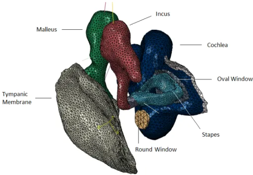
2.10 Ear Model . . . 25
3.1 Ear model - Bone . . . 28
3.2 Ear model - TM . . . 29
3.3 Ear model - Malleus . . . 30
3.4 Ear model - Incus . . . 31
3.5 Ear model - Stapes . . . 32
3.6 Ear model - Middle Ear . . . 33
3.7 Small tumour . . . 36
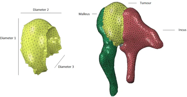
3.8 Medium tumour . . . 36
3.9 Large tumour . . . 37
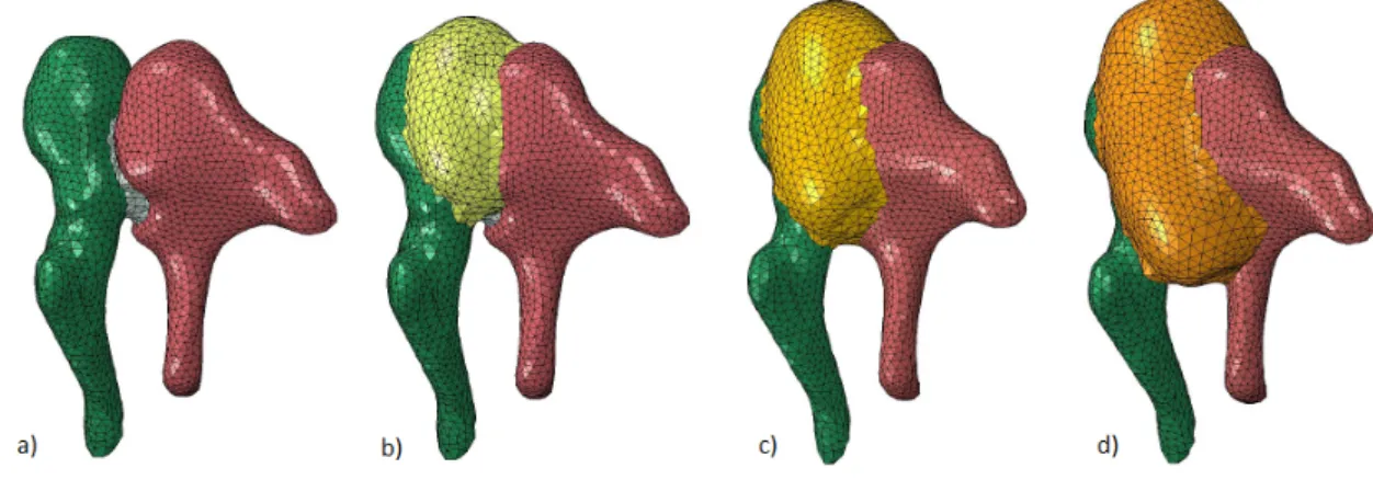
3.10 Tumour growth . . . 37
3.11 Chorda Tympani Nerve - Gray’s Anatomy . . . 39
3.12 Ear model - Chorda Tympani Nerve . . . 39
3.13 Ear model - Chorda Tympani Nerve . . . 40
3.14 Ear model - Chorda Tympani Nerve and Medium Tumour . . . 41
3.15 Ear model - Chorda Tympani Nerve and Large Tumour . . . 42
4.1 Model validation . . . 44
4.2 Comparison 60 dB and 80 dB . . . 44
4.3 Displacements 60 dB . . . 45
4.4 Displacements 80 dB . . . 46
4.5 Displacements 60 dB new tumour properties . . . 47
4.6 Displacements 80 dB new tumour properties . . . 47
4.7 Displacements 60 dB ossicle degradation small tumour . . . 48
4.8 Displacements 60 dB ossicle degradation medium tumour . . . 48
4.9 Displacements 60 dB ossicle degradation large tumour . . . 49
4.10 Displacements 80 dB ossicles degradation small tumour . . . 50
4.11 Displacements 80 dB ossicles degradation medium tumour . . . 50
xii LIST OF FIGURES
4.12 Displacements 80 dB ossicles degradation large tumour . . . 50
4.13 Phase angle for healthy case and three tumours . . . 51
4.14 Phase angle for tumour with different properties . . . 52
4.15 Phase angle for ossicles degradation . . . 52
4.16 Stress in chorda tympani nerve caused by the medium tumour . . . 53
4.17 Stress in chorda tympani nerve caused by the large tumour . . . 54
A.1 Chorda Tympani Literature Measures . . . 59
List of Tables
3.1 Ear model - Bone Mesh . . . 28
3.2 Ear model - Bone Properties . . . 28
3.3 Ear model - TM Dimensions . . . 29
3.4 Ear model - TM Mesh . . . 29
3.5 Ear model - TM Properties . . . 30
3.6 Ear model - Malleus Dimensions . . . 30
3.7 Ear model - Malleus Mesh . . . 30
3.8 Ear model - Malleus Properties . . . 31
3.9 Ear model - Incus Dimensions . . . 31
3.10 Ear model - Incus Mesh . . . 31
3.11 Ear model - Incus Properties . . . 32
3.12 Ear model - Stapes Dimensions . . . 32
3.13 Ear model - Stapes Mesh . . . 32
3.14 Ear model - Stapes Properties . . . 32
3.15 Ear model - Joints Mesh . . . 33
3.16 Ear model - Ossicle Joints . . . 33
3.17 Ear model - Ligaments . . . 34
3.18 Ear model - Muscles . . . 34
3.19 Modeled Tumours Dimensions . . . 35
3.20 Modeled Tumours Mesh . . . 35
3.21 Chorda Tympani Nerve Dimensions . . . 39
3.22 Chorda Tympani Nerve Mesh . . . 40
3.23 Chorda Tympani Nerve Properties . . . 40
4.1 Stress in chorda tympani nerve caused by the medium tumour . . . 53
4.2 Stress in chorda tympani nerve caused by the large tumour . . . 54
4.3 Stress in chorda tympani nerve when pressure is directly applied on it . . . 55
Abbreviations and Symbols
AOM Acute Otitis Media COM Chronic Otitis Media CT Computed Tomography CTN Chorda Tympani Nerve FEM Finite Elements Method FP Facial Palsy
MRI Magnetic Resonance Imaging OM Otitis Media
SPL Sound Pressure Level TM Tympanic Membrane WHO World Health Organization
Chapter 1
Introduction
1.1
Motivation
The ear is an important human organ, having essential functions in the day-to-day life. Re-sponsible for one of the five human senses, it is a valuable system, used to grasp stimulus and information that surround people everyday. The ear is then of the utmost importance for a daily well-being.
Considering that the human body is such a complex system, some disorders may occur. Dis-eases associated to the auditory system may result in hearing loss or other nefarious consequences. Otitis media, an inflammatory disease of the middle ear, is a common illness of the auditory sys-tem. If this inflammation becomes chronic, it can lead to the development of a cholesteatoma, a benign ear tumour. This mass can appear in several locations, interfering with the regular functions of the internal ear structures, leading to hearing problems. If it grows in such a site that pressures the chorda tympani nerve (CTN), as one of the facial nerve branches that crosses the ear ossicular chain in the middle ear, it can result in facial palsy (FP), the inability to move a part of the face. Tumour development around this area can cause lesions that may be irreparable.
Hearing loss and FP can bring adaptation problems. Patients suffering from this condition would greatly benefit from a study to evaluate the risk and influence of the presence of different sized tumours in the middle ear as well as these bodies pressuring the CTN. Therefore, a study on the effect of tumour growing in the surroundings of this nerve is of major interest and pertinence. The results of this work may be used to better understand this, leading to further studies on the best approaches to treat this condition or minimize its effects.
1.2
Context
The human ear is the organ responsible for hearing and equilibrium. The peripheral auditory system can be divided into external, middle and inner ear, subsystems that work together with the same final goal. In spite of the well established coordinated functioning of the ear structures, several types of diseases can occur in the ear, compromising its behaviour. Otitis media (OM) is
2 Introduction
an infection in the middle ear, being one of the most frequent conditions associated to this organ [1]. This disease can include the appearance of fluid in the middle ear and it can present itself as acute, associated with intense pain and lasting one to two weeks, or, if it lasts for more than three months, chronic, leading to the creation of the necessary conditions to hearing loss and tumour development.
Cholesteatoma is the most frequent type of tumour that can result from chronic otitis media (COM), being composed of an accumulation of skin debris [2]. Although this mass is usually a benign tumour, its surveillance is very important, since it grows and wears out the surrounding structures [3].
The effect of benign tumours on different sites of the middle ear was already a matter of study before, by Savaris et al., in the scope of the investigation in which this project is inserted [4]. Tumours connected to the incus, stapes and the medial surface of the tympanic membrane (TM) were simulated and their effects were analysed. Depending on its size and location, these tumours can affect the ear function in several ways. However, there is no study on the influence of a cholesteatoma that pressures the CTN. This structure is a facial nerve branch that is directly involved in the taste sensation of the anterior two thirds of the tongue as well as saliva production. The growth of these tumours between the malleus and the incus may pressure the CTN, causing ultimately FP. According to Ikeda et al., this problem can only be completely reverted if it is treated while the nerve is under pressure but has not suffered lesion, otherwise there is no full recovery [2]. This way, a study on the influence of these tumours in the CTN would be helpful to better understand the relation between the size and location of the tumour and the effects on hearing and on the nerve itself.
1.3
Goals
The goal of this work is to study the consequences of the growth of a cholesteatoma near CTN in what concerns hearing function and FP. With this in mind, Finite Element Method (FEM) will be used to simulate the desired conditions. A computational ear model has already been developed in former projects and will be used in this work. This model consists of TM, middle ear ossicles, ligaments and tendons, cochlear fluid, involving temporal bone, skin and ear cartilage, jaw and air in the external auditory canal and tympanic cavity. However, the CTN itself is not modeled yet. Thus, the CTN must be constructed and incorporated in the existing model. After that, different sized tumours associated to the ossicular chain will be simulated. The area of interest is the connection between the malleus and the incus, since that is the location of the middle ear crossed by CTN. The effects of the tumour growth around the CTN will be studied, in order to better understand for which tumour sizes the CTN is under pressure and from which tumour dimensions injuries in CTN occur.
Tumour resection is a delicate process and it should be performed as early as possible, since cholesteatomas wear out the structures to which they are connected to. This way, when applying pressure in the CTN, these masses will also erode this nerve. Thus, this project aims to evaluate
1.4 Expected Contribution 3
the consequences of this condition in order to better understand it and so that, in the future, further studies can be conducted to avoid middle ear structures damage.
Overall, this dissertation is relevant to allow a better comprehension of CTN and its position in the human body, as well as the influence of a cholesteatoma in human hearing and the conse-quences of pressuring this nerve.
1.4
Expected Contribution
In this project, the CTN structure was modeled. This model was added to the current ear biomechanical model, contributing to a more complete and accurate model. After this, the effects of cholesteatoma growth associated to the ossicle chain on hearing were assessed. Additionally, the influence of the appearance and development of a cholesteatoma in CTN surroundings was studied, providing more information on this structure as well as its weaknesses.
It is possible to summarize this project main contributions as:
• Computational development of an ear structure and biomechanical analysis of its behaviour, • Better knowledge and study of CTN,
• Study of the effects of tumour (cholesteatoma) development in the surroundings of CTN regarding hearing and facial repercussions.
1.5
Structure
This dissertation is organized in five chapters. It starts with an introduction, where motivation and context, as well as main goals and expected contributions are presented.
Chapter 2 includes the anatomy and physiology of the auditory system with the main focus being CTN. Some middle ear pathologies are also described, with focus on OM and consequent cholesteatoma development. Finnaly, some works on CTN and cholesteatoma development are presented as well as the current biomechanical model of the ear and some previous related projects. The followed methodology is presented in Chapter 3 and the results and their discussion in Chapter 4.
Chapter 2
The ear
The human ear is a complex biomechanical system that plays an essential role in people’s lives. In spite of much information being captured by the eyes, a lot of content is only perceived by the auditory system. Hearing is then an important skill to perform daily activities.
The ear is capable of tracking air pressure variations, connected to sound waves propagation, and transform them into electrical variations to be identified by the brain. It is responsible for both hearing and balance.
2.1
Anatomy and Physiology
The auditory system is located in the temporal bone and it can be divided in central auditory system and peripheral auditory system.
The central auditory system is formed by both the auditory nerve and auditory cortex. The peripheral auditory system can be split into three subsystems: external, middle and inner ear, as presented in Figure2.1, with specific functions, that will be detailed next.
2.1.1 External Ear
The external ear is the most outer part of the ear and it goes from the atrium up to the TM, also called eardrum. When sound waves reach the external ear, they proceed through the exter-nal auditory caexter-nal. This caexter-nal has different properties along itself. Its most outer third part is fibrocartilaginous while the two internal thirds are made out of bone. This is important as the fibrocartilaginous part is covered by skin and one of the skin’s layers, the dermis, is responsible for cerumen production, which avoids the passage of foreign bodies.
In the end of the external auditory canal there is the TM. Sound waves cause the vibration of the TM, making sound information follow to the next phase, the propagation in the middle ear. The TM is capable of transforming sound energy into mechanical energy. This membrane is a circular structure that can be split into pars tensa and pars flaccida. It has three tissue layers. The most external one is connected to the external auditory canal coating, being a thin layer. The inner layer is formed by epithelial tissue and connected to the middle ear coating. The portion
6 The ear
Figure 2.1: Representation of the human ear [5].
standing between these two layers is the one responsible for this structure function, being fibrous, with radial, parabolic and transverse fibers [6]. This layer holds the TM, providing it its vibratory ability. Pars flaccida has a low fibers content, as it only includes the internal and external layers, which explains its softness, while pars tensa has all three layers, being firm and elastic. The umbo is a salient part on the medial side of this membrane, connected to the malleus extremity [6].
2.1.2 Middle Ear
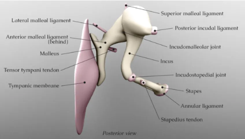
The middle ear is composed by the tympanic box, formed by the ossicular chain and the tympanic cavity. This part of the ear is filled with air, being the Eustachian tube responsible for the middle ear ventilation. The ossicular chain is formed by three small bones named ossicles that can be seen in Figure 2.2. The one that contacts with the TM is the malleus. This ossicle can be divided into head, neck, anterior and external apophysis and manubrium. The malleus neck is connected to the TM pars flaccida and the manubrium to the superior part of the TM, dragging it inwards. In the anterior apophysis there is the insertion of tensor tympani muscle. The malleus mass varies between 23 and 27 mg and its length between 7.6 to 9.1 mm [6,7,8,9,10]. The head of the malleus articulates with the next ossicle, the incus, which is formed by its body and three apophysis, long, short and lenticular. It is the only ossicle that is not connected to a muscle, being the heaviest of the three ossicles [6]. Its mass can go from 25 to 32 mg [7, 9]. Incus lenticular apophysis articulates with the stapes, the most internal and smallest ossicle, with only around 2 mg mass and 2.5 to 4 mm length [10]. It can be divided into head, neck, footplate and two crus, anterior and posterior, that connect the neck to the footplate. The posterior crus has a broader curvature than the anterior. In the posterior part of the neck there is the insertion of the stapedius muscle. The footplate is connected to the inner ear through the oval window.
2.1 Anatomy and Physiology 7
Figure 2.2: Middle ear ossicular chain representation1.
As mentioned during the ossicles description, two muscles connected to these ossicles exist, tensor tympani and stapedius, being their main function to damp excessively loud noises.
Tensor tympani is a muscle located in the bony canal above the Eustachian tube that is inserted in the anterior apophysis of the malleus. It is innervated by the V cranial nerve (trigeminal nerve). Its main function is to damp loud sounds, like chewing or shouting. When under tension, this muscle pulls the malleus medially. This will lead to tension in the TM, causing the damping of vibration in the ossicles and thereby reducing the perceived amplitude of sounds [11].
The stapedius is the smallest skeletal muscle in the human body, being innervated by the VII cranial nerve (facial nerve). Its main functions are to stabilize the stapes and to reduce the pressure in the inner ear. This muscle controls sound waves intensity, protecting the inner ear. This is called esteatoacoustic reflex. People with injuries in stapedius muscle can develop hyperacusia, less tolerance to regular sounds.
Damping is more effective for low frequency sounds. Damages caused by sudden noises are hard to avoid, since the reflex is not fast enough. The articular surfaces of the ossicles are covered with cartilage, in order to avoid producing noise when moving. The same happens for the connection between the stapes footplate and the oval window.
The middle ear ossicles are hanging through six ligaments. Three of them are attached to the malleus (superior, anterior and lateral), two to the incus (posterior and superior) and one to the stapes (annular). In Figure2.3it is possible to see the mentioned muscles as well as some of the ligaments.
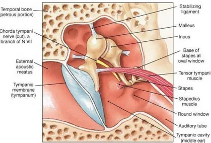
Also in the middle ear there is the CTN, a nerve that crosses the tympanic cavity between the malleus and the incus. This structure is described in detail in Section2.1.2.1.
1Middle ear anatomy, Author:Skalski, Matthttps://radiopaedia.org/images/30697108Accessed on:
8 The ear
Figure 2.3: Middle ear muscles and CTN localization.
2.1.2.1 Chorda Tympani Nerve
The facial nerve arises from the brainstem proceeding to the facial area of the fundus of the internal acoustic meatus, finally entering the geniculum of the facial canal [12,13]. It is respon-sible for the command of the muscles related to facial expression, for the external ear sensitivity, for fiber transportation for submandibular and sublingual glands and also for taste sensation in the anterior two thirds of the tongue.
This nerve has two paths, an intratemporal and an extratemporal one. The intratemporal seg-ment can be divided into three parts, labyrinthic, tympanic and mastoid. The first corresponds to the facial nerve portion that passes inside the inner auditory canal. When it reaches the genicu-late ganglion, the greater petrosal nerve arises, proceeding its way to innervate the lacrimal gland. The tympanic part, that has around 8 to 11 mm, starts in the geniculate ganglion, where the nerve bends around 40 to 80 degrees, proceeding through the tympanic segment until reaching the lateral semicircular canal. The nerve to stapedius, nerve that is related to the stapedius muscle, emerges from this part of the facial nerve. Finally, the mastoid segment has a 90 degrees curvature and goes down until the stylomastoid foramen, proceeding to the parotyd gland. From this point on, the extratemporal path of the facial nerve begins. In the parotid gland, the nerve splits into several branches, temporal, zygomatic, buccal, mandibular and cervical. This branch network is known as pes anserinus [15,16,17].
CTN, the structure of interest in the present work, is one of the facial nerve branches. Ac-cording to Henry Gray, CTN arises from the facial nerve, around 6 mm above the stylomastoid foramen [18]. The course of CTN can be divided into three portions, as it is shown in Figure2.4. The first portion, from the connection with the facial nerve to the tympanic cavity, is found in the mastoid process. The central section crosses the tympanic cavity. The final segment is located in the submandibular fossa and it goes from the tympanic cavity to the connection with the lingual nerve [15,14].
2.1 Anatomy and Physiology 9
Figure 2.4: CTN portions between the facial nerve and the lingual nerve. Portion 1: mastoid process, portion 2: tympanic cavity, portion 3: submandibular fossa [14].
Inside the tympanic cavity, the chorda goes sideways with the stapes and crosses the ossicles between the malleus and the incus, on the medial surface of the neck of the malleus, as one can see in Figure 2.3. It then runs posteriorly and descending, close to the TM, and exits the tympanic box through the petrotympanic fissure. CTN provides parasympathetic innervation to submandibular and sublingual glands as well as special sensory taste fibers for the anterior two thirds of the tongue, through the lingual nerve [19,20]. Besides being connected to taste sensation, this structure is also responsible for saliva secretion, as CTN is responsible for efferent innervation of the glands responsible for saliva production.
Some studies have been conducted along time in order to better understand the facial nerve and its branches, among which CTN, with several studies on diseases affecting such important nerves. Smith et al. studied FP as a consequence of facial nerve tumor [21]. They studied a case of a man with FP and taste loss in the right side of the face and tongue, that had a tumour behind the TM. This schwannoma was affecting the facial nerve, but the patient recovered well after surgery. The facial nerve is an extremely important nerve that is involved in facial expression, tears, taste and even hearing. Bell’s palsy is a condition that can lead to facial nerve disorder. Other diseases, such as trauma, infection, tumours or genetic factors can also affect this nerve.
Several studies on the impact of CTN injuries on taste have been conducted. Most of these lesions were connected to taste aberrations and dry mouth. These symptoms are related to the main functions of this nerve, taste sensation transportation in the two anterior thirds of the tongue and facilitation of salivary production. Another of these studies was performed by Gopalan et al., that found that the stretch of CTN resulted in most significant symptoms of taste disturbance than CTN transection [22]. This kind of injuries is most of the times iatrogenic, meaning that they result from lesions during surgery. Michael and Raut (2007) reached the same conclusion, when they assessed operative findings on CTN [23]. Clark and O’Malley (2007) conducted an
inves-10 The ear
tigation including 42 patients that would undergo ear surgery, in order to study the influence of iatrogenic CTN injuries in taste sensation comparing cholesteatoma, a benign ear tumour, surgery with other middle ear surgeries [24]. Of all these patients, 21 had cholesteatoma, while the rest was having procedures as myringoplasty or stapedectomy to treat other ear condition. A total of 16 patients had their CTN completely sectioned, with only 5 (31%) showing symptoms of so. These symptomatic patients showed altered taste after surgery. However, symptoms had disappeared 6 months after. The same did not happen for myringoplasty and stapedectomy patientes that had CTN compressed or streched during the procedure, showing symptoms even 6 months after the intervention. Although in 5 of the 21 cholesteatoma cases CTN was slightly touched or stretched during surgery, none of these patients showed altered taste postoperatively. The results of this study support former research that states that taste disturbance can be quicker resolved when CTN is sectioned than in cases the nerve is compressed or stretched. The explanation may be that the conditions in which injuries due to surgery happen are different than those in which cholesteatoma affects this nerve, and so different outcomes will be observed when these lesions occur. This inves-tigation also indicates that cholesteatoma leads to damages in CTN, as patients with cholesteatoma show less symptoms alteration after nerve injury during surgery, which indicates that the tumour leads to CTN hypofunctioning, as suggested in previous studies. This way, Clark and O’Malley findings also sustain the theory that symptoms are less intense and less frequent in patients that had chronic inflammation, once their nerves and ear structures were already working poorly. CTN recovery can be a slow process, taking up to 2 years. This way, a careful follow up must be done to fully assess its rehabilitation [25].
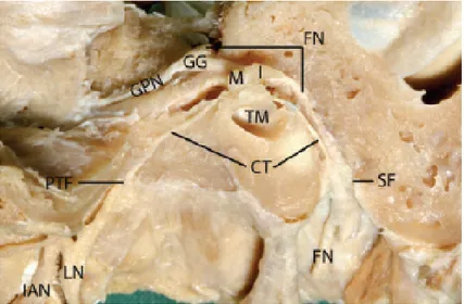
CTN was described in literature as having a diameter of between 0.3 and 0.5 mm, showing wide inter patient variation [26]. Nevertheless, there was a lack of information regarding CTN and its anatomy. Thus, some studies to better understand CTN position in the human head have been conducted. Trost et al. carried out a study to better assess the spatial relation of the CTN in the intratemporal fossa [27]. They considered that teaching CTN anatomy was a hard task due to the little spatial information there was about it. This way, CTN and other nerves were catheterized and X-ray and computed tomography (CT) scans examinations were carried out. The course of CTN was better studied and these findings contributed to raise the knowledge of head nerves. With the same goal in mind, and in order to better understand their spacial position to have better surgery outcome, Liu et al. studied the whole course of some deep nerves in human head [14]. Regarding CTN, dimensions showed wide inter patient variation. The obtained values for the part of the CTN that crosses the tympanic cavity were a diameter around 0.44 mm and a length of (9.83 ± 1.24) mm. The total length of CTN, from the facial to the lingual nerve, was about 54 mm. As already described in literature, CTN crosses the middle ear close to the ossicles and the TM, as shown in Figure 2.5. Literature states CTN arises from the facial nerve approximately 6 mm above the stylomastoid foramen. Nevertheless, the distance from CTN and the stylomastoid foramen was assessed by Liu et al., obtaining values from 5.93 to 21.63 mm, being the average 13.32 mm [14]. As there is no cover involving the CTN, this structure is highly vulnerable to the events that happen in its surroundings [28,29].
2.1 Anatomy and Physiology 11
Figure 2.5: Dissection of the chorda tympani nerve from facial nerve, passing through the tympanic cavity and joining the lingual nerve. FN: facial nerve, I: incus, M: malleus, CT: chorda tympani nerve, GG: geniculate ganglion, GPN: greater petrosal nerve, CN: cochlear nerve, PTF: petrotympanic fissure, LN: lingual nerve, TM: tympanic membrane, SF: stylomastoid foramen, IAN: inferior alveolar nerve.
In 2015, a study was conducted to assess the degeneration of CTN, the major taste nerve, during COM [29]. As this structure runs uncovered in the middle ear, this investigation aimed to evaluate ultrastructural changes of CTN in different inflammatory middle ear diseases, namely simple COM and cholesteatoma. In order to do so, 10 CTN were analysed, 5 being controls and 5 being associated to COM or cholesteatoma, in which CTN was affected. The different data was analysed using electron microscopy. In the cases of unhealthy CTN, there were records of a higher portion of axon and myelin degeneration, which were associated to taste disturbance. Nevertheless, 3 of the 5 analysed damaged CTN showed signals of sprouting. This suggests that CTN has the ability to regenerate, which can justify taste recovery in the majority of cases.
2.1.3 Inner Ear
In the inner ear, the bone forms canals that are called bony labyrinth. This labyrinth is covered by connective tissue, periosteum, and in its inner part a similar but smaller structure is present, the membranous labyrinth. Two fluids fill the inner ear structures, endolymph and perilymph. The first fills the membranous labyrinth while the second fills the space between the bony and the membranous labyrinth.
The bony labyrinth is present in the main structures that belong to the inner ear, the cochlea and the vestibular system. While the first is directly connected with hearing, the latter, that comprises the semicircular canals and the vestibule, is involved in equilibrium function.
The stapes vibrations are transmitted to the inner ear through the round window. As the di-ameter of this structure is smaller than the one of the TM, when sound information travels from the middle to the inner ear, there is an amplification of the signal, important to overcome the impedance related to the cochlear fluid [30]. The round window is connected to the vestibule,
12 The ear
that communicates with the vestibular ramp, one of the cochlear chambers. The vestibular ramp goes from the oval window to the helicotrema, the peak of the cochlea. The tympanic ramp, other cochlear chamber, goes from the helicotrema to the round window, side by side with the vestibular ramp.
The portion of the cochlea next to the oval window is rigid and related to high frequency vibrations while the portion around the helicotrema is flexible and answers best to low frequency oscillations. Inside the cochlea it is possible to find the organ of Corti. It has more than thirteen thousand hair cells, that are very particular cells as they are able to transform pressure variations into electrical impulses.This way, it is possible to send the appropriate information to the brain, through the vestibulocochlear nerve.
2.2
Acoustics
Acoustic is the science field that dedicates to the study of sound. Sound can be described as a disturbance in the environment caused by a longitudinal mechanical wave, that makes the environ-ment particles oscillate. This disturbance is caused by an emitter, that generates the sound, travels through the environment and reaches the receptor, the entity that receives the sound. Acoustic studies the phenomena involved in the generation, transmission and reception of sound waves. Since these are mechanical waves, they need a material so they can propagate, that can either be a solid, a liquid or a gas. When the emitter generates a sound, the particles around it oscillate, conveying the information to the surrounding particles. This movement goes on and on, making sound a pressure wave, meaning that there are compression areas, where there is a greater density of particles, and rarefaction areas, where one can find less particles. These areas alternate with each other along time, resulting in a sequence of compression and rarefaction, if one analyses a certain point for a period of time.
The main attributes of sound are pitch, intensity and timbre. Although sound is perceived differently by different people, the analysis of a sound is done with specific devices that quantify accurately the measured properties [6].
2.2.1 Frequency and Period
The frequency ( f ) is defined as the number of cycles during one second, being measured in cycles per second or Hertz (Hz) . On the other hand, the period (T ) is the duration of a complete cycle, measured in seconds, in other words, it is the inverse of frequency, which is showed in Equation2.1.
f= 1
T (2.1)
The human ear is capable of distinguish sounds with frequencies between 16 Hz and 20 kHz. Sounds which frequency is below 16 Hz are called infrasounds while the ones with frequency
2.2 Acoustics 13
higher than 20 kHz are ultrasounds. Frequencies up to 256 Hz are considered low, from 256 Hz to 1 kHz are considered medium, and above 1 kHz are considered high frequencies [6].
The way the ear grasps a sound frequency is called pitch. Thus, low frequencies are heard as low sounds whilst high frequencies are interpreted as high sounds, being this phenomenon connected to the area of the basilar membrane that suffers most excitation when a sound reaches the ear.
As mentioned before, sound waves are longitudinal mechanical waves. Wavelength (λ ) is, by definition, the length a wave can travel in a period of time equal to T , and can be expressed through Equation2.2, where C is the sound speed in the air and f is the wave frequency.
λ =C
f (2.2)
This way, high sounds have low wavelength while low sounds have high wavelength.
2.2.2 Intensity and Pressure
Sound intensity is connected to the amplitude of a sound wave. It is defined as the sound power, in watts, per unit of area and its unit is W /m2. Sound intensity level (LI) establishes the
relation between sound intensity (I) and an intensity reference value (Io), equal to 10−12W/m2,
through the expression presented in Equation2.3[31]. LI= 10 × log(
I
I0) (2.3)
Sound pressure is related to the pressure oscillation suffered in a certain point due to a sound wave. As mentioned before, sound waves are pressure waves. That being said, when a sound wave reaches a certain point, the pressure in that point, that was previously the atmospheric pressure, will suffer an alteration, increasing and decreasing its value.
Sound pressure level (SPL) relates pressure variations with a reference pressure value. The decibel is used, as the ear reacts with a logarithmic behaviour and so, a logarithmic expression is easier to address. The SPL decibel scale establishes a sound level by comparing sound pressure (p) with a reference pressure value (p0), equal to 2 × 10⁻⁵Pa, that indicates the audibility threshold.
Equation2.4establishes this relation.
SPL= 20 × log( p p0
) (2.4)
The human ear can detect sounds between 0 and 120 dB SPL, which correspond to 20 µPa and 20 Pa if one is referring to pressure [32].
2.2.3 Timbre
Timbre is the sound attribute that allows to distinguish sounds with the same intensity and pitch coming from different sources. It is an intricate sound characteristic hard to quantify.
14 The ear
2.2.4 Impedance
The acoustic system present in humans can be interpreted as an electrical circuit, where the fluid movement is the electric current and the pressure variation is the equivalent tension in that part of the circuit. This way, the impedance (Z) of a fluid through an area can be obtained through the quotient of the pressure exerted in that area (p) and the volumetric speed (U ), expressed in Equation2.5.
Z= p
U (2.5)
Specific acoustic impedance (z) can be obtained through the quotient between pressure (p) and speed of a certain environment (v), as shown in Equation2.6. This physical quantity can be useful for calculations where transmission between environments happens.
z= p
v (2.6)
2.3
Middle Ear Diseases
Hearing loss is considered to have a significant impact on the Global Burden of Disease, states the World Health Organization (WHO). This happens mainly in industrialized countries [33].
Different conditions may be associated with the auditory system. According to their location and severity, they will present different repercussions to one’s health. Although deafness can happen due to a genetic condition or pregnancy complication, hearing loss may occur, among others, by virtue of virus, bacteria or trauma [1].
Great intensity sounds may be responsible for ear damage. Environments with long lasting high intensity sounds like concerts or certain jobs are, therefore, nefarious for people’s hearing health. These kinds of sounds can lead to structural and functional alterations in the ear structures. Hearing damage may result in deafness, which can be total or partial, permanent or temporary. Deafness can be associated to problems with sound transmission, due to damage in the external or middle ear. However, it may also be connected to injuries in the most inner part of the auditory system and so related to sensorineural hearing loss.
Some ear disorders can also be responsible for hearing loss. As the part of the ear under study in the current work is the middle ear, some diseases associated to this part of the ear are briefly described.
Middle ear diseases can be caused by virus, bacteria or some other ear diseases, such as tym-panosclerosis or otosclerosis. OM includes acute OM (AOM) and COM, with and without effusion [1]. It consists in an infection and consequent inflammation in the middle ear. It can start due to the Eustachian tube blocking and its malfunction hinders the otitis treatment. Most of the times, it can be treated through antibiotics.
Myringosclerosis is a hyalin or sclerotic modification, a change in the hardness properties of the mucous membrane of the TM that results from an accumulation of calcium phosphate [34]. If
2.3 Middle Ear Diseases 15
it does not reach an advanced stage of development, it can be treated through a tympanoplasty. If this condition proceeds, spreading to the ossicles, it becomes a tympanosclerosis [35,36]. This condition is characterized by an intense fibroblast activity, which leads to collagen deposition. Tympanomastoidectomy is normally the surgery that can treat this disease.
Otosclerosis is a disorder of the stapes that influences the fixation of this ossicle. This disease is characterized by an absorption of bone by osteoclasts and consequently substitution with a denser and harder bone, which results in stapes footplate fixation, leading to stapes immobilization [37]. This phenomenon will be an obstacle to sound propagation in the middle ear, causing conductive hearing loss, as the sound will have trouble in propagation. This disease can lead to a hearing loss between 0 and 50 dB [38]. Stapedectomy, a surgery in which this bone is replaced by a prosthesis, is the typical solution.
Ossicular chain discontinuity can also happen 2. A separation of the middle ear ossicles can occur after a chronic infection in the ear, as it can deteriorate parts of the ossicles, or as a con-sequence of a trauma. In the majority of the cases, the problems occur in the joint between the incus and the stapes. This discontinuity will lead to hearing loss as sound information will have to struggle hard to proceed to the inner ear.
TM perforation is another injury that can lead to hearing loss or even vertigo. It consists of a hole in the TM and it makes the middle ear susceptible to infection. It may need surgery to be treated. The reason behind a TM perforation may be a middle ear infection, unbalanced external ear and middle ear air pressure or even a loud sudden sound.
In the following subsection, OM and its consequences will be further discussed.
2.3.1 Otitis Media
OM is one of the most common childhood infections and the one responsible for the majority of children’s hospital visits, antibiotics consumption and surgeries in developed countries [39,40]. In the US, it is estimated that 3 to 5 billion dollars are spent yearly in OM related expenses. How-ever, in reality this amount should be higher, once indirect costs may be underestimated [41]. It is estimated that almost every children will experience at least one OM episode during its childhood [1].
As mentioned before, this condition may manifest in different ways, as an acute disease or as a chronic one.
AOM is children’s major cause of morbidity, being a middle ear infection characterized by an intense acute pain and, sometimes, escorted by fluid in the middle ear, which is called OM with effusion [39]. Studies show that, on average, until the age of three, children will experience one episode of AOM each year [1]. Despite being much more frequent in children, AOM incidence is decreasing for all ages [42]. AOM incidence was determined in 2005. Records show an incidence between 45% and 60% for children under five years old, between 19% and 22% for children from
2Ossicular descontinuity. https://ent.keckmedicine.org/condition/
16 The ear
five to fourteen, 3.1% to 3.5% from fifteen to twenty four years old and between 1.5% and 2.3% for adults aged 25 and older [43].
When the infection proceeds for more than 3 months, it is considered a COM. Usually, this condition is associated with a TM perforation unable to heal or to an OM without recovering. While AOM appears much more commonly in young children, COM and problems derived from it are more frequent in older children and adults [44]. According to WHO, COM has a prevalence of around 1% in general population in developed countries [45].
COM can present itself as a simple COM or cholesteatomatous COM. Simple COM can be non-infected or with effusion. The first is caracterized by the existence of a hole in the TM with no fluid accumulation in the middle ear. People are able to live with this condition for an indefinite period, as far as the ear stays dry. Surgery to repair the hole may be needed as prevention or to improve hearing. COM with effusion is characterized by the existence of fluid inside the middle ear, due to an infection. The fluid can be serous, mucous or even pus [30]. The standard treatment for this type of COM is antibiotics. A concerning possibility is the cholesteatomatous COM. In this case, there is the development of a cholesteatoma, a benign pouch like lesion formed by skin cells and exfoliated keratin debris in the middle ear [3]. Cholesteatoma development can happen either if the TM is perforated or if the Eustachian tube is blocked despite the TM not being perforated. This can happen due to Eustachian tube malfunction, since this way a good ventilation of the middle ear does not happen, hampering the otitis treatment. In addiction, Eustachian tube malfunctioning may also lead to partial vacuum in the middle ear, that can pull the TM inwards, creating a cyst that can develop to a cholesteatoma, as showed in Figure2.6.
Figure 2.6: Example of cholesteatoma development due to Eustachian tube malfunction3.
COM may be the source of severe complications. If the infection is capable of spreading itself outside the middle ear, it can lead to critical situations. If the infection is able to make its way to the mastoid bone, a mastoiditis can occur. It can also disseminate to the inner ear, leading to an infection that may cause dizziness. If it arrives to the brain, it can origin meningitis or even
2.3 Middle Ear Diseases 17
brain abscess. Although these complications are uncommon, COM surveillance is of the utmost importance.
In order to assess ossicular chain degradation and discontinuity caused by COM, Haidar et al. conducted a study including 279 ears that underwent surgery due to COM [46]. A part of the patients had cholesteatoma. In their study, they found that the ossicular chain showed erosion in 23,66% of the analysed patients. It was also found that erosion is correlated with cholesteatoma, as 69,3% of the patients with this disease had their ossicular chain eroded. The incus was, of the three ossicles, the most affected by osteoclastic activity. It is then necessary to be alert to this condition as this phenomenon may highly compromise hearing.
2.3.1.1 Cholesteatoma
A cold can lead to the Eustachian tube blocking. If this happens, infection due to fluid ac-cumulation in the middle ear may happen, as the ear ventilation is not proper and so it becomes harder to treat the otitis, that can develop to a more critical condition, as it is a cholesteatoma.
A cholesteatoma is a keratinized squamous epithelium that can grow within the middle ear. It is a benign tumor, that can either be congenital - due to genetic factors, embryologic phenomenon or pregnancy complications - or, in the majority of the cases, acquired [47].
It is estimated that cholesteatoma has an incidence of 9.2 per 100 000 habitants a year in northern Europe countries [48]. Thus, considering that a primary care practitioner sees around 2 500 patients in its working life, he is expected to have one new cholesteatoma case each four to five years [48]. The incidence of cholesteatoma is higher in children from 5 to 15 yearls old, being the average age for acquired cholesteatoma diagnosis 9.7 years. Men are more likely to develop acquired cholesteatoma, being the ratio men/women for acquired cholesteatoma around 1.4 [47].
Despite being benign lesions, cholesteatomas should not be ignored as they are connected to infections and hearing loss. The development of a cholesteatoma will possibly lead to the ero-sion of the nearby structures due to its expanding nature and surrounding inflammatory reaction [3]. This inflammatory reaction will release several molecules, namely lytic enzymes, cytokines and growth factors, that can recruit osteoclasts, capable of destroying bone cells [47]. This can result in permanent damage in the middle ear ossicles, culminating in severe and conductive hear-ing loss, once the ossicle chain can even be interrupted due to ossicle degradation. Moreover, a cholesteatoma that grows attached to the ossicles will also contribute to damping phenomena, leading to hearing loss, as there will be an extra mass that will affect sound transmission through the ossicles. Furthermore, tumour properties evolve along time, making the tumour a harder and denser structure as it grows. This way, the larger the tumour, the more sound damping.
The WHO conducted a study on global costs of unaddressed hearing loss, in 2017, also as-sessing the cost-effectiveness of preventive measures and interventions [49]. Unaddressed hearing loss results in significant costs to the health-care system. This way, WHO considers that hearing
3Cholesteatoma, Author: Dr. Harrison Lin https://harrisonlinmd.com/conditions/
18 The ear
loss must be considered a public health matter. With this in mind, public health measures for pre-vention and early diagnosis of hearing loss would be cost-effective. Hearing loss implies several costs. Direct costs are normally the ones incurred by health-care systems and also the costs of special support for deaf people adaptation. Indirect costs comprise the incapacity of individuals to contribute to the economy. In addiction, there is also the stigma experienced by patients that suffer from hearing loss, as well as the problems associated with the loss of all the experiences that involve sound. Costs associated with adaptation of children with moderately severe hearing loss to educational environment and support for communication also have to be considered. The global cost associated with unaddressed hearing loss in the world is estimated as 750 to 790 billion dollars. Despite being a value challenging to obtain, informal care and intangible costs, namely social exclusion, may increase this value in about 104 billion dollars, in a global scale [49]. Thus, prevention and early diagnosis of ear diseases such as cholesteatoma would be helpful to reduce these costs.
COM together with a cholesteatoma may lead to damages in CTN, the facial nerve branch that crosses the middle ear. Considering that nerves are fragile structures, if a middle ear cholesteatoma grows near the CTN, it can affect this nerve. The CTN may be pressured and stretched or it can even be torn due to cholesteatoma growth. According to the lesions caused by this mass, CTN may be able to recover its functions when pressure fades away or, in more concerning cases, lesions in the nerve may be permanent, which can possibly result in FP.
The connection between CTN and cholesteatoma development in its surrounding was studied by Hu and Wang (2001), who carried out a research of the CTN related to the appearance of a cholesteatoma [50]. In their study, 19 cholesteatoma cases were studied in terms of taste and facial nerve functioning. CTN damages have been registered in all cases, namely swelling and disarrangement. Two patients suffered from taste alteration after surgery and FP has not occured in any patient. The study conclusions suggested that cholesteatoma leads to ultrastructural changes of the CTN.
Cholesteatoma development in the middle ear is still an intriguing process in some aspects. The origin and growth pattern of a cholesteatoma has been a matter of study for a long time. In 1873, Wendt [51] suggested that a cholesteatoma could develop due to squamous metaplasia fis-sure in the middle ear. Later on, Palva et al. (1968) [52] and Sade et al. (1982) [53] indicated that squamous epithelium of the middle ear mucosa could be the source of acquired cholesteatoma. Wells and Michaels (1991) studied four temporal bones of patients with cholesteatoma, propos-ing that an acquired cholesteatoma resulted from TM retraction pockets due to Eustachian tube malfunction. They also concluded that inflammation is an important factor for the growth of cholesteatoma [54]. Nowadays, there are several reasonable theories for the origin of cholesteatomas, being the latter one of the most acceptable.
The link between FP and OM and cholesteatoma was also a matter of interest. With this in mind, Takahashi et al. (1985) conducted a study to assess this connection. In the hospital from which the data was obtained, 1 638 patients were examined due to problems of FP. From all these cases, only 50 people also showed presence of OM, which corresponds to 3.1% [55]. Although
2.3 Middle Ear Diseases 19
some of these patients showed AOM, the majority was experiencing COM, as there were 17 cases of COM with effusion and 22 cases of COM with cholesteatoma. These investigators concluded that only 2% to 4% of the FP cases occurred due to OM. They also concluded that FP caused by COM usually occurred together with cholesteatoma or acute increase of inflammation in the middle ear, being the paralysis mild and its progression slow. This research group also considers that a FP that results from a COM must be treated through surgery. They state the tympanic portion of the facial nerve is the most susceptible to inflammation as its cover is very thin in this segment. Savic and Djeric (1989) conducted a study including 1 261 patients that were subjected to a surgery due to COM. Of all these patients, 64 showed FP before surgical intervention, 42 (66%) of them being considered complete paralysis and 22 (34%) of them incomplete. When looking for a cholesteatoma presence, they found it in 52 (80%) of these patients. According to this data, one can see that FP is highly connected to cholesteatoma following COM. After surgery, complete recovery was managed in 45 (70%) of the patients, partial recovery in 15 (24%) and failure happened in 4 (6%) of the patients that suffered from FP [56]. That being said, surgery can be considered an efficient approach to restore facial function after incomplete or complete paralysis. Also with the aim of studying FP caused by COM and understanding the effects of a cholesteatoma and surgery outcome, Yetiser et al. (2002) conducted a research with 24 patients with FP [57]. During surgery, doctors found cholesteatoma in 17 patients (70.8%). The latter and other 3 patients with no evidence of tumour also showed bone destruction. After the intervention, 14 patients (58.3%) showed recovery within 3 months. This way, it was concluded that FP caused by COM appears most of the times as a consequence of a cholesteatoma.
It was also found that if a cholesteatoma is positioned in the anterior epitympanum or in the petrous apex there is an increased danger of FP. Peron and Schuknecht reported some cases of petrous apex cholesteatoma where a fifth of the patients showed FP. Atlas et al. discovered nerve dysfunction in 7 of the 14 studied cases where a petrous apex cholesteatoma was present [28].
Former studies show that there is a close connection between pressure and the induction of osteoclastic bone resorption that is typically associated to cholesteatomas. Thus, Orisek and Chole conducted a research on the pressure exerted by cholesteatomas on nearby structures [58]. This way, they used pressure gauges to measure the static pressure employed by the tumours. The results pointed to pressures between 1.31 mm Hg and 11.88 mm Hg being the ones that lead to greater osteoclastic activity. Another important aspect was the size of a cholesteatoma. This way, cholesteatomas were also studied in terms of dimension and proportion in the middle ear [3]. Their maximum length in transverse and coronal planes was measured, through magnetic resonance imaging (MRI). In this study, 164 ears were analysed, being 102 (62%) male and 62 (38%) female, with ages from 6 to 77 years old. The values for cholesteatoma diameter obtained in this research fluctuate between 4 and 27 mm, being the median value 9 mm. As scarce information was found regarding reference values for the pressure exerted by a cholesteatoma as well as their size, these two studies were the ones considered in the current work.
Some of the cholesteatoma’s symptoms may include a sense of pressure in the ear, headache and fluid drainage. Swelling, redness and pain behind the ear can also happen. In more
concern-20 The ear
ing cases, hearing loss, vertigo and FP are also possible to be experienced by patients with this condition4.
As cholesteatomas may occur as a consequence of a simple COM, these symptoms may, at the beginning, be mistaken as a simple OM. In other cases, this mass can also appear as a silent condition, with no significant symptoms in the beginning. However, as it develops, symptoms should start to intensify.
A cholesteatoma can present a diagnostic challenge. Although it can be diagnosed in an early stage, as it can be associated with a COM that may be surveilled by a doctor, it can also develop with no clear symptoms nor cause.
Cholesteatomas can be identified during a visit to the doctor, through ear examination and symptoms analysis. Using specific devices, doctors can properly analyse the ear behaviour. An otoscope, that allows to look into the ear, can be used for examination. With this device, doctors can look for the presence of a cholesteatoma, searching aggregations of skin cells or a large mass of blood vessels in the ear.
Through devices that allow obtaining high quality images and procedures that provide infor-mation on the ear functioning, doctors can perform accurate diagnosis.
An audiometry is a test that can determine patients hearing levels as well as their acoustic reflex and ability to discriminate between different sound intensities. The results are useful to diagnose hearing loss. Approaches like audiometry may help to diagnose the extent of a lesion.
CT and MRI are also important techniques that enable a careful assessment. These are imag-ing procedures, that allow to obtain 2D slices of the analysed structures. The first uses several X-ray measurements captured from different angles combined using computational processes. The second makes use of strong magnetic fields, not involving X-rays or other ionizing radia-tion. CT of the temporal bone without contrast is a widely used diagnostic imaging technique within the otolaryngology field. MRI can also be helpful in diagnosis. These techniques are of major significance for surgery planning and provide doctors with essential information on the area of interest [47]. This is extremely important as surgery is the main treatment option in case of cholesteatoma [59].
Although antibiotics treatment may be enough for a simple COM, the infection might remain, leading to the appearance and development of a cholesteatoma. In this case, the situation asks for a surgery to remove the infected tissue and repair the TM perforation and other lesions that may have occurred in the ear during the tumour growth. This is a delicate procedure due to the affected structures dimensions. Tympanomastoidectomy is, usually, the selected surgery for cholesteatoma removal. Two main different approaches can be chosen, canal wall up or canal wall down, being the main difference that, in the latter, the posterior part of the ear canal is removed. This way, the most frequently used of these procedures is canal wall up, while canal wall down mastoidectomy is more commonly used when the cholesteatoma is already in an advanced stage [3,47]. Tumor
4Cholesteatoma Ear Cysts: Symptoms, Diagnosis, Treatmenthttps://www.webmd.com/cold-and-flu/
2.4 Biomechanical models of the ear 21
resection is usually performed by the time the patient visits the doctor showing symptoms, which differs from patient to patient.
Hearing ability may or may not be fully recovered after surgery as cholesteatomas are able to recruit osteoclasts, which may lead to the erosion of the ossicles in such a way that the chain is interrupted, leading to conductive hearing loss.
Patients that suffer from FP due to the pressure exerted by a cholesteatoma usually recover the facial functions after cholesteatoma removal, as the pressure on the nerve vanishes. The time period for this recovery can go up to some months. However, if for some reason the cholesteatoma causes irreparable damages in the nerve, facial function may never return to normal.
After surgery, it is important to do a careful follow-up as the cholesteatoma may reappear. Usually, a second surgery is planned 6 to 18 months after the cholesteatoma removal, in order to assess if there is any chance of recurrence or if any residues were left behind. Alternatives to surgery, as MRI and some otoscopic and auditory exams, become more frequent, in order to avoid surgical intervention. This is extremely useful as surgical intervention for the first cholesteatoma removal may deform the ear anatomy, in case a canal wall down is performed. If a canal wall up technique is used, the cholesteatoma may reappear in difficult areas to access and inspect, usually requiring a second-look surgery. New MRI techniques arise, namely diffusion-weighted MRI, that provides image contrast in regard to the capability of water molecules to diffuse through tissues [60]. This reliable approach was proven to be efficient as a substitute for a second-look procedure in former studies, as besides avoiding surgery, it does not comprise any risks from radiation expo-sure. Diffusion-weighted MRI is considered sensitive as well as specific for recurrent or residual cholesteatoma detection.
The most critical period when referring to cholesteatoma recurrence are the first two years after surgery, as the majority of reported cases happens during this term [3].Recurrence of 26.9% was obtained in a study conducted by Syms and Luxford in 2003 [61].
2.4
Biomechanical models of the ear
A technological approach for medical problems is to develop models of the part of the human body that one wants to study in detail and use them to assess the desired conditions. Previously developed computational models have been used to study the ear. Some of these projects are mentioned in this section. Furthermore, some projects that led to the current biomechanical ear model that was used in this thesis are presented.
With the purpose of better studying the auditory system, several computational models of the ear have been developed over time. This is of major importance and a benefit to students as these models enable them to better understand the intricate anatomy of the ear and to better relate the two dimensional structures present in the literature to the real three dimensional shapes and positions. Some of these projects are briefly described below.
Wang et al. (2006) created a three dimensional virtual model of the temporal bone to promote a better knowledge of this bone’s anatomy among science and, specifically, medicine students
![Figure 2.1: Representation of the human ear [5].](https://thumb-eu.123doks.com/thumbv2/123dok_br/19179484.944623/26.892.196.652.142.484/figure-representation-of-the-human-ear.webp)
![Figure 2.4: CTN portions between the facial nerve and the lingual nerve. Portion 1: mastoid process, portion 2: tympanic cavity, portion 3: submandibular fossa [14].](https://thumb-eu.123doks.com/thumbv2/123dok_br/19179484.944623/29.892.271.667.152.456/figure-portions-lingual-portion-mastoid-process-tympanic-submandibular.webp)
![Figure 2.7: Ear model developed by Gan et al. (2006) [63].](https://thumb-eu.123doks.com/thumbv2/123dok_br/19179484.944623/42.892.222.613.479.768/figure-ear-model-developed-by-gan-et-al.webp)