Letters to the Editor
Radiol Bras. 2016 Nov/Dez;49(6):406–413
412
http://dx.doi.org/10.1590/0100-3984.2015.0139 the lesions by the paramagnetic contrast agent. Examination of
the cerebrospinal fluid showed that there was no infection with
Cryptococcus or any other agents, indicating an effective response to the treatment. The combination of clinical and radiological worsening, despite an effective treatment response, in a patient with evidence of immunosuppression (probably due to nutritional restriction), strongly suggested immune reconstitution inflam-matory syndrome (IRIS). The antifungal therapy was maintained, and corticosteroid therapy was started, resulting in significant clini-cal improvement and regression of the lesions seen on follow-up imaging studies (Figure 1D).
In immunocompromised patients, cryptococcosis is common and is mainly caused by inhalation of Cryptococcus neoformans
or Cryptococcus gattii(1–4). Many patients with cryptococcosis develop pulmonary colonization, which is often asymptomatic. The symptoms become pronounced when they evolve to meningoen-cephalitis, presenting high tropism for the leptomeninges(1–5). The presentations include masses and pulmonary consolidations(1,3), gelatinous cystic formations in the brain, especially in the thala-mus and basal nuclei(2). The differential diagnoses include lung cancer with brain metastasis(3,4). The diagnosis is made by iden-tifying the fungus in sputum samples, bronchoalveolar lavage fluid, cerebrospinal fluid, histological sections, or culture(4). Fluconazole and amphotericin-B are therapeutic options(2,4).
Malnutrition is a known cause of immunodeficiency, affect-ing the mechanisms of adaptive immunity(5,6). Some patients with acquired immunodeficiency syndrome or immunosuppression develop IRIS, in which the immune system begins to recover, re-sponding to a previously acquired opportunistic infection with an exuberant inflammatory response that paradoxically causes a worsening of the symptoms(2,7,8). Here, we have described an unusual case suggestive of cryptococcosis-related IRIS in an
HIV-negative patient, probably developing immunosuppression due to nutritional restriction for weight loss.
REFERENCES
1. Fox DL, Müller NL. Pulmonary cryptococcosis in immunocompetent patients: CT findings in 12 patients. AJR Am J Roentgenol. 2005;185: 622–6.
2. Saigal G, Post MJD, Lolayekar S, et al. Unusual presentation of central nervous system cryptococcal infection in an immunocompetent patient. AJNR Am J Neuroradiol. 2005;26:2522–6.
3. McAdams HP, Rosado-de-Christenson ML, Templeton PA, et al. Tho-racic mycoses from opportunistic fungi: radiologic-pathologic correla-tion. Radiographics. 1995;15:271–86.
4. Severo CB, Gazzoni AF, Severo LC. Chapter 3 – Pulmonary cryptococcosis. J Bras Pneumol. 2009;35:1136–44.
5. Fontana MH, Coutinho MF, Camargo ES, et al. Neurocryptococcosis in childhood. Report of three cases in the first decade of life. Arq Neuro-psiquiatr. 1987;45:403–11.
6. Chinen J, Shearer WT. Secondary immunodeficiencies, including HIV infection. J Allergy Clin Immunol. 2010;125(2 Suppl 2):S195–203. 7. Murdoch DM, Venter WDF, Van Rie A, et al. Immune reconstitution
inflammatory syndrome (IRIS): review of common infectious manifesta-tions and treatment opmanifesta-tions. AIDS Res Ther. 2007;4:9.
8. Somerviller LK, Henderson AP, Chen SCA, et al. Successful treatment of Cryptococcus neoformans immune reconstitution inflammatory syn-drome in an immunocompetent host using thalidomide. Med Mycol Case Rep. 2014;7:12–4.
Rodolfo Mendes Queiroz1, Lara Zupelli Lauar1, Marcus Vinicius Nascimento Valentin1, Cecília Hissae Miyake1, Lucas Giansante Abud1
1. Documenta – Hospital São Francisco, Ribeirão Preto, SP, Brazil. Mailing Address: Dr. Rodolfo Mendes Queiroz. Documenta – Centro Avançado de Diagnóstico por Imagem. Rua Bernardino de Campos, 980, Centro. Ribei-rão Preto, SP, Brazil, 14015-130. E-mail: [email protected].
Neurocutaneous melanosis
Dear Editor,
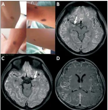
A 12-year-old male patient presented with a delay in neuropsychomotor development that had been diagnosed in the first year of life. Two years prior (at 10 years of age), he had under-gone ventricular shunt placement because of hydrocephalus. Six
months prior to the visit reported here, he had experienced epi-sodes of seizures. Physical examination revealed multiple cutane-ous nevi (Figure 1A). Cerebrospinal fluid examination showed an elevated level of protein (1359.7 mg/dL; reference range: 15.0– 45.0 mg/dL) and revealed the presence of epithelioid cells. Mag-netic resonance imaging (MRI) of the brain (Figures 1B, 1C, and 1D) showed extensive, bilateral, asymmetric leptomeningeal thick-Figure 1. A: Axial computed tomography of the chest showing consolidation with air bronchogram and partially rounded hilar opacity, both presenting areas of low signal intensity, in the upper lobe of the left lung. B: Initial MRI of the brain. Scan with T2-weighted turbo spin-echo sequence showing multiple rounded cystic formations of varying dimensions scattered throughout the cerebral and cerebellar parenchyma, as well as in the left thalamus and nucleocapsular regions, having a discrete compressive effect and no signs of significant perilesional edema. C: Follow-up MRI of the brain. Scan with fluid-attenuated inversion recovery sequence, performed five weeks after the start of fluconazole therapy and the restoration of adequate nutrition, showing intense perilesional vasogenic edema indicating a reactive inflam-matory process that was nearly undetectable at the beginning of treatment, due to immunosuppression. D: Follow-up MRI of the brain. Scan with fluid-attenuated inversion recovery-weighted sequence, performed five weeks after the start of corticosteroid therapy, when the patient was still under treatment with fluconazole, showing significant regression of the edema around the brain lesions, as well as a reduction in the size of the lesions.
Letters to the Editor
Radiol Bras. 2016 Nov/Dez;49(6):406–413
413
ening, mainly in the cortical sulci of the right cerebral hemisphere, with spontaneous T1 hyperintense signal (presumably melanin) and diffuse enhancement after intravenous administration of gadolinium contrast. The MRI scan also showed involvement of the brain parenchyma, characterized by spontaneous T1 hyperin-tense signal in the amygdaloid nuclei and in the cortex, probably due to melanin/melanocyte deposits. Although meningeal biopsy was not performed, a presumptive diagnosis of neurocutaneous melanosis (NCM) was considered.
NCM is a rare sporadic neuroectodermal syndrome, first de-scribed by Rokitansky in 1861, and characterized by congenital cutaneous nevi (one large nevus or multiple nevi) associated with benign or malignant central nervous system (CNS) proliferation of melanocytes(1–4). The diagnostic criteria, which were first de-scribed by Fox and later revised by Kadonaga and Frieden(1) in 1991, include the combination of all of the following: – a single giant congenital nevus (measuring at its greatest diameter ≥ 20 cm in adults, or ≥ 9 cm on the head or ≥ 6 cm on the trunk in neonates and infants) or multiple (three or more) congenital nevi, accompanied by meningeal melanosis or CNS melanoma; – the absence of cutaneous melanoma, except in patients with a meningeal lesion histologically proven to be benign; – the ab-sence of meningeal melanoma, except in patients with cutane-ous lesions histologically proven to be benign.
Approximately 60% to 70% of all individuals with NCM de-velop symptoms, which usually appear before five years of age(2). Clinically, patients can experience seizures, hydrocephalus, de-velopmental delays, psychiatric disorders, cranial nerve palsies,
intracranial hemorrhage, and myelopathy(1,2,5–7). Seizures are the most common initial neurological manifestation(2). Involvement of the CNS can include parenchymal or leptomeningeal lesions, such as melanosis (aggregation of benign melanocytic cells) or melanomas(5).
In NCM, the MRI findings can include hyperintense areas in the temporal lobes on T1-weighted images, diffuse leptom-eningeal enhancement of the brain and spine, and mass of ma-lignant melanoma(4). Parenchymal melanosis typically occurs in the temporal lobes (amygdaloid nuclei), cerebellum, or pons; and its lesions usually exhibit a high signal intensity on T1-weighted images and do not commonly show enhancement after contrast administration(5,7,8). The leptomeningeal lesions usually present intermediate to high signal intensity on T1-weighted images, low to intermediate signal intensity on T2-weighted images, high sig-nal intensity on fluid-attenuated inversion recovery (FLAIR) se-quence and diffuse enhancement after gadolinum administra-tion. Mass effect, edema, hemorrhage, and necrosis favor the possibility of melanoma and make benign melanocytic lesion less likely(5). Abnormalities in the spine, especially cystic malforma-tions (mainly arachnoid cysts), are relatively common in patients with NCM(2).
The differential diagnoses that can be based on MRI images of the brain include subarachnoid hemorrhage, meningitis, lep-tomeningeal carcinomatosis, other melanin-containing lesions, and non-melanocytic hemorrhagic tumors. The clinical context and the imaging characteristics will aid in making that differen-tiation(4,9).
Regardless of the treatment instituted, the prognosis is usu-ally poor, especiusu-ally in cases with diffuse leptomeningeal involve-ment(2,5,6).
REFERENCES
1. Kadonaga JN, Frieden IJ. Neurocutaneous melanosis: definition and review of the literature. J Am Acad Dermatol. 1991;24:747–55. 2. Ramaswamy V, Delaney H, Haque S, et al. Spectrum of central nervous
system abnormalities in neurocutaneous melanocytosis. Dev Med Child Neurol. 2012;54:563–8.
3. Scattolin MA, Lin J, Peruchi MM, et al. Neurocutaneous melanosis: fol-low-up and literature review. J Neuroradiol. 2011;38:313–8. 4. Hayashi M, Maeda M, Maji T, et al. Diffuse leptomeningeal hyperintensity
on fluid-attenuated inversion recovery MR images in neurocutaneous melanosis. AJNR Am J Neuroradiol. 2004;25:138–41.
5. Oliveira RS, Carvalho AP, Noro F, et al. Neurocutaneous melanosis. Arq Neuropsiquiatr. 2013;71:130–1.
6. Sabat SB. Teaching NeuroImages: neurocutaneous melanosis. Neurol-ogy. 2010;74:e82.
7. Demirci A, Kawamura Y, Sze G, et al. MR of parenchymal neurocutane-ous melanosis. AJNR Am J Neuroradiol. 1995;16:603–6.
8. Fu YJ, Morota N, Nakagawa A, et al. Neurocutaneous melanosis: surgi-cal pathologisurgi-cal features of an apparently hamartomatous lesion in the amygdala. J Neurosurg Pediatr. 2010;6:82–6.
9. Pont MS, Elster AD. Lesions of skin and brain: modern imaging of the neurocutaneous syndromes. AJR Am J Roentgenol. 1992;158:1193– 203.
Bruno Lima Moreira1, Thiago Grunewald1, Auro Augusto Junqueira Côrtes1, Victor Hugo Rocha Marussi1, Lázaro Luís Faria do Amaral1
1. Hospital Beneficência Portuguesa de São Paulo, São Paulo, SP, Brazil. Mailing address: Dr. Bruno Lima Moreira. Med Imagem – Unidade São Joaquim. Rua Maestro Cardim, 769, Bloco 3, 1º subsolo, Bela Vista. São Paulo, SP, Brazil, 01323-001. E-mail: [email protected].
Figure 1. A: Multiple cutaneous nevi seen on physical examination. B,C: Gado-linium-enhanced axial T1-weighted MRI sequence with fat suppression showing hyperintense signals along the cortical sulci of the cerebral hemispheres, pre-sumably due to diffuse leptomeningeal lesion with melanin content, together with areas of high signal intensity in the amygdaloid nuclei (arrows) and in the cerebral cortex, which likely correspond to parenchymal involvement by melanin/melano-cyte deposits in the context of NCM. D: Gadolinium-enhanced axial T1-weighted MRI sequence with fat suppression showing diffuse enhancement of the leptom-eninges along the cortical sulci of both cerebral hemispheres, especially on the