Arquivos Brasileiros de Cardiologia - Volume 82, Nº 4, Abril 2004
398
Image
Hypert rophic Cardiom yopat hy in M onozygot ic Tw ins
Aloir Q. Araujo, Edmundo Art eaga, Charles M ady
São Paulo, SP - Brazil
Cardiomyopathies Division - Heart Institute (InCor) São Paulo University Medical School
Mailing adress: Aloir Q. Araujo - InCor - Av. Dr. Enéas de Carvalho Aguiar, 44 - 05403-900 - São Paulo, SP Brazil
E-mail: [email protected] Received: 3/3/04
Accepted 25/3/04
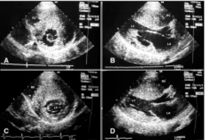
Male monozygotic twins (MT), age 19 years, with obstructive hypertrophic cardiomyopathy (HCM), symptoms (exertional dysp-nea) started around age 13 years, and similar clinical presentation (a grade 3/6 systolic murmur heard in the left lower sternal border, typical ECG signs of left ventricular hypertrophy). In both patients, there were echocardiographic moderate left atrial enlargement, massive septal hypertrophy (>30mm) and severe resting outflow tract gradient, without significant differences betweem them. Some discordances were observed in two-dimensional images (Figure 1). Color Doppler showed marked septal coronary branchs in twin G (Figure 2), which was not found in twin V. Twin G evoluted to medical refractory NYHA III/IV functional class and was referred to a surgical myectomy.
Hypertrophic cardiomyopathy is a primary disease of the myo-cardium caused by mutations in genes coding for sarcomeric pro-teins1, and the phenotype can be influenced by modifiers genes2
and environmental factors. Then, the clinical and morphological manifestations of the disease are variable, from absence of symp-toms to heart failure, even among mutation carriers of a given family. MT are considered genetically identical (causing and mo-difiers genes), and phenotypic discordances have been credited to the action of environmental factors. In MT with HCM, it would be expected similar expression of the disease, as observed by Maron et al3 , with limited discordances. However, the cases reported in
the literature do not support this view. There are reports of MT pairs with concordant3-6 and discordant4,7 phenotypes. In general
terms, our patients showed similar manifestations of the disease, but there were some diverse echocardiographic and clinical findings. The cases reported here and the literature review bring more questions than answers about the pathogenetic mechanisms of hypertrophic cardiomyopathy. There is not a reasonable expla-nation to understand the contrasting situation existing between MT pairs with very similar findings and pairs with absolutely different expression of the disease, including symptoms, extent and distribution of the hypertrophy, outflow obstruction, age of presentation and outcome.
Can we assume that the genetic background is absolutely identical in MT? they develop from a separation of the embryonic
cells of a single embryo at the two-cell stage, but human twinning can also occur at a developmental stage as late as 7 days of gestation, and this timing can determine placental anastomoses and umbilical differences, exposing the developing twins to variable intrauterine environments8. In addition, there are studies of genetic
mechanisms that may result in phenotypic, genotypic, and epige-netic differences between MT9, but those phenomena are rare
and cannot be assumed as a major cause of the discordances under discussion.
Fig. 1 - A and B) dimensional echocardiogram of twin V; C and D) two-dimensional echocardiogram of twin G. In A and C (short axis), the LV hypertrophy is more difuse in twin G. Note a displasy in the inferior wall of twin V (arrow). In B and D (long axis), similar IVS thicknesses are showed but the PW is more hypertrophied in twin G. RV- right ventricle cavity; LA- left atrium; AO- aorta; LV-left ventricle cavity; IVS- interventricular septum; PW- posterior wall.
Arquivos Brasileiros de Cardiologia - Volume 82, Nº 4, Abril 2004
399
Hypertrophic Cardiomyopathy in Monozygotic Twins1. Marian AJ, Roberts R. The molecular genetic basis for hypertrophic cardiomyopa-thy. J Mol Cell Cardiol 2001; 33: 655-70.
2. Marian AJ. Modifiers genes for hypertrophic cardiomyopathy. Curr Opin Cardiol 2002;17:242-52.
3. Maron BJ, Casey AS, Almquist AK. Hypertrophic cardiomyopathy in monozygotic twins. Circulation 2002; 105: 22-9.
4. Littler WA. Twin studies in hypertrophic cardiomyopathy. British Heart Journal 1972; 34: 1147-51.
5. Ciró E, Nichols PF, Maron BJ. Heterogeneous morphologic expression of genetical-ly transmitted hypertrophic cardiomyopathy. Two-dimensional echocardiographic analysis. Circulation 1983; 67: 1227-33.
References
6. Wylie L, Ramage A, MacLeod DC. Hypertrophic cardiomyopathy with shared mor-phology in identical twins: a case report. Scott Med J 2002; 47: 64-5 . 7. Clarck CE, Henry WL, Epstein SE. Familial prevalence and genetic transmission of
idiopathic hypetrophic subaortic stenosis. N Engl J Med 1973; 289: 709-14. 8. Singh SM, Murphy B, OReilly R. Epigenetic contributors to the discordance of
monozygotic twins. Clin Genet 2002; 62: 97-103.