Zbornik Matice srpske za prirodne nauke / Proc. Nat. Sci, Matica Srpska Novi Sad, ¥110, 55—64, 2006
UDC 633.63:631.53.01:632.3
M i r j a n a B. M i l o š e v i ã, M a j a V. I g n j a t o v, S l a ð a n a S. M e d i ã - P a p
National laboratory for seed testing
Maksima Gorkog 30, Novi Sad, Serbia and Montenegro
THE MOST IMPORTANT PATHOGENS
TRANSMITTED BY SUGAR BEET*
ABSTRACT: Pathogenic fungi and viruses transmitted by sugar beet seed represent a complex group of organisms. Detection of these pathogens is an important issue in sugar beet protection. Their identification is a difficult task because the most available methods rely on the growth characteristics, morphological and biochemical criteria. Three domestic and eight foreign sugar beet varieties, from Germany, Italy and Greece were included in the investigation. Seed health testing was performed in laboratory and in field conditions. Du-ring the trials, the following methods were used: blotter method, agar plate method and ELISA test for viruses. Seeds were incubated in “Conviron" aparatus at 22°C which is sui-table for sporulation of different kind of fungi (light and temperature were adjussui-table). The appereance of following fungi was noted during incubation:Pleospora bjoerlingii (Phoma betae),Fusariumspp.,Pythiumspp.Aphanomyces cochlioidesandCercospora beticola. Vi-ruses tested by ELISA test werebeet necrotic yellow vein virus(BNYVV) andbeet yellows virus (BYV). Viruses were tested in sugar beet seedlings grown in laboratory conditions and on leaves of individual plants from the field. The disease index was calculated on the basis of intensity of infection of plants forCercospora beticolaandPhoma betaeaccording to Mc Kinney's formula. Results were presented by graphs, tables and original photos.
KEY WORDS: ELISA test, leasf spot, seed-borne, sugar beet, diseases, viruses
INTRODUCTION
More than 90% of food in the world is produced from the seed. Seed it-self often presents the basic source of parasite inoculum. The reasons for gi-ving such attention to pathogens are ever more increasing exchange of seed material and the danger of spreading of new pathogens to those parts of the world where they were not found previously. The exchange of seed material contributed to increased number of seed-borne pathogens. Viruses found in field and vegetable crops only recently gained importance. Economic
tance of seed-borne parasites led to altered attitude of developed countries to-wards phytopathology and the seed-borne pathogens. Now, special attention is paid to problems related to quarantine, seed quality determination and chemi-cal protection. Seed certification has become the part of integral management system in plant protection (M i l o š e v i ã, 2001).
Regular control of seed health on the presence of quarantine and econo-mically harmful organisms, in the laboratory and in the quarantine field is ne-cessary due to the increased import of sugar beet seed from different parts of the world. Continual quarantine and post-quarantine supervision is the basis for well developed and stable production as well as for the protection of do-mestic varieties from uncontrolled import.
Sugar beet seed plays an important role in seed certification scheme. Due to economic importance of this plant species, it is important to be familiar with harmful organisms attacking its seed. The most common sugar beet seed-borne parasites are Pleospora bjoerlingii B y f o r d (an. Phoma betae B j o e r l i n g) — Phoma leaf spot and damping off; Peronospora farinosa (Fr.) Fr. f. sp. betae B y f — downy mildew;Cercospora beticola S a c c. — Cercospora leaf spot; Ramularia beticola F a u t r. et L a m b. — Ramularia leaf spot; Uromyces betae (P e r s.) L e v. — sugar beet rust; Alternaria tenuis N e e s. — Alternaria leaf spot; Fusarium oxysporum — Fusarium yellows; beet necrotic yellow vein virus (BNYVV); beet yellows virus (BYV).
Beet necrotic yellow vein virus is a quarantine parasite listed on the A2 EPPO list No. 160 (OEPP/EPPO, 1988). It is also listed in Rules on health examination of crops and objects for production of seed, seedlings and plan-ting material and health examination of seed, seedling and planplan-ting material (Official register of SRJ, 66/99). Beet yellows virus, Peronospora farinosaand Phoma betae are also listed as economically significant parasites.
Effective protection of crops from those above mentioned diseases could be achived by complex mesaures in which cultivation practice, the introduction of tolerant varieties into production and the application of fungicides are inclu-ded (M a r i ã and J e v t i ã, 2001).
MATERIAL AND METHODS
The appearance of quarantine and economically harmful parasites of su-gar beet on domestic and foreign varieties was observed. Three domestic and eight foreign sugar beet varieties from Germany, Italy and Greece were inclu-ded in the investigation. Domestic varieties were marked as S1, S2 and S3; those from Germany as N1, N2, N3 and N4; those from Italy as I1, I2 and from Greece as G1 and G2. Testing was done both in the field and in the la-boratory.
Laboratory testing
trade and import. Blotter method, method of nutritive media and ELISA test were used as laboratory tests.
Pelleted seed was previously washed in order to remove preparation and initiate the development of seed-borne parasites. The seed for testing on blotter and nutritive media were initially prepared by immersing in 1% NaOCl solu-tion for 5 minutes. After that, the seed was washed (3 times) in sterile water, dried and placed into previously prepared Petri dishes. Ten seeds, from 400 were placed into each Petri dish. The incubation of seed on blotter and potato dextrose agar was done in sterile conditions in Petri dishes for 7 days at 22°C (ISTA Rules, 2002) using alternating light cycle (12h NUV/12h of dark, “Con-viron" apparatus). Upon incubation, each seed was observed under a stereo microscope and present pathogens were determined (M a t h u r and K o n g s -d a l, 2003).
Viruses were identified using serological method (Enzyme immuno-ad-sorbtion ELISA test). Samples were tested in two stages. In the first stage, the seed was germinated in sterile boxes and ELISA test was done using obtained sugar beet seedlings. In the second stage, the samples of leaves from field were used. Forty-five seedlings from one variety and 45 leaves from individual plants were tested for both viruses.
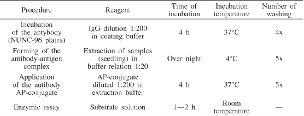
Table 1. — ELISA test for BNYVV and BYV
Procedure Reagent incubationTime of temperatureIncubation Number ofwashing Incubation
of the antybody (NUNC-96 plates)
IgG dilution 1:200
in coating buffer 4 h 37°C 4x
Forming of the antibody-antigen
complex
Extraction of samples (seedling) in
buffer-relation 1:20 Over night 4°C 5x Application
of the antibody AP-conjugate
AP-conjugate diluted 1:200 in
extraction buffer 4 h 37°C 5x
Enzymic assay Substrate solution 1—2 h temperatureRoom —
Reagents of beet necrotic yellow vein virus BNYVV andbeet yellows vi-rus (BYV) (LOEWE Biochemica GmbH, Germany), Kit Complete consisting of antibody, conjugate, positive and negative control were used. Automatic ELISA reader, Multiskan Ascent at 405 nm was used for reading of Nunc pla-tes with 96 wells.
Field testing
with 70 cm distance between the rows. Fungicide treatments were not used du-ring vegetation period. Disease intensity was evaluated by exemination of indi-vidual plants (each fifth plant) according to scale of 0—9. Disease index was calculated on the basis of the intensity of leaf spot diseases according to Mc Kinney's formula.
I — disease index
a — number of exeminated plants b — number of categories (0—9) k — total number of plants 10 — number of categories
I axb
kx x
( )
10 100
RESULTS
Results of laboratory testing
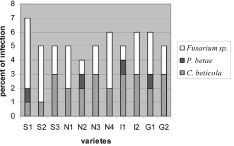
Results of laboratory testing on blotter and nutritive media are shown in graphs 1 and 2. Total percentage of seed infection ranged from 2—5% on blotter, and from 4—7% on nutritive media. Aphanomyces cochlioides and Pythium sp. were not found in sugar beet samples.
According to obtained results of ELISA test (Table 2) for sugar beet see-dlings, all tested samples were healthy.
Table 2 — Range of absorption values obtained by DAS ELISA test forbeet necrotic yellow vein virus (BNYV) and beet yellows virus (BYV) (from seedlings)
Variety Beet necrotic yellow vein virus(BNYV) Beet yellows virus(BYV)
S1 0,074—0,092 0,055—0,111
S2 0,072—0,137 0,041—0,131
S3 0,093—0,134 0,043—0,132
N1 0,076—0,127 0,052—0,121
N2 0,076—0,116 0,051—0,109
N3 0,089—0,133 0,062—0,100
N4 0,083—0,134 0,044—0,110
I1 0,085—0,125 0,055—0,121
I2 0,070—0,122 0,066—0,133
G1 0,072—0,124 0,067—0,119
G2 0,072—0,103 0,058—0,128
Positive control 1,295 0,768
Negative control 0,137 0,118
Results of laboratory testing
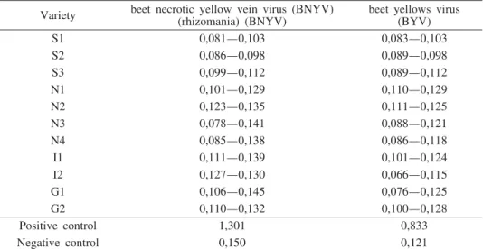
According to ELISA test (Table 3) results obtained from sugar beet lea-ves, all tested samples were healthy.
Table 3 — Range of absorption values obtained by DAS ELISA test forbeet necrotic yellow vein virus (BNYV) andbeet yellows virus (BYV) (from leaves of individual plants from field)
Variety beet necrotic yellow vein virus (BNYV)(rhizomania) (BNYV) beet yellows virus(BYV)
S1 0,081—0,103 0,083—0,103
S2 0,086—0,098 0,089—0,098
S3 0,099—0,112 0,089—0,112
N1 0,101—0,129 0,110—0,129
N2 0,123—0,135 0,111—0,125
N3 0,078—0,141 0,088—0,121
N4 0,085—0,138 0,086—0,118
I1 0,111—0,139 0,101—0,124
I2 0,127—0,130 0,066—0,115
G1 0,106—0,145 0,076—0,125
G2 0,110—0,132 0,100—0,128
Positive control 1,301 0,833
Negative control 0,150 0,121
The high percentage of infection caused by Cercospora beticola ranging from 19—60% was noticed based on the results of index of diseases (Graph. 3), while Phoma betae was present in much smaller percentage, from 10— 25%
Symptoms of sugar beet leaf spot (Cercospora beticola and Phoma be-tae) are shown on figure 1 and 2.C. beticolacauses appearance of tiny, round, grey spots with dark red tissue zone on the edge of sugar beet leaf (Figure 1). P. betae forms large round concentrically zoned spots, often with cracked tis-sue in the centre (Figure 2).
DISCUSSION
Sugar beet seed health testing (blotter and nutritive media) revealed the presence of parasitic fungi Phoma betae and Cercospora beticola, and fungi fromFusarium genus. Total percentage of infection ranged from 2—5%. From obtained results, it can be seen that Fusarium was present on most of the vari-eties except on N2 and I2. Phoma betae was found on two tested varieties: I2 and G2 and the percentage of infection did not exceed 1%.Cercospora betico-lawas not found in domestic variety S1 and the percentage of infection in ot-her varieties ranged from 1—3%.
Nutritive media method revealed the presence of seed parasites in appro-ximately the same percentage as in the method on blotter. Obtained percentage of infection is the result of chemical treatment (pelleted seed) and does not significantly influence disease level on plants in the field (R i c h a r d s o n, 1990).
spite of different opinions on seed virus transmission, those in charge of sugar beet import should check the seed on the presence of beet yellows virus and beet necrotic yellow vein virus in a laboratory and check crops prior to main aphids flight in the field (group of authors, 1980).
ELISA test was also used for samples of individual plants from the field (parts of leaf were taken). Viruses were not found in analyzed samples. Con-centration of viruses is usually very unequal and content of viruses and their concentration is higher in older leaves (A g r i o s, 1997). Beet necrotic yellow vein virus and beet yellows virus are mainly found in phloem, although they can be found in other plant parts too (leaf) (Š u t i ã, 1995). These viruses can be transmitted during vegetation period from overwintered infection sources, such as some weeds, mangel or fodder rape pits, self seeded plants etc. Virus vectors are aphids Myzus persicae and Aphis fabae. Intensity of infection de-pends on density of aphid population and sources of infection, especially those close to sugar beet field.
Field trials were observed during vegetation period. Seedling decline was not found in the stage of sugar beet emergence due to fungicide treatment (pelleted seed). Mini-pelleting and pelleting of sugar beet seed is a technologi-cal procedure in seed processing when seed is covered with several different micro and macro substances, growth stimulators, fungicides and insecticides ( K a w a k a t s u et al., 1998).
Meteorological conditions during vegetation period and especially quan-tity and schedule of precipitation influenced intensity of appearance of leaf di-seases during 2005. No chemical fungicide treatment was used during sugar beet vegetation period. Sugar beet in isolation was sown on the bordering plot (0,1 ha) in the previous year (2003/04) which could be the source of initial pa-rasite inoculum. High percentage of appearance of leaf diseases is the result of combination of above mentioned factors. The greatest values of disease index for Cercospora beticola (60%) were found for I1 and G2 varieties, while the smallest index was found for domestic varieties S1 and S2 (20%). Phoma be-tae was found in much lesser percentage, ranging from 10—25%.
Optimal conditions for plant infection are temperature at around 25°C and relative humidity exceeding 95%. By cultivating tolerant variety and using re-gular agrotechnical measures and chemical protection, the intensity of appea-rance of sugar beet leaf diseases is decreased. Infected seed could be a poten-tial source of inoculum, but infected residual debris, poor agrotechnical measu-res and irregular crop rotations make even greater threat. Infected leaves left on field are the most significant source of infection, so regular crop rotation has great influence on disease development.
REFERENCES
A g r i o s, N. G. (1997): Plant pathology 4th, Academic Press, USA.
ISTA Rules, (2002):International for Seed Testing, Annexe to Chapter 7. Seed Health Testing, Zürich, Switzerland.
K a w a k a t s u, M., S e k i m u r a, K., O q a t a, N., T a n a k a, M. (1998): Investiga-tion of qualities of sugar beet (Beta vulgaris) seed: 1. Effect of seed growing con-dition on qualities of seeds, 2. Effect of processing and storing on germination of
seeds, Proceedings of the Sugar Beet Research Accociation, Japan, 39: 48—45,
42—47.
Kolektiv autora (1980): Priruånik o karantenskim biljnim bolestima i štetoåinama SFR Jugoslavije, Fakultet poljoprivrednih znanosti, Zagreb.
M a c h a d o, J. C., L a n g e r a k, C. J., J a c c o u d - F i l h o, D. S. (2002):
Seed-Bor-ne Fungi. A Contribution to RutiSeed-Bor-ne Seed Health Analysis, ISTA, Bassersdorf, CH
— Switzerland.
M a r i ã, A., Å a m p r a g, D. (1982):Štetoåine i bolesti šeãerne repe, Nolit, Beograd. M a r i ã, A., J e v t i ã, R. (2001):Atlas bolesti ratarskih biljaka, Poljoprivredni fakultet
i Školska knjiga, Novi Sad.
M a t h u r, S. B., K o n g s d a l, O. (2003): Common Laboratory Seed Health Testing
Methods for Detecting Fungi, ISTA, Bassersdorf, CH — Switzerland.
M i l o š e v i ã, M. (2001): Biljni karantin, Feljton d.o.o., Novi Sad.
N e e r g a a r d, P. (1979): Seed Pathology, The Macmillan Press. Ltd., Grande-Bre-tagne.
OEPP/EPPO, (1988): Data sheets on quarantine organisms No 162, Beet necrotic
yellow vein virus, Bulletin OEPP/EPPO Bulletin 18, 527—532.
R i c h a r d s o n, M. J. (1990): An annoted list of seed-borne diseases, ISTA, Bassers-dorf, CH — Switzerland.
Š u t i ã, D. (1995): Viroze biljaka, Institut za zaštitu bilja i ÿivotnu sredinu, Beograd.
EKONOMSKI NAJZNAÅAJNIJI PARAZITI KOJI SE PRENOSE SEMENOM ŠEÃERNE REPE
Mirjana B. Miloševiã, Maja V. Igwatov i Slaðana S. Mediã-Pap Nacionalna laboratorija za ispitivawe semena, Maksima Gorkog 30,
21000 Novi Sad, Srbija i Crna Gora
Rezime
podešavawa svetlosti i temperature, potrebnih za sporulaciju razliåitih gqi-va. Tokom inkubacije praãena je pojava sledeãih parazitnih gqiva: Pleospora
bjoerlingii (Phoma betae), Fusarium spp., Pythium spp., Aphanomyces cochlioides i
Cercospora beticola. Virusi su ispitivani ELISA testom i to: Virus