143
CLINICS 2008;63(1):143-6
LETTER TO THE EDITOR
Gastroenterology Departament, Surgical Division, Hospital das Clinicas, Faculdade de Medicina da Universidade de São Paulo - São Paulo/SP, Brazil. [email protected]
ATYPICAL PERIANAL HERPES SIMPLEX INFECTION
IN HIV-POSITIVE PATIENTS
Marcelo Simonsen, Sergio Carlos Nahas, Edesio Vieira da Silva Filho, Sergio Eduardo Alonso Araújo, Desiderio Roberto Kiss, Caio Sergio Rizkallah Nahas
INTRODUCTION
Anal lesions affect up to 34% of patients with Acquired Immunodeficiency Syndrome (AIDS),1 and are more
fre-quent in males who have sex with males (MSM).2 The most
common anal infection in human immunodeficiency virus (HIV)-positive patients is caused by human papillomavirus (HPV).2,3 It is suggested that the appearance of this illness
is related to the conversion of HIV into AIDS.2,4,5 Even with
the introduction of highly-active antiretroviral therapy (HAART), the prevalence of HPV infection in HIV-posi-tive patients has not decreased, nor has anal cancer inci-dence.6 It is well-known that HPV infection in
HIV-posi-tive patients is a risk factor for the development of squa-mous cell cancer of the anus. Routine screening is strongly recommended in this population in order to identify pre-malignant lesions. 6
HPV infection frequently manifests as verrucous lesions (warts) that cause pruritus, discomfort, and, more rarely, pain or bleeding.2,3 However, some other infectious agents
such as varicella-zoster virus (VZV), cytomegalovirus (CMV), molluscum contagiosum (MC), and particularly herpes simplex virus (HSV),can also cause verrucous skin lesions in HIV-positive patients.7
Herpes simplex virus is found in 29% of MSM with symptomatic anorectal disease,3 although the majority of
confirmed herpes simplex cases are reported in asympto-matic individuals.3,8 The most frequently encountered
find-ings are ulcerated aphthous lesions, vesicles and inguinal lymphadenopathy.3,9 Additional signs and symptoms include
pain, pruritus, lymphadenopathy, superficial ulcers, vesicu-lar erosion,3,9,10 urinary retention11,12 and constipation.12,13
A very small number of cases of verrucous herpes of the perineum have been reported in the English medical literature and treatment options varied, including the use of acyclovir,4,14 valacyclovir15 and surgical resection.16 We
report a case of a perianal verrucous lesions that was ini-tially suspected to be anal neoplasia, but was revealed to be a herpes infection. The patient required surgical resec-tion after antiviral therapy failure. This article reviews the literature in order to describe the main characteristics of patients with verrucous perineal herpes infection and the outcomes of different treatment modalities.
CASE REPORT
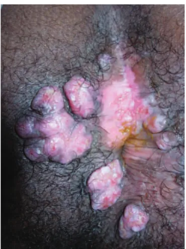
A 39-year-old male was referred to our service who complained of having had verrucous and painful perianal nodules for 3 months. He had a 10-year history of HIV in-fection and had been using HAART for the last two years. His CD4 T cell count was 400/¼l and his HIV viral load was 60.000 copies per ml.
The proctologic exam revealed painful 2-3 cm verru-cous perianal nodules (Figure 1). Endoscopy of the rectum and sigmoid was normal. Bilateral inguinal lymphadenopa-thy was detected during the physical exam. An anal pap smear revealed high-grade anal dysplasia, while a conven-tional pathologic exam of perianal lesions revealed no dys-plasia but demonstrated cytoarchitectural alterations com-patible with HSV infection. Immunohistochemical tests
144
CLINICS 2008;63(1):143-6 Atypical perianal herpes simplex infection in hiv-positive patients
Simonsen M et al.
b114® for HSV type 1 and b116® for HSV type 2 from Dako Denmark A/S (Glostrup - Denmark) revealed the pres-ence of HSV type 2. Fungal and acid-alcoholresistant ba-cilli screening were negative.
The patient was administered oral acyclovir for four weeks (2.4 g/d). Because there was minimal or no regres-sion of the leregres-sions and persistence of the pain, we chose to resect all lesions. After surgery, the patient was admin-istered prophylactic acyclovir. Disease recurrence was not detected after a 9-month follow-up (Figure 2).
DISCUSSION
Our review of the English language medical literature published within the last 15 years identified eight cases of
Table1 - Patients with anal hypertrophic herpes.
Author and Year Case HSV Age Gender CD4+ T Treatment Recurrence Follow-up Prophylaxy
Number type lymphocyte (mo) (mo)
count (cells/µL)
Tong et al. (1996) 1 (HSV2) 32 M 13 Acyclovir No NA NR
(6 wks)
Saramatunga et al. (2001) 2 (HSV2) 46 M 470 LE alone No 11 Valacyclovir
(oral)
Gubinelli et al. (2003) 3 (HSV1) 46 M 500 Acyclovir No 4 Valacyclovir
(oral, failure) + (oral)
valacyclovir (oral, 2 mo)
Nadal et al. (2005) 4 (HSV1) 41 M 73 LE alone 12 NA Not used
Nadal et al. (2005) 5 (HSV2) 46 M 370 LE alone resolution 24 Not used
Nadal et al. (2005) 6 (HSV2) 49 M 186 Acyclovir 10 NA Not used
(oral, topical & IV) + LE
Nadal et al. (2005) 7 (HSV2) 42 M 251 Acyclovir 3 NA Not used
(oral, topical & IV) + LE
Nadal et al. (2005) 8 (HSV2) 54 F 116 Acyclovir resolution 12 Not used
(oral, topical & IV) + LE
Present case 9 (HSV 2) 39 M 400 Acyclovir resolution 9 Acyclovir
(oral) + LE (oral)
HSV = herpes simplex virus; NA = not applicable; NR = not reported; LE = local excision; IV = intravenous; M = male; F = female
Figure 2 - Proctologic exam after surgery.
verrucous perineal herpes infection. The patient character-istics are summarized and compared to the findings from our case in Table 1. Data from the literature demonstrate that this atypical presentation of herpes infection has a pre-dilection for middle-aged males infected with HIV.4,15,16,17
The features of the present case were found to be similar to those reviewed in the literature.
The patient characteristics reported in the literature sug-gest risk factors for this uncommon presentation. An immunocompromised status, which is related to HIV in-fection, was reported in all cases of perianal verrucous her-petic lesions, which suggests that it may be a significant factor. Additionally, all patients had CD4 T cell counts lower than 500 cells/µL, despite the use of HAART. A his-tory of receptive anal intercourse may also play an impor-tant role in this affliction. Although this issue was not al-ways addressed in previous reports, two of the patients were MSM.4,15
HSV, as well as VZV, CMV, MC and HPV infections, can manifest as hyperkeratosis5 and verrucous4 lesions in
HIV-positive patients. The cause for this manifestation of herpes simplex infection is unknown,4,14 although many
hy-potheses have been postulated. According to Smith et al.,5
there is an increased number of dendritic cells that are posi-tive for XIIIa factor, which might be related to the pathogenesis of HIV18,19 as these cells can work as an
145
CLINICS 2008;63(1):143-6 Atypical perianal herpes simplex infection in hiv-positive patients
Simonsen M et al.
can produce TNF-alpha in certain conditions, which might increase the growth index of keratinocytes22,23 and generate
acanthosys and hyperkeratosis. In uninfected people, vari-ous stimuli of keratinocytes are inhibited by IFN-gamma, which is produced by cytotoxic T cells and T helper cells. This mechanism is diminished in HIV-infected patients.23
Interestingly, the majority (7/9) of the lesions in the re-ported cases were caused by HSV2 (cases 1, 2, and 5-9). However, due to the small number of cases, an association between HSV type and verrucous lesions cannot be as-sumed.
Regarding the treatment modalities and outcomes, it is difficult to compare the results from previous reports to our case. As seen in Table 1, there was significant treatment diversity among these nine cases and relatively short fol-low-up. However, it must be emphasized that although two cases were successfully treated with antiviral therapy alone (cases 1 and 3), the majority of cases (7/9) received local excision as part of their treatment. Local excision alone was performed in three cases (cases 1, 4 and 5) and in combi-nation with upfront antiviral therapy in four cases (cases 6-9). Among these seven surgically treated patients, three of them presented with disease recurrence (cases 4, 6 and 7) despite the administration of oral and topical acyclovir to two of them (cases 6 and 7). The use of prophylactic acyclovir after surgical treatment effectively prevented
dis-ease recurrence in our patient during the nine-month fol-low-up period.
According to the literature, patients with HSV infec-tions resistant to acyclovir, which usually occurs after ir-regular use of the drug,7 could be treated with foscarnet24
and beta-interferon.10 Because valacyclovir is more
bioavailable than acyclovir, it was more effective in the resolution of cutaneous HSV infections.25,26
Despite the relatively short follow-up period in these reports, they suggest that prophylactic use of oral acyclovir or valacyclovir may prevent recurrence. In addition, there is evidence which indicates that the survival of patients with AIDS and previous exposure to herpes virus infections may increase with chronic use of suppressive therapy with acyclovir.27 Moreover, the evidence seems to support
pro-phylactic use of these drugs.
CONCLUSION
Atypical presentation of herpes simplex infection should be considered as a differential diagnosis of perianal neo-plasia in HIV-positive patients, because the nature of this presentation may be related to an immunocompromised sta-tus. Surgical resection followed by acyclovir prophylactic treatment appears to be an effective therapy.
REFERENCES
1. Wexner SD, Smithy WB, Milsom JW, Dailey TH. The surgical management of anorectal diseases in AIDS and pre-AIDS patients. Dis Colon Rectum. 1986;29:719-23.
2. Yuhan R, Orsay C, DelPino A, Pearl R, Pulvirenti J, Kay S, et al. Anorectal disease in HIV-infected patients. Dis Colon Rectum. 1998;41:1367-70.
3. Quinn TC, Corey L, Chaffee RG, Schuffler MD, Brancato FP, Holmes KK. The etiology of anorectal infections in homosexual men. Am J Med. 1981;71:395-406.
4. Tong P, Mutasim DF. Herpes simplex virus infection masquerading as condyloma acuminata in a patient with HIV disease. Br J Dermatol. 1996;134:797-800.
5. Smith KJ, Skelton HG 3rd, Frissman DM, Angritt P. Verrucous lesions secondary to DNA viruses in patients infected with the human immunodeficiency virus in association with increased factor XIIIa-positive dermal dendritic cells. The Military Medical Consortium of Applied Retroviral Research Washington, D.C. J Am Acad Dermatol. 1992;27:943-50. Erratum in: J Am Acad Dermatol. 1993;28:411. 6. Heard I, Palefsky JM, Kazatchkine MD. The impact of HIV antiviral
therapy on human papillomavirus (HPV) infections and HPV-related diseases. Antivir Ther. 2004;9:13-22.
7. Beasley KL, Cooley GE, Kao GF, Lowitt MH, Burnett JW, Aurelian L. Herpes simplex vegetans: atypical genital herpes infection in a patient with common variable immunodeficiency. J Am Acad Dermatol. 1997;37:860-3.
8. Goldmeier D. Proctitis and herpes simplex virus in homosexual men. Br J Vener Dis. 1980;56:111-4.
9. Jacobs E. Anal infections caused by herpes simplex virus. Dis Colon Rectum. 1976;19:151-7.
10. Waugh MA. Anorectal Herpesvirus hominis infection in men. J Am Vener Dis Assoc. 1976;3:68-70.
11. Goldmeier D. Herpetic proctitis and sacral radiculomyelopathy in homosexual men. Br Med J. 1979;1;2:549.
12. Samarasinghe PL, Oates JK, MacLennan IP. Herpetic proctitis and sacral radiomyelopathy—a hazard for homosexual men. Br Med J. 1979;11;2:365-6.
146
CLINICS 2008;63(1):143-6 Atypical perianal herpes simplex infection in hiv-positive patients
Simonsen M et al.
14. Carrasco DA, Trizna Z, Colome-Grimmer M, Tyring SK. Verrucous herpes of the scrotum in a human immunodeficiency virus-positive man: case report and review of the literature. J Eur Acad Dermatol Venereol. 2002;16:511-5.
15. Gubinelli E, Cocuroccia B, Lazzarotto T, Girolomoni G. Nodular perianal herpes simplex with prominent plasma cell infiltration. Sex Transm Dis. 2003;30:157-9.
16. Nadal SR, Calore EE, Manzione CR, Horta SC, Ferreira AF, Almeida LV. Hypertrophic herpes simplex simulating anal neoplasia in AIDS patients: report of five cases. Dis Colon Rectum. 2005;48:2289-93. 17. Samaratunga H, Weedon D, Musgrave N, McCallum N. Atypical
presentation of herpes simplex (chronic hypertrophic herpes) in a patient with HIV infection. Pathology. 2001;33:532-5.
18. Gendelman HE, Orenstein JM, Baca LM, Weiser B, Burger H, Kalter DC, et al. The macrophage in the persistence and pathogenesis of HIV infection. AIDS. 1989;3:475-95.
19. Mellert W, Kleinschmidt A, Schmidt J, Festl H, Emler S, Roth WK, et al. Infection of human fibroblasts and osteoblast-like cells with HIV-1. AIDS. 1990;4:527-35.
20. Horner PJ, Harris JR. A herpes simplex skin ulcer in a patient with AIDS—an unusual presentation. Int J STD AIDS. 1990;1:288-9.
21. Mahoney SE, Duvic M, Nickoloff BJ, Minshall M, Smith LC, Griffiths CE, et al. Human immunodeficiency virus (HIV) transcripts identified in HIV-related psoriasis and Kaposi’s sarcoma lesions. J Clin Invest. 1991;88:174-85.
22. Saiag P, Coulomb B, Lebreton C, Bell E, Dubertret L. Psoriatic fibroblasts induce hyperproliferation of normal keratinocytes in a skin equivalent model in vitro. Science. 1985;230:669-72.
23. Nickoloff BJ, Karabin GD, Barker JN, Griffiths CE, Sarma V, Mitra RS, et al. Cellular localization of interleukin-8 and its inducer, tumor necrosis factor-alpha in psoriasis. Am J Pathol. 1991;138:129-40.
24. Wagstaff AJ, Bryson HM. Foscarnet. A reappraisal of its antiviral activity, pharmacokinetic properties and therapeutic use in immunocompromised patients with viral infections. Drugs. 1994;48:199-226.
25. Baker DA. Valacyclovir in the treatment of genital herpes and herpes zoster. Expert Opin Pharmacother. 2002;3:51-8.
26. Bras AP, Sitar DS, Aoki FY. Comparative bioavailability of acyclovir from oral valacyclovir and acyclovir in patients treated for recurrent genital herpes simplex virus infection. Can J Clin Pharmacol. 2001;8:207-11.