Major Article
Corresponding author: Dr. Caio Fernando de Oliveira. e-mail: [email protected]
Received 11 January 2016 Accepted 14 June 2016
INTRODUCTION
Coagulase-negative staphylococci (CoNS) are currently the most prevalent microorganisms responsible for nosocomial infections related to indwelling medical devices(1). In prospective
surveillance studies, CoNS were identiied as the most common pathogens in nosocomial bloodstream infections of pediatric patients(2) (3). Among CoNS, the main isolated species from
nosocomial infections is Staphylococcus epidermidis, in particular in relation with indwelling devices(1).
Coagulase-negative staphylococci infections are often dificult to treat due to multidrug resistance (MDR) and bioilm
Coagulase-negative staphylococci in Southern Brazil:
looking toward its high diversity
Caio Fernando de Oliveira
[1], Jorunn Pauline Cavanagh
[2], [3],
Elizabeth G. Aarag Fredheim
[2],[3], Keli Cristine Reiter
[4], Alexandre Rieger
[5],
Claus Klingenberg
[3], Pedro Alves d’Azevedo
[4]and Johanna Ericson Sollid
[6][1]. Programa de Pós Graduação em Promoção da Saúde, Universidade de Santa Cruz do Sul, Santa Cruz do Sul, Rio Grande do Sul, Brasil. [2]. Paediatric Research Group, Dept. of Clinical Medicine, Faculty of Health Sciences, UiT-The Artic University of Norway, Tromsø, Norway.
[3]. Department of Paediatrics, University Hospital of North Norway, Tromsø, Norway.
[4]. Departamento de Microbiologia e Parasitologia, Universidade Federal de Ciências da Saúde de Porto Alegre, Porto Alegre, Rio Grande do Sul, Brazil. [5]. Laboratório de Biotecnologia e Genética, Universidade de Santa Cruz do Sul, Santa Cruz do Sul, Rio Grande do Sul, Brasil.
[6]. Research Group for Host-Microbe Interactions, Department of Medical Biology, Faculty of Health Sciences, UiT-The Artic University of Norway, Tromsø, Norway.
Abstract
Introduction: Coagulase-negative staphylococci (CoNS) are the most prevalent pathogens in nosocomial infections and may serve as a reservoir of mobile genetic elements such as the staphylococcal cassette chromosome mec (SCCmec) encoding methicillin resistance. Molecular characterization of SCCmec types combined with advanced molecular typing techniques may provide essential information for understanding the evolution and epidemiology of CoNS infections. We therefore aimed to investigate the SCCmec distribution, multidrug-resistance (MDR), and bioilm formation in CoNS blood culture isolates from a hospital in Southern Brazil. Methods: We analyzed 136 CoNS blood culture isolates obtained during 2002-2004 from patients admitted to a tertiary care hospital in Brazil. SCCmec types I to V were determined using multiplex PCR. The clonal relationship of Staphylococcus epidermidis was determined using pulsed ield gel electrophoresis (PFGE) and multilocus sequence typing (MLST). Molecular epidemiological data were interpreted along with data on bioilm formation, presence of the icaD gene, and MDR. Results: The most prevalent species were S. epidermidis, Staphylococcus haemolyticus, and Staphylococcus hominis harboring mainly SCCmec types II, III, and V. Overall, the presence of multiple SCCmec was associated with non-MDR, except for S. epidermidis. S. epidermidis isolates showed a high prevalence of icaD, but had low phenotypic bioilm formation. PFGE and MLST revealed high genetic diversity in the S. epidermidis population. Conclusions: Our results suggest a major shift in SCCmec types within a short period and reveal a different behavior of S. epidermidis with regard to the association between the presence of multiple SCCmec types and MDR proile.
Keywords: Coagulase-negative staphylococci. SCCmec. Multidrug-resistance. Bioilm. Molecular typing.
formation(4) (5) (6). This may result in infections with limited
therapeutic options, increased risk of treatment failure, and high cost(6). Moreover, their high genetic diversity and constant
presence on the human body makes CoNS a permanent reservoir of genetic material for more virulent staphylococcal species including Staphylococcus aureus(7).
The staphylococcal cassette chromosome (SCC) is a family of mobile genetic elements found in the genus Staphylococcus. SCCmec harbors the mec genes that encode for resistance to methicillin and almost all β-lactam antibiotics(8). Furthermore,
the SCCmec element frequently harbors integrated insertion sequences, plasmids, and transposons that often encode additional resistance determinants(9). According to the current SCCmec
genetic diversity within SCCmec elements carried by CoNS makes the identiication of SCCmec types in CoNS challenging and relects a high degree of genetic lexibility(11) (14).
In addition to SCCmec classification, other molecular techniques are currently used to characterize CoNS. Pulsed ield gel electrophoresis (PFGE) is considered the most discriminatory method to explore local epidemiological outbreaks(10) (15) (16), and
multilocus sequence typing (MLST) is a more robust tool to identify population structure and global epidemiology(17) (18) (19).To
date, there are few studies evaluating the molecular epidemiology of CoNS from Brazilian hospitals(16) (20) (21).
The aim of this study was to investigate the SCCmec distribution, MDR, and bioilm formation in CoNS blood culture isolates from a hospital in Southern Brazil. We also compared the SCCmec distribution with data from other studies from the same institution in order to observe possible modiications in the prevalence of this mobile genetic element. Furthermore, for S. epidermidis, we investigated the molecular epidemiology to reveal the possible spread of successful clones.
METHODS
Setting
We used a collection of 136 CoNS isolated from the blood culture of patients admitted at Santa Casa de Misericórdia de Porto Alegre Hospital, a tertiary care hospital in South Brazil, during the period of 2002 to 2004. The isolates were stored at -80°C on skimmed milk (Difco Skim Milk, Becton Dickinson), and for this study, each isolate was grown on blood agar 24 hours prior to each test procedure.
Multidrug-resistant deinition criteria
The antimicrobial susceptibility proile of each isolate was determined as described before.(22) MDR was deined as
a resistant phenotype to at least one agent in three or more antimicrobial categories(23).
Bioilm formation in Staphylococcus epidermidis
Semi-quantitative determination of biofilm formation for S. epidermidis was performed as described previously(4) (24). All the
S. epidermidis isolates were tested in polystyrene microtiter plates with three parallel runs using tryptic soy broth (TSB) with 1% glucose to induce bioilm formation. We determined the optical density (OD) of the crystal violet-stained adherent bioilm using a spectrophotometer (model MR580; Dynatech Laboratories, Inc.) at 570nm. For each parallel, the highest and the lowest values of OD were removed to exclude outliers, and the remaining values were averaged. The isolates were considered bioilm producers if they had an OD ≥ 0.12. The cutoff value was chosen to distinguish between isolates that produced signiicant amounts of bioilm and those that did not, taking into account the OD values for the negative controls included in each experiment. The presence of the icaD gene was determined by polymerase chain reaction (PCR) as described previously(4). In
both assays, S. epidermidis RP62A was used as a positive control and S. epidermidis ATCC 12228 and Staphylococcus haemolyticus 51-03 were included as negative controls.
Determination of the SCCmec types
among all the CoNS
Deoxyribonucleic acid (DNA) for PCR was extracted using the QIAamp DNA mini kit (Qiagen) as described by Oliveira et al.(25).The SCCmec types and subtypes I, II, III, IVa, IVb, IVc,
IVd, and V and mecA were investigated using the multiplex PCR designed by Zhang et al.(26) with the following modiications: the
PCR reaction mixtures contained 1.5mM MgCl2 and 1.15 unit of Platinum Taq DNA polymerase (Invitrogen Inc., Carlsbad, CA). The inal concentrations of each pair of primers were I (0.096µM), II (0.064µM), III (0.08µM), IVa (0.208µM), IVb (0.184µM), IVc (0.156µM), IVd (0.56µM), and V (0.12µM). The positive control strain for each gene was as follows: type I (NCTC10442), type II (N315), type III (85/2082), type IVa (CA05), type IVb (8/6-3P), type IVc (MR108), type IVd (JCSC4469), and type V [WIS (WBG8318)-JCSC3624](5).
Methicillin-resistant isolates that did not produce PCR products using any of the primers tested were considered non-typable.
Pulsed ield gel electrophoresis of the Staphylococcus
epidermidis isolates
Agarose plugs (Certified Megabase Agarose, BioRad) containing chromosomal DNA were prepared as previously described(10); Lysozyme (Omega) and Lysostaphin (Sigma) were
used in the lysis step. Restriction digestion of chromosomal DNA was performed using SmaI (New England Biolabs), and the fragments were separated using PFGE employing a CHEF-DRIII device (Bio-Rad) as previously described(10).The
PFGE types were deined after analysis using the BioNumerics software version 7.1 (Applied Maths). Clustering was performed using the Dice similarity coeficient and the unweighted pair group method with arithmetic means (UPGMA) with 1.3% of tolerance and 0.8% optimization. The PFGE types were automatically assigned using a cutoff similarity value of 79%; the types obtained were represented by numbers.
Multilocus sequence typing of the Staphylococcus
epidermidis isolates
Fragments of seven housekeeping genes were ampliied by conventional PCR using the MLST scheme published by Thomas et al.(27).The alleles and sequence type (ST) numbers
were assigned using the S. epidermidis MLST database (http:// sepidermidis.mlst.net). The most likely patterns of evolutionary descent in the collection were assessed using the eBurst algorithm (http://eburst.mlst.net).
Statistical analysis
Comparisons between the prevalence of independent samples were performed using the binomial test. The associations of different variables were assessed using the Pearson Chi-square test, and when necessary, Fischer’s exact test. Eventually adjusted standardized residual analysis(28) was
TABLE 1 - Distribution of SCC
mec
* types in MDR-CoNS isolated fr
om a hospital in Southern Brazil fr
om 2002 to 2004.
SCC mec type Species Number I II III IV a IVb IVc IVd V
I + III
I +V
II +V
III +IV
a
III +IVd
II +III +
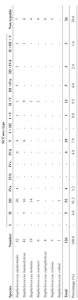
V Non-typable Staphylococcus epidermidis 52 - - 20 4 - - 6 4 - 1 1 5 3 1 7 Staphylococcus haemolyticus 41 - 5 16 - - - - 6 - - 9 - - 1 4 Staphylococcus hominis 19 - - 14 - - - - - - - 1 - - - 4 Staphylococcus warneri 6 - - 1 - - - - - - - 1 - - - 4 Staphylococcus sapr ophyticus 4 - - - - - - - - - - - - - - 4 Staphylococcus xylosus 3 - - - - - - - - - - - - - - 3 Staphylococcus cohnii 1 - - 1 - - - - - - - - - - - -Total 126 - 5 52 4 - - 6 10 - 1 12 5 3 2 26 Percentage (%) 100.0 - 4.0 41.2 3.2 - - 4.8 7.9 - 0.8 9.5 4.0 2.4 1.6 20.6 mec: Staphylococcal cassette chromosome mec ; MDR-CoNS:
multidrug-resistant coagulase-negative staphylococci.
*
SCC
mec
type was determined using multiplex PCR as described by Zhang et al.(2005).
RESULTS
Staphylococcal cassette chromosome mec
The species and SCCmec distribution among all the methicillin-resistant CoNS (MR-CoNS) isolates are presented in Table 1. The 10 methicillin-sensitive isolates were all S. epidermidis species. One hundred (79%) of the 126 methicillin-resistant isolates harbored at least one of the SCCmec types tested in this study.
Overall, MDR was seen among 106/136 (78%) isolates. There was a significant association between the presence of SCCmec and MDR (p = 0.005). However, an association between individual SCCmec types and positive MDR was signiicant only for type III (p < 0.001), and type V (p = 0.011). As the frequency of some SCCmec types were too low to perform solid statistical tests, and since we observed several combinations of SCCmec types in our isolates (Table 1), the types were divided into three categories: single, multiple, and non-typable. The association between the different categories and MDR is presented in Table 2. For CoNS as a whole and S. haemolyticus as single species, there was an association between the presence of single SCCmec and MDR, while S. epidermidis showed similar results but without signiicance. For Staphylococcus hominis and other less prevalent species, there were not enough isolates for reliable statistical calculation.
Bioilm formation
Twenty-eight (45%) of the 62 S. epidermidis isolates had an OD of ≥ 0.12 and were considered bioilm producers. Of these, 26 (93%) were icaD positive and 2 (7%) were icaD negative. On the other hand, of the 34 non-bioilm producers, 24 (71%) were icaD positive and 10 (29%) were icaD negative. The overall prevalence of the icaD gene was 81% (50/62) and that of bioilm formation was 45% (28/62). No signiicant associations were found between bioilm production or the presence of icaD and the MDR phenotype (p = 0.494 and p = 0.389, respectively).
Pulsed ield gel electrophoresis
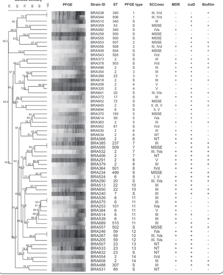
PFGE analysis of 62 S. epidermidis isolates clustered 40 (64%) isolates in 14 groups. The remaining 22 (36%) isolates were considered sporadic strains (Figure 1). The largest cluster (PFGE type 11; n = 7) showed a positive association with MDR (p = 0.037); 4 of these 7 isolates harbored SCCmec type III and were identiied as ST6. The second largest cluster (PFGE type 7; n = 4) showed a positive association between bioilm formation and the presence of icaD (both p = 0.034). For the remaining PFGE types, there was no difference between the clustered and sporadic strains with regard to MDR (p = 0.055), bioilm formation (p = 0.223), or the presence of icaD (p = 0.741). PFGE types 5, 6, 8, and 12 were triplets, and the remaining 8 types found (1, 2, 3, 4, 9, 10, 13, 14) comprised only 2 isolates. Two pairs of isolates showed identical banding patterns (isolates BRA526/575 and BRA246/267).
Multilocus sequence typing
FIGURE 1 - Dendrogram of the PFGE proiles and main characteristics of the 62 Staphylococcus epidermidis isolates from Southern Brazil. Numbers
in the horizontal upper bar and connection lines indicate similarity (percent). Isolates showing similarity ≥ 79% were considered genetically related. PFGE: pulsed-ield gel electrophoresis; ID: identiication; ST: sporadic strain; SCCmec: staphylococcal cassette chromosome mec; MDR: multidrug-resistant; IcaD: icaD gene; NT: non-typable; MSSE: methicillin susceptible Staphylococcus epidermidis.
Strain ID ST PFGE type SCCmec MDR icaD Bioilm
BRA538 340 1 III, IVd - - +
BRA544 506 1 III, IVd - +
-BRA510 340 S III - +
-BRA359 53 S MSSE - + +
BRA582 340 S IVa + -
-BRA258 500 S MSSE - -
-BRA555 500 S MSSE - +
-BRA553 507 2 MSSE - + +
BRA556 508 2 III, IVd + + +
BRA509 504 S MSSE - + +
BRA543 505 S IVd - -
-BRA373 2 S III + + +
BRA378 503 S IVd + + +
BRA496 2 S III + + +
BRA354 2 3 III + +
-BRA389 23 3 V + +
-BRA618 2 S III + + +
BRA208 2 4 V - -
-BRA320 2 4 V - +
-BRA641 20 S III, IVa - + +
BRA372 17 S III + +
-BRA602 72 S MSSE - + +
BRA645 2 S II, III, V + + +
BRA694 6 S II, V - +
-BRA370 193 5 MSSE - +
-BRA614 59 5 IVa - +
-BRA363 2 5 III + + +
BRA542 81 S IVd - -
-BRA530 2 6 III + +
-BRA634 2 6 NT + +
-BRA566 2 6 NT + +
-BRA385 237 7 III + + +
BRA595 509 7 MSSE - + +
BRA532 2 7 III, IVa + + +
BRA406 2 7 NT + + +
BRA291 2 8 V + + +
BRA379 2 8 III + + +
BRA364 501 8 IVd + +
-BRA234 499 9 MSSE - + +
BRA524 6 9 I, V + + +
BRA290 20 S III, IVa + + +
BRA513 22 10 III + +
-BRA650 22 10 III + + +
BRA240 7 S III + + +
BRA526 6 11 III + -
-BRA575 6 11 III + -
-BRA253 101 11 IVa + -
-BRA384 6 11 V + -
-BRA514 6 11 III + +
-BRA539 6 11 III + - +
BRA689 510 11 III + +
-BRA557 502 S MSSE - + +
BRA246 59 12 IVa + +
-BRA267 59 12 III, IVa + +
-BRA205 59 12 III, IVa - +
-BRA507 23 13 NT + +
-BRA533 23 13 NT + +
-BRA632 23 S NT + +
-BRA554 2 14 IVd - +
-BRA608 2 14 III + + +
BRA488 307 S III - + +
BRA531 89 S NT - -
-50 60 70 80 90 100
Genetic similary
PFGE
72.7 60.7
78.3 60.0
76.2 63.7
87.0 49.2
70.0 74.1 64.2 57.8 80.0
74.6 70.2
96.0 75.5
55.2
66.7 59.3
72.7 49.0
83.3 80.7 78.7 65.4
87.0 81.7
84.0 80.5 72.4
88.9 82.3 63.1 54.0
68.3
92.3 77.0
85.7 72.0 65.9
92.0 91.4 89.6 88.8 86.4 70.8 61.6
68.374.1
58. 2 84.6 80.0 74.5 68. 2
503 499 510
501
FIGURE 2 - eBurst V3 analysis of Staphylococcus epidermidis CC2 after adding our MLST data to all the isolates available in the MLST database on
April 2016. Each ST is represented by a dot, and lines connect single locus variants. The blue dot represents the founder of CC2, and the yellow dots represent subgroup founders. Green numbers represent new STs found in this study. Red squares represent STs reported in previous studies that were also found in this study. eBurst V3: algorithm eBurst version 3; CC2: clonal complex 2; MLST: multilocus sequence typing; ST: sequence type.
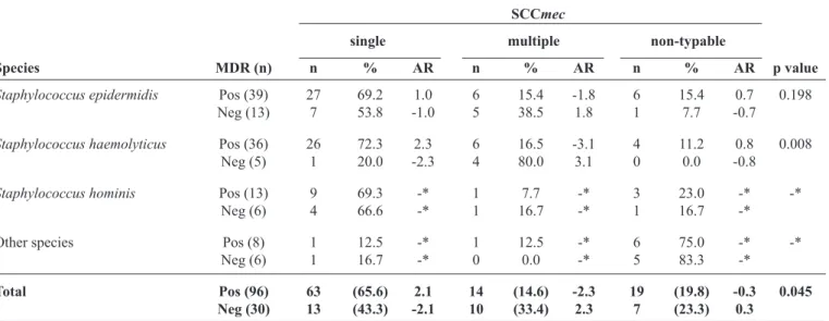
TABLE 2 - Association between MDR and SCCmec category in Brazilian CoNS nosocomial isolates from 2004.
SCCmec
single multiple non-typable
Species MDR (n) n % AR n % AR n % AR p value
Staphylococcus epidermidis Pos (39) 27 69.2 1.0 6 15.4 -1.8 6 15.4 0.7 0.198
Neg (13) 7 53.8 -1.0 5 38.5 1.8 1 7.7 -0.7
Staphylococcus haemolyticus Pos (36) 26 72.3 2.3 6 16.5 -3.1 4 11.2 0.8 0.008
Neg (5) 1 20.0 -2.3 4 80.0 3.1 0 0.0 -0.8
Staphylococcus hominis Pos (13) 9 69.3 -* 1 7.7 -* 3 23.0 -* -*
Neg (6) 4 66.6 -* 1 16.7 -* 1 16.7 -*
Other species Pos (8) 1 12.5 -* 1 12.5 -* 6 75.0 -* -*
Neg (6) 1 16.7 -* 0 0.0 -* 5 83.3 -*
Total Pos (96) 63 (65.6) 2.1 14 (14.6) -2.3 19 (19.8) -0.3 0.045 Neg (30) 13 (43.3) -2.1 10 (33.4) 2.3 7 (23.3) 0.3
MDR: multidrug-resistant;SCCmec: staphylococcal cassette chromosome mec; CoNS: coagulase-negative staphylococci; AR: adjusted
residual; Pos: positive; Neg: negative. AR value ≥ [2.0] was considered statistically signiicant; *Unreliable results due to low n.
(CC2). Fifty-two percent (32/62) of the isolates were identiied as ST2 (n = 17), ST6 (n = 7), ST59 (n = 4), and ST23 (n = 4). The majority of these most frequent STs were MDR (84%), icaD positive (87%), and bioilm negative phenotypically (59%). STs 2 and 6 were associated with SCCmec type III, and ST59 was found to be associated with SCCmec III and IVa (p < 0.05). The remaining isolates were assigned as singletons, pairs, or triplets
of different STs. The new STs were mainly positive for icaD and bioilm formation, but not for MDR (Figure 1).
DISCUSSION
We found that the types and combinations of SCCmec within the CoNS population had changed dramatically in this period. Upon comparing the results from the different periods, we observed an increase in the number of different SCCmec types and non-typable isolates and a decrease in the presence of multiple cassettes in the same isolate. Typing of the S. epidermidis isolates revealed a great genetic diversity, but some similarities in ST distribution were observed when compared with S. epidermidis isolates from other institutions in Brazil and around the world.
We conducted the same SCCmec multiplex PCR as that used in a former study(5) to enable comparisons between CoNS
isolates collected from the same hospital. Statistically signiicant differences were found in the prevalence of all the SCCmec types except for the subtype IVd. Overall, we observed a decreasing prevalence of SCCmec types II, III, and V over the years (p< 0.0001) and an increasing prevalence of SCCmec types I and IV (p < 0.0001). It is noteworthy that just one isolate harbored SCCmec type I and none of the isolates harbored types IVb and IVc (Table 1). A previous study in the same institution(5)
reported the emergence of 2 and 4 isolates harboring SCCmec types IVb and IVc, respectively, and the presence of 14 isolates harboring type I. We consider the appearance of new SCCmec types that were previously not found in this hospital especially concerning because this relects the ongoing horizontal gene transfer between species. Unfortunately, the SCCmec typing scheme used failed to classify 26 out of the 126 (20.6%) isolates, which is a large proportion of the CoNS isolates.
The acquisition and accumulation of resistance genes through mobile elements like SCCmec is enabled by integration into regions of the SCCmec element called the joining (J) regions(29).
Antibiotic resistance determinants like tet (tetracycline), aacA-aphD (aminoglycosides), or ermA (erythromycin) genes may be carried within J regions originating in MDR strains(14) (30). In our
isolates, there was a strong association between the presence of SCCmec and MDR. However, when we grouped our results to analyze the presence of more than one SCCmec type in the same isolate (multiple), we observed that the presence of multiple SCCmec types is associated with non-MDR and the presence of a single SCCmec type is associated with MDR. Interestingly, no such associations were found when the S. epidermidis isolates were analyzed separately (Table 2).
The ability to form bioilms is considered the most important pathogenic factor for S. epidermidis, and the icaD genes are recognized as one of the most important genes involved in bioilm formation(31). In our S. epidermidis isolates, despite the high
prevalence of icaD (81%), we did not ind a high prevalence of in vitro bioilm formation (45%). In addition, there was no statistical association between icaD presence and biofilm formation (data not shown). As suggested by Mertens and Ghebremedhin(32),
the natural occurrence of insertion sequence elements like IS256 might be one of the reasons for this divergence. Likewise, the presence of two isolates producing bioilms in absence of the icaD gene reinforces the fact that although icaD is important for the development of bioilm, other factors(31) and
genes like aap and bhp(30) could be involved in the process.
As reported in other studies(15) (17), analysis of our
S. epidermidis strains using two reliable typing methods revealed a high degree of genetic diversity; our isolates showed high Simpson’s indexes of diversity (SID) in PFGE (SID = 97.7%), and MLST (SID = 91.1%) (data not shown). PFGE analysis revealed several small clusters as well as a considerable number of sporadic strains, and MLST presented a range of different STs and several new alleles and STs (Figure 1
and Figure 2). The proportion of different STs observed in this
study (29 STs among 62 isolates) was higher than that previously reported by Mendes et al.(19) in 2012 (27 STs among 71 isolates)
and Miragaia et al.(17) in 2007 (74 STs among 217 isolates).
SCCmec typing also ampliied all the SCCmec types searched and some different combinations of SCCmec types in individual strains, thus conirming the high variability of SCCmec types in S. epidermidis(10) (17).Finally, despite the presence of 4 closely
related isolates revealed by PFGE, no large cluster indicating an epidemic outbreak was detected.
The only two major clusters found in our study were assigned by MLST and match two very common MLST types detected worldwide: ST2 (n = 17) and ST6 (n = 6). Furthermore, our MLST results agree with the increasing high prevalence of STs 20, 22, 23, and 89 detected in several countries(10) (17) (19) (32) (34) (35).
Interestingly, we did not ind any ST5 isolates, which is a very frequent type worldwide(17) (36), but we found two ST5 single
locus variants (ST7 and ST17) (Figure 2). However, the second most common ST worldwide (ST23)(17) was among the four
most prevalent ST in our collection. As suggested previously, the genetic diversity of S. epidermidis nosocomial isolates may be caused by the need to adapt to different environments in hospital settings, leading to increased frequency of horizontal gene transfer and dissemination of mobile genetic elements(12).
A comparison of the overall results generated by PFGE with the results of MDR, bioilm formation, and icaD presence revealed no difference between the clustered and sporadic strains. The high diversity of our isolates might be the reason for this result, since other studies have shown cluster isolates identiied by PFGE with higher rates of antibiotic resistance and bioilm formation than non-cluster isolates(18) (32). Finally, there
is currently a lack of data concerning the epidemiology of both nosocomial and community S. epidermidis isolates from Brazil. Further studies need to be conducted in order to determine if there exist two different populations in these two settings, as previously reported for S. epidermidis in Europe(33) (34).
This study has limitations. The multiplex PCR employed is easy to use, but the detection of only a single locus of each SCCmec type gives less discriminatory power to this assay when compared with that of the current recommended methodology (http://www.sccmec.org). Moreover, the isolates were not stratiied according to the place of origin or period, and it is not possible to perform comparison between hospital wards. Novel typing studies with more recent isolates from the same institution and/or from the local community could be very useful in the elucidation of several epidemiological aspects of CoNS from South Brazil.
institution. As a novelty, we observed a different behavior of S. epidermidis with regard to the association between the presence of multiple SCCmec types and a MDR proile when this specie was compared with other CoNS. In addition, we did not ind an epidemic clone of this species using well-established molecular tools. A rapid shift in the prevalence of the SCCmec types from our study and to a previous study from the same hospital(5)
performed 6-8 years after indicate a high degree of horizontal gene transfers, which conirm the hypothesis that CoNS is a permanent reservoir of genetic material that can be exchanged within and between Staphylococcal species.
Acknowledgements
The authors thanks to Mariana Preussler Mott, Bruna Gerardon Batista, Maria
Sangvik and Umaer Naseer for help provided.
Conlicts of Interest
The authors declare that there is no conlict of interest.
Financial Support
This study was supported by grants from Brazilian government by the Coordination for the Improvement of Higher Education Personnel [Coordenação de Aperfeiçoamento de Pessoal de Nível Superior (CAPES)] and from Research
Support Foundation of Rio Grande do Sul [Fundação de Amparo à Pesquisa do Estado do Rio Grande do Sul (FAPERGS)]. Furthermore, this study was also
supported by grants from the Northern Norway Regional Health Authority and the Paediatric Research Group, Tromsø, Norway.
REFERENCES
1. Rogers KL, Fey PD, Rupp ME. Coagulase-negative staphylococcal
infections. Infect Dis Clin North Am 2009; 23:73-98.
2. Pereira CAP, Marra AR, Camargo LFA, Pignatari ACC, Sukiennik
T, Behar PRP, et al. Nosocomial bloodstream infections in Brazilian pediatric patients: microbiology, epidemiology, and clinical features. PLoS ONE 2013; 8:e68144.
3. Mitt P, Metsvaht T, Adamson V, Telling K, Naaber P, Lutsar I, et al. Five-year prospective surveillance of nosocomial bloodstream
infections in an Estonian paediatric intensive care unit. J Hosp Infect 2014; 86:95-99.
4. Fredheim EGA, Klingenberg C, Rohde H, Frankenberger S,
Gaustad P, Flӕgstad T, et al.Bioilm formation by Staphylococcus haemolyticus. J Clin Microbiol 2009; 47:1172-1180.
5. Reiter KC, Paim TGS, de Oliveira CF, d’Azevedo PA. High bioilm
production by invasive multirresistant staphylococci. APMIS 2011; 119:776-781.
6. Batistão DW, Campos PA, Camilo NC, Royer S, Araujo BF, Naves
KS, et al. Bioilm formation of Brazilian MRSA strains: prevalence
of bioilm determinants and clonal proiles. J Med Microbiol 2016; doi: 10.1099/jmm.0.000228. [Epub ahead of print].
7. Méric G, Miragaia M, de Been M, Yahara K, Pascoe B, Mageiros L, et al. Ecological Overlap and Horizontal Gene Transfer in Staphylococcus aureus and Staphylococcus epidermidis. Genome
Biol Evol 2015; 7:1313-1328.
8. Ito T, Okuma K, Ma XX, Yuzawa H, Hiramatsu K. Insights on antibiotic resistance of Staphylococcus aureus from its hole
genome: genomic island SCC. Drug Resist Updat 2003; 6: 41-52.
9. Shore AC, Coleman DC. Staphylococcal cassette chromosome
mec: advances and new insights. Int J Med Microbiol 2013;
303:350-359.
10. Hanssen A, Kjeldsen G, Sollid JUE. Local variants of staphylococcal cassette chromosome mec in sporadic methicillin-resistant staphylococcus aureus andmethicillin-resistant coagulase-negative staphylococci: evidence of horizontal gene
transfer? Antimicrob Agents Chemoter 2004; 48:285-296.
11. Rolo J, de Lencastre H, Miragaia M. High frequency and diversity of cassette chromosome recombinases (ccr) in methicillin-susceptible Staphylococcus sciuri. J Antimicrob Chemother 2014; 69:1461-1469.
12. Bouchami O, Achour W, Mekni MA, Rolo J, Hassen AB. Antibiotic resistance and molecular characterization of clinical isolates of methicillin-resistant coagulase-negative staphylococci isolated
from bacteremic patients in oncohematology. Folia Microbiol 2011; 56:122-130.
13. Svensson K, Hellmark B, Söderquist B. Characterization of SCCmec elements in methicillin-resistant Staphylococcus epidermidis isolated from blood cultures from neonates during
three decades. APMIS 2011; 119:885-893.
14. Miragaia M, Carriço JA, Thomas JC, Couto I, Enright MC, Lencastre H. Comparison of molecular typing methods for characterization of Staphylococcus epidermidis: proposal for clone
deinition. J Clin Microbiol 2008; 46:118-129.
15. Rolo J, de Lencastre H, Miragaia M. Strategies of adaptation of Staphylococcus epidermidis to hospital and community:
ampliication and diversiication of SCCmec. J Antimicrob
Chemother 2013; 67:1333-1341.
16. Martins A, Riboli DFM, Camargo CH, Pereira VC, Sampaio
RA, Cunha MLRS. Antimicrobial resistance and persistence
of Staphylococcus epidermidis clones in a Brazilian university
hospital. Diagn Microbiol Infect Dis 2013; 77:164-168.
17. Miragaia M, Thomas JC, Couto I, Enright MC, Lencastre H. Inferring a population structure for Staphylococcus epidermidis from
multilocus sequence typing data. J Bacteriol 2007; 189:2540-2552.
18. Klingenberg C, Rønnestad A, Anderson AS, Abrahamsen TG, Zorman J, Villaruz A, et al. Persistent strains of coagulase-negative staphylococci in a neonatal intensive care unit: virulence
factors and invasiveness. Clin Microbiol Infect 2007; 13:1100-1111.
19. Mendes RE, Deshpande LM, Costello AJ, Farrell DJ. Molecular epidemiology of Staphylococcus epidermidis clinical isolates from
U.S. Hospitals. Antimicrob Agents Chemother 2012;
56:4656-4661.
20. Almeida LM, Lincopan N, Araújo MRE, Mamizuka EM. Dissemination of the linezolid-resistant Staphylococcus epidermidis clone ST2 exhibiting the G2576T mutation in the
23S rRNA gene in a tertiary-carehospital, Brazil. J Antimicrob Chemother 2012; 67:768-784.
21. Iorio NLP, Caboclo RF, Azevedo MB, Barcellos AG, Neves FPG,
Domingues RMCP, et al. Characteristics related to antimicrobial resistance and bioilm formation of wide spread
methicillin-resistant Staphylococcus epidermidis ST2 and ST23 lineages in
Rio de Janeiro hospitals, Brazil. Diagn Microbiol Infect Dis 2012;
72:32-40.
22. d’Azevedo PA, Siquiera I, Gugel J, Antunes ALS, Secchi C, Pasternak J, et al. Evaluation of the automated system vitek2 for
identiication and antimicrobial susceptibility testing of Grazilian Gram-positive cocci strains. Braz J Infect Dis 2009; 13:107-110.
23. Magiorakos A-P, Srinivasan A, Carey RB, Carmeli Y, Falagas
drug-resistant and pan drug-drug-resistant bacteria: an international expert
proposal for interim standard deinitions for acquired resistance. Clin Microbiol Infect 2012; 18:268-281.
24. Christensen GD, Simpson WA, Younger JJ, Baddour LM,
Barrett FF, Melton DM, et al. Adherence of coagulase-negative staphylococci to plastic tissueculture plates: a quantitative model for the adherence of staphylococci to medical devices. J Clin
Microbiol 1985; 22:996-1006.
25. Oliveira CF, Paim TGS, Reiter KC, Rieger A, d’Azevedo PA.
Evaluation of four different DNA extraction methods in coagulase-negative staphylococci clinical isolates. Rev Inst Med Trop São Paulo 2014; 56:29-33.
26. Zhang K, McClure J, Elsayed S, Louie T, Conly JM. Novel
multiplex PCR assay for characterization and concomitant
subtyping of staphylococcal cassette chromosome mec types I to V in methicillin-resistant Staphylococcus aureus. J Clin Microbiol
2005; 43:5026-5033.
27. Thomas JC, Vargas MR, Miragaia M, Peacock SJ, Archer GL,
Enright MC. Improved multilocus sequence typing scheme for
Staphylococcus epidermidis. J Clin Microbiol 2007; 45:616-619. 28. Sharpe D. Your Chi-square test is statistically signiicant: now
what? Practical Assessment, Research & Evaluation 2015; 20:1-10.
29. International Working Group on the Staphylococcal Cassette
Chromosome Elements (IWG-SCC). http://www.sccmec.org/ Pages/SCC_ClassiicationEN.html. 2016
30. Semmler T, Harrison EM, Lübke-Becker A, Ulrich RG, Wieler LH, Guenther S, et al. A look into the melting pot: the mecC-Harboring
Region is a recombination hot spot in Staphylococcus stepanovicii.
PLoS One 2016; 11:e0147150.
31. Khodaparast L, Khodaparast L, Shahrooei M, Stijlemans B,
Merckx R, Baatsen P, et al. The Possible Role of Staphylococcus epidermidis LPxTG Surface Protein SesC in Bioilm Formation. PLoS One 2016; 11: e0146704.
32. Mertens A, Ghebremedhin B. Genetic determinants and bioilm formation of clinical Staphylococcus epidermidis isolates from
blood cultures and indwelling devices. Eur J Microbiol Immunol 2013; 3:111-119.
33. Soroush S, Jabalameli F, Taherikalani M, Amirmozafari N, Imani Fooladi AA, Asadollahi K, et al. Investigation of bioilm formation ability, antimicrobial resistance and the staphylococcal cassette chromosome mec patterns of methicillin resistant Staphylococcus epidermidis with different sequence types isolated from children.
Microb Pathog 2016; 93:126-130.
34. Li M, Wang X, Gao Q, Lu Y. Molecular characterization of Staphylococcus epidermidis strains isolated from a teaching
hospital in Shanghai, China.J Med Microbiol 2009; 58:456-461.
35. Hellmark B, Söderquist B, Unemo M, Nilsdotter-Augustinsson A. Comparison of Staphylococcus epidermidis isolated from prosthetic joint infections and commensal isolates in regard
to antibiotic susceptibility, agr type, bioilm production, and epidemiology. Int J Med Microbiol 2013; 303:32-39.
36. Cherii S,Byl B, Deplano A, Nhonhoff C, Denis O, Hallind M. Comparative epidemiology of Staphylococcus epidermidis isolates from patients with catheter-related bacteremia and from healthy