ABSTRACT
Sao Paulo Med J. 2007;125(4):246-9. CONTEXT AND OBJECTIVE: Following
hematopoi-etic stem cell transplantation (HSCT), karyotyping is a valuable tool for monitoring engraftment and disease status. Few studies have examined the prognostic signifi cance of karyotypes in pa-tients who underwent HSCT for chronic myeloid leukemia (CML). The objective of this study was to evaluate the signifi cance of pretransplantation cytogenetic status in relation to outcomes follow-ing HSCT in CML patients.
DESIGN AND SETTING: Case series study at Instituto Nacional do Câncer (INCA), Rio de Janeiro, Brazil.
METHODS: Cytogenetic analysis was performed by G banding on 39 patients treated with HSCT.
RESULTS: Thirty-one patients were in the chronic phase and eight were in the accelerated phase. Prior to HSCT, additional chromosomal abnor-malities on the Philadelphia (Ph) chromosome were found in 11 patients. The most frequent additional abnormality was a double Ph, which was observed in four cases. Following HSCT, full chimeras were observed in 31 patients (79.5%). Among these, 23 (82.3%) had presented Ph as the sole abnormality. Mixed chimeras were observed in seven patients, of which three had additional abnormalities. Only one case did not present any cytogenetic response. Five patients presented cytogenetic relapse associated with clinical relapse following HSCT. Twenty-seven patients are still alive and present complete hematological and cytogenetic remission. CONCLUSION: In our study, the presence of additional abnormalities was not associated with worse outcome and relapse risk. Also, no differences in survival rates were observed. Our study supports the view that classical cytogenetic analysis remains an important tool regarding HSCT outcome.
KEY WORDS: Hematopoietic stem cell trans-plantation. Chronic myeloid leukemia. Chromo-some aberrations. Philadelphia chromoChromo-some. Prognosis.
shor
t communic
a
tio
Luize Otero
Maria Helena Ornellas
Alexandre Mello de Azevedo
Rita de Cássia Tavares
Virgínia Pires
Eliana Abdelhay
Luis Fernando Bouzas
Teresa de Souza Fernandez
Karyotype abnormalities and their
clinical signifi cance in a group of
chronic myeloid leukemia patients
treated with hematopoietic stem
cell transplantation
Cytogenetic Laboratory, Bone Marrow Transplantation Center, Instituto
Nacional do Câncer (INCA)
INTRODUCTION Chronic myeloid leukemia (CML) is a myeloproliferative disorder that is ge-netically characterized by the translocation t(9;22)(q34;q11), which results in a BCR-ABL gene fusion on the derivative chromosome 22, called the Philadelphia chromosome (Ph).1,2
Additional cytogenetic abnormalities are generally considered to be an important step in the evolution of CML from the chronic phase (CP) to the terminal blast crisis (BC). Additional chromosomal changes are detected in 70-80% of BC cases3 and in approximately
10% of Ph-positive CML in the CP at the time of the diagnosis.4 Patients with karyotypic
clonal evolution have generally been reported to have a worse clinical outcome.5
The current therapies include hemato-poietic stem cell transplantation (HSCT) and drug regimens like interferon alpha and imatinib mesylate. HSCT is associated with substantial morbidity and mortality and is limited to patients for whom a suitable donor is available.6 The results are better for
patients who are allografted in the CP than in the accelerated phase (AP) or BC.7
Following HSCT, karyotyping is a valuable tool for monitoring engraftment and disease status. However, few studies have examined the prognostic signifi cance of karyotyping fi ndings among patients who underwent HSCT for CML.
OBJECTIVE The objective of this study was to evalu-ate the signifi cance of pretransplantation cytogenetic status in relation to outcomes following HSCT.
METHODS Type of study
Type of study
This was a case series study.
Patients
Patients
We analyzed cytogenetic data from 39 patients with CML who underwent HSCT from an identical sibling (n = 35) or from unrelated volunteer donors (n = 4) between January 2000 and May 2005. Of these pa-tients, 31 (79.5%) were in the CP and eight (20.5%) were in the AP. The criteria for CP and AP were those of the International Bone Marrow Transplant Registry.8 There were
27 males and 12 females and their median age was 39 years (range: 17-57 years). All the patients received conditioning consisting of cy-clophosphamide and busulphan. Graft-versus-host disease (GVHD) prophylaxis was provided by cyclosporin and methotrexate. The median observation period was 27 months (range: 6-48 months). This study was approved by the Ethics Review Committee of Instituto Nacional do Câncer (INCA) (Protocol 58/05).
Cytogenetic studies
Cytogenetic studies
Chromosomal analysis on bone mar-row cells was carried out before HSCT and one, three and six months subsequent to HSCT, and every six months thereafter, using standard G banding.9 The
chromo-somes were classifi ed according to the Inter-national System for Human Cytogenetic No-menclature (ISCN).10 At least 20 metaphases
were analyzed per patient. The cytogenetic response after HSCT was defi ned according to the chimerism level: full chimeras (100% donor metaphases), mixed chimeras (% do-nor metaphases/% patient metaphases) and no response (100% patient metaphases).
Statistical analysis
Statistical analysis and survival
and survival
The association between the cytogenetic status prior to HSCT and the cytogenetic response was analyzed using the χ2 test.
247
Sao Paulo Med J. 2007;125(4):246-9.
curves were produced using the Kaplan-Meier method (SPSS software, SPSS Inc., Chicago, United States).
RESULTS Cytogenetic fi ndings
Cytogenetic fi ndings prior to HSCT
prior to HSCT
Additional chromosomal abnormalities were found in 11 patients (28.2%): three patients in the CP (27.3%) and eight in the AP (72.7%). The most frequent additional abnormalities prior to HSCT were a dou-ble Ph, which was observed in four cases (36.4%), and trisomy 8 in two cases (18%). The other additional chromosomal abnor-malities are described in Table 1. Patients 8 and 11 did not show the Ph chromosome, but were bcr-abl-positive according to the reverse transcription-polymerase chain reac-tion (RT-PCR).
Cytogenetic studies
Cytogenetic studies following HSCT following HSCT
Full chimerism was observed in 31 patients (79.5%). Among these, eight (25.81%) had presented additional ab-normalities prior to HSCT. In these pa-tients, karyotyping prior to HSCT showed one case of each of trisomy 8, t(6;13), t(10;15), del(3q), i(17q) and del(3p) and two cases of double Ph. Mixed chimerism was observed in seven patients, of whom three had had additional abnormalities: add(19p), i(9q)/del(22q) and double Ph.
One case (2.5%) showed no response, and this case only showed the Ph chromosome prior to HSCT. The difference between the patients with a single Ph chromosome and the patients with additional abnormalities was not statistically signifi cant (p = 0.51). In fi ve patients (12.8%), cytogenetic relapse associated with clinical relapse after HSCT was observed, and two of these patients had had additional abnormalities: double Ph and add(19)(q13). The treatment
adminis-tered in cases of relapse was transfusion of donor lymphocytes (DLI) and/or imatinib mesylate. Our results showed no differences between patients with the Ph chromosome and patients with additional chromosomal abnormalities in relation to relapse following HSCT (p = 0.91).
Survival
Survival
Twenty-seven patients are still alive and present complete hematological and
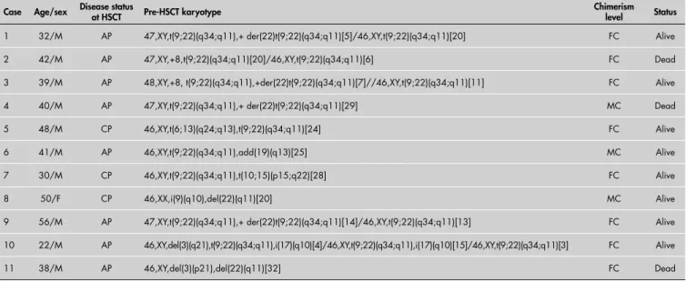
Table 1. Characteristics and responses to hematopoietic stem cell transplantation (HSCT) among the enrolled patients who had
additional chromosomal abnormalities
Case Age/sex Disease status at HSCT Pre-HSCT karyotype Chimerism level Status
1 32/M AP 47,XY,t(9;22)(q34;q11),+ der(22)t(9;22)(q34;q11)[5]/46,XY,t(9;22)(q34;q11)[20] FC Alive
2 42/M AP 47,XY,+8,t(9;22)(q34;q11)[20]/46,XY,t(9;22)(q34;q11)[6] FC Dead
3 39/M AP 48,XY,+8, t(9;22)(q34;q11),+der(22)t(9;22)(q34;q11)[7]//46,XY,t(9;22)(q34;q11)[11] FC Alive
4 40/M AP 47,XY,t(9;22)(q34;q11),+ der(22)t(9;22)(q34;q11)[29] MC Dead
5 48/M CP 46,XY,t(6;13)(q24;q13),t(9;22)(q34;q11)[24] FC Alive
6 41/M AP 46,XY,t(9;22)(q34;q11),add(19)(q13)[25] MC Alive
7 30/M CP 46,XY,t(9;22)(q34;q11),t(10;15)(p15;q22)[28] FC Alive
8 50/F CP 46,XX,i(9)(q10),del(22)(q11)[20] MC Alive
9 56/M AP 47,XY,t(9;22)(q34;q11),+ der(22)t(9;22)(q34;q11)[14]/46,XY,t(9;22)(q34;q11)[13] FC Alive
10 22/M AP 46,XY,del(3)(q21),t(9;22)(q34;q11),i(17)(q10)[4]/46,XY,t(9;22)(q34;q11),i(17)(q10)[15]/46,XY,t(9;22)(q34;q11)[3] FC Alive
11 38/M AP 46,XY,del(3)(p21),del(22)(q11)[32] FC Dead
AP = accelerated phase; CP = chronic phase; FC = full chimeras; MC = mixed chimeras; M = male; F = female.
Figure 1. Overall survival according to karyotyping prior to hematopoietic stem cell
transplantation (HSCT).
1.0
.9
.8
.7
.6
.5
.4
.3
.2
.1
.0
0 250 500 750 1000 1250 1500 1750 2000
Sur
vival
Time (days)
Only Ph chromosome Additional abnormalities
248
Sao Paulo Med J. 2007;125(4):246-9.
cytogenetic remission. Eleven patients died from GVHD and/or severe infection and one patient died from relapse with disease evolution (lymphoid blast crisis) with the karyotype complex: 45,XY,dic(3;9)(q11; q11),del(5)(q31q35),-7,t(9;22)(q34;q11),-1 7 , + 2 d e r ( 2 2 ) t ( 9 ; 2 2 ) ( q 3 4 ; q q11),del(5)(q31q35),-7,t(9;22)(q34;q11),-1 q11),del(5)(q31q35),-7,t(9;22)(q34;q11),-1 ) [ q11),del(5)(q31q35),-7,t(9;22)(q34;q11),-1 8 ] / 5,XY,t(9;22)(q34;q11),dic(17;20)(q11;q11),-18,+der(22)t(9;22)(q34;q11)[4]. Among the patients who died, three (18.2%) had additional chromosomal abnormalities. The patients with the Ph translocation alone showed survival similar to that of patients with additional abnormalities (p = 0.53) (Figure 1).
DISCUSSION Additional chromosomal abnormalities prior to therapy such as interferon alpha or imatinib mesylate are associated with poor
response and worse outcome.11,12 However,
few studies have examined the effect of pre-HSCT cytogenetics on pre-HSCT outcome. Przepiorka and Thomas13 examined 126
pa-tients in the AP or BC and found additional cytogenetic abnormalities in 84% and variant Ph in 14%. The patients with variant Ph, and those with +8 or +Ph, showed a higher risk of relapse. Slovak et al.14 examined 21
patients in the AP and found that 10 showed additional cytogenetic abnormalities. No dif-ference was found between those with and without additional abnormalities. Nevertheless, Konstantinidou et al.15 studied 418 patients
in the pre-blastic phase who had undergone HSCT and observed that patients with stand-ard Ph translocation, variant Ph translocation and negative for Ph may have different out-comes: Ph-negative patients showed a better outcome, and patients with variant Ph had
1. Shet AS, Jahagirdar BN, Verfaillie CM. Chronic myelogenous leukemia: mechanisms underlying disease progression. Leuke-mia. 2002;16(8):1402-11.
2. Johansson B, Fioretos T, Mitelman F. Cytogenetic and molecular genetic evolution of chronic myeloid leukemia. Acta Haematol. 2002;107(2):76-94.
3. Kantarjian HM, Keating MJ, Talpaz M, et al. Chronic myel-ogenous leukemia in blast crisis. Analysis of 242 patients. Am J Med. 1987;83(3):445-54.
4. Kantarjian HM, Smith TL, McCredie KB, et al. Chronic myelogenous leukemia: a multivariate analysis of the associations of patient charac-teristics and therapy with survival. Blood. 1985;66(6):1326-35. 5. Singh S, Wass J, Vincent PC, Young GA, Gunz FW. Signifi cance
of secondary cytogenetic changes in patients with Ph-positive chronic granulocytic leukemia in the acute phase. Cancer Genet Cytogenet. 1986;21(3):209-20.
6. Druker BJ, Talpaz M, Resta DJ, et al. Effi cacy and safety of a specifi c inhibitor of the BCR-ABL tyrosine kinase in chronic myeloid leukemia. N Engl J Med. 2001;344(14):1031-7.
7. Clift RA, Anasetti C. Allografting for chronic myeloid leukae-mia. Baillieres Clin Haematol. 1997;10(2):319-36. 8. Speck B, Bortin MM, Champlin R, et al. Allogeneic
bone-mar-row transplantation for chronic myelogenous leukaemia. Lancet. 1984;1(8378):665-8.
9. Seabright M. A rapid banding technique for human chromo-somes. Lancet. 1971;2(7731):971-2.
10. Mitelman F. ISCN 1995. An International System for Human Cytogenetic Nomenclature. Basel: Karger; 1995. Available from: http://www.iscn1995.org/. Accessed in 2007 (Jun 4). 11. Farag SS, Ruppert AS, Mrózek K, et al. Prognostic
sig-nifi cance of additional cytogenetic abnormalities in newly diagnosed patients with Philadelphia chromosome-positive chronic myelogenous leukemia treated with interferon-alpha: a Cancer and Leukemia Group B study. Int J Oncol. 2004;25(1):143-51.
12. Schoch C, Haferlach T, Kern W, et al. Occurrence of additional chromosome aberrations in chronic myeloid leukemia patients treated with imatinib mesylate. Leukemia. 2003;17(2):461-3.
13. Przepiorka D, Thomas ED. Prognostic signifi cance of cytoge-netic abnormalities in patients with chronic myelogenous leukemia. Bone Marrow Transplant. 1988;3(2):113-9. 14. Slovak ML, Kopecky KJ, Wolman SR, et al. Cytogenetic
correlation with disease status and treatment outcome in advanced stage leukemia post bone marrow transplantation: a Southwest Oncology Group study (SWOG-8612). Leuk Res. 1995;19(6):381-8.
15. Konstantinidou P, Szydlo RM, Chase A, Goldman JM. Cytoge-netic status pre-transplant as a predictor of outcome post bone marrow transplantation for chronic myelogenous leukaemia. Bone Marrow Transplant. 2000;25(2):143-6.
Sources of funding: None
Confl ict of interest:None
Date of fi rst submission:July 3, 2006
Last received: June 13, 2007
Accepted: June 14, 2007
REFERENCES a worse outcome than did the patients with standard Ph translocation. Patients with the additional changes of +8, +Ph and i(17q) do not necessarily show a worse outcome than do those with no additional changes, whereas those with other additional changes may fare worst of all. Although the number of cases was small, our results suggest that the presence of additional abnormalities was not associated with worse outcome and relapse risk, nor was it associated with any differences in survival rates.
249
Sao Paulo Med J. 2007;125(4):246-9.
AUTHOR INFORMATION
Luize Otero.Biologist,Bone Marrow Transplantation Center, Instituto Nacional do Câncer (INCA). Postgraduate student of School of Medical Sciences, Universidade do Estado do Rio de Janeiro (UERJ), Rio de Janeiro, Rio de Janeiro, Brazil.
Maria Helena Ornellas, MD, PhD.Hematologist and Research-er, Bone Marrow Transplantation CentResearch-er, Instituto Nacional do Câncer (INCA). Professor, Department of Pathology, School of Medical Sciences, Universidade do Estado do Rio de Janeiro (UERJ), Rio de Janeiro, Rio de Janeiro, Brazil.
Alexandre Mello de Azevedo, MD, MSc. Hematologist, Bone Marrow Transplantation Center, Instituto Nacional do Câncer (INCA),Rio de Janeiro, Rio de Janeiro, Brazil.
Rita de Cássia Tavares, MD.Hematologist,Bone Marrow Transplantation Center,Instituto Nacional do Câncer (INCA),Rio de Janeiro, Rio de Janeiro, Brazil.
Virgínia Pires, PhD. Biologist and Researcher, Bone Marrow Transplantation Center,Instituto Nacional do Câncer (INCA),Rio de Janeiro, Rio de Janeiro, Brazil.
Eliana Abdelhay, PhD. Head of Laboratory Unit, Bone Mar-row Transplantation Center, Instituto Nacional do Câncer (INCA),Rio de Janeiro, Rio de Janeiro, Brazil.
Luiz Fernando Bouzas, MD, MSc. Head of Bone Marrow Transplantation Center, Instituto Nacional do Câncer (INCA),Rio de Janeiro, Rio de Janeiro, Brazil.
Teresa de Souza Fernandez, PhD. Biologist and Researcher, Bone Marrow Transplantation Center, Instituto Nacional do Câncer (INCA), Professor, School of Medical Sciences, Universidade do Estado do Rio de Janeiro (UERJ), Rio de Janeiro, Rio de Janeiro, Brazil.
Address for correspondence:
Luize Otero
Laboratório de Citogenética — Centro de Transplante de Medula Óssea (CEMO) Instituto Nacional de Câncer (INCA)
Praça da Cruz Vermelha, 23 — 6o andar
Rio de Janeiro (RJ) — Brasil CEP 20230-130 E-mail: [email protected]
Copyright © 2007, Associação Paulista de Medicina
RESUMO Alterações cariotípicas e seu signifi cado clínico em um grupo de pacientes portadores de leucemia mielóide crônicas tratados com transplante de células tronco-hematopoéticas
CONTEXTO E OBJETIVO: Após o transplante de células tronco-hematopoéticas (TCTH), o cariótipo é uma ferramenta valiosa para monitorar o status do enxerto e da doença. Poucos estudos investigaram o signifi cado prognóstico do cariótipo nos pacientes que se submeteram ao TCTH para leucemia mielóide crônica (LMC). O objetivo desse estudo foi verifi car o signifi cado dos achados citogenéticos pré-TCTH em pacientes portadores de LMC.
TIPO DE ESTUDO E LOCAL: Série de casos. Instituto Nacional do Câncer (INCA), Rio de Janeiro, Brasil. METODOLOGIA: Foram realizados estudos citogenéticos por bandeamento G em 39 pacientes submetidos ao TCTH.
RESULTADOS: Trinta e um pacientes estavam em fase crônica e oito em fase acelerada. Pré-TCTH, alterações cromossômicas adicionais ao cromossomo Philadelphia (Ph) foram observadas em 11 pacientes. A mais freqüente foi o duplo Ph observado em quatro casos. Após o TCTH, quimerismo total foi observado em 31 pacientes (79,5%). Desses, 23 (82,3%) apresentavam somente o cromossomo Ph. Quimerismo misto foi observado em sete pacientes, sendo três com alterações adicionais ao Ph. Um caso não apresentou resposta ao TCTH. Recaída citogenética associada com recaída clínica foi observada em cinco pacientes. Após o TCTH, 27 pacientes permanecem vivos e com remissão clínica e citogenética.
CONCLUSÃO: Em nosso estudo a presença de alterações cromossômicas adicionais ao Ph, prévias ao TCTH, não foi associada com pior evolução, com risco de recaída, bem como não foi observada diferença entre as taxas de sobrevida. Nosso estudo sugere que a citogenética clássica permanece uma grande ferramenta no monitoramento do TCTH.