Selenium deficiency and the effects of supplementation on preterm infants
Deficiência de selênio e os efeitos da suplementação em prematurosDeficiencia de selenio y los efectos de la suplementación en prematuros
Renata Germano B. O. N. Freitas1, Roberto José N. Nogueira2, Maria Ângela R. G. M. Antonio1, Antonio de Azevedo Barros-Filho1,
Gabriel Hessel1
Instituição: Universidade Estadual de Campinas (Unicamp), Campinas, SP, Brasil
1Faculdade de Ciências Médicas da Unicamp, Campinas, SP, Brasil 2Hospital de Clínicas da Unicamp, Campinas, SP, Brasil.
ABSTRACT
Objective: This study aimed to review the literature
about blood concentrations of selenium associated with ges-tational age, feeding, supplementation and related clinical features in preterm infants.
Data sources: Systematic review in the following
da-tabases: MEDLINE, PubMed, Google academics, SciELO. org, ScienceDirect (Elsevier) and CINAHL-Plus with Full Text (EBSCO). Articles published up to January 2013 with the keywords “selenium deficiency”, “selenium supple-mentation”, “neonates”, “infants”, “newborn” and “preterm infants” were selected.
Data synthesis: The studies reported that low blood
selenium levels are associated with increased risk of respi-ratory diseases. Preterm infants, especially with low birth weight, presented lower selenium levels. Selenium deficiency has also been associated with the use of oral infant formula, enteral and parenteral nutrition (with or without selenium addition). The optimal dose and length of selenium supple-mentation is not well-established, since they are based only on age group and selenium ingestion by breastfed children. Furthermore, the clinical status of the infant affected by con-ditions that may increase oxidative stress, and consequently, selenium requirements is not taken into account.
Conclusions: Prematurity and low birth weight can
contribute to low blood selenium in premature infants. Selenium supplementation seems to minimize or prevent clinical complications caused by prematurity.
Key-words:review; selenium; supplementation; infant,
newborn; infant, premature.
RESUMO
Objetivo: Revisar os trabalhos que analisaram as
concentra-ções sanguíneas de selênio associadas à idade gestacional, à ali-mentação, à suplementação e ao quadro clínico de prematuros.
Fontes de dados: Revisão sistemática da literatura por
meio de buscas eletrônicas nas seguintes bases de dados: MEDLINE PubMed, Google acadêmico, SciELO.org, Scien-ceDirect (Elsevier) e CINAHL-Plus with Full Text (EBSCO). Buscaram-se trabalhos publicados até janeiro de 2013 com as seguintes palavras-chave: “selenium deficiency”, “selenium supple-mentation”, “neonates”, “infants”, “newborn” e “preterminfants”.
Síntese dos dados: Os estudos relataram que os baixos
níveis selênio associam-se ao risco aumentado para doenças respiratórias. Os prematuros, principalmente com baixo peso ao nascer, apresentam os menores níveis de selênio. A deficiência do mineral tem sido associada ao uso de fórmula infantil oral, nutrição enteral e parenteral (com e sem adição de selênio). A dosagem e o tempo ideal para a suplementação de selênio ainda não estão bem estabelecidos, visto que se baseiam apenas na faixa etária e na ingestão do mineral por crianças amamentadas no peito. Além disso, não se considera o quadro clínico do recém-nascido, que pode ser acometido de doenças que aumentam o estresse oxidativo e, consequen-temente, elevam as necessidades de selênio.
Conclusões: A prematuridade e o baixo peso ao nascer
podem contribuir para reduzir as concentrações sanguíneas de selênio em prematuros. A suplementação parece minimizar ou prevenir as complicações clínicas causadas pela prematuridade.
Palavras-chave: revisão; selênio; suplementação;
recém-nascido; prematuro.
Endereço para correspondência: Renata Germano B. O. N. Freitas
Rua Tessália Vieira de Camargo, 126 – Barão Geraldo CEP 13083-887 – Campinas/SP
E-mail: renatagbonfreitas@yahoo.com.br
Conflito de interesse: nada a declarar
RESUMEN
Objetivo: Revisar los trabajos que analizaron las
con-centraciones sanguíneas de selenio asociadas con la edad gestacional, alimentación, suplementación y cuadro clínico de prematuros.
Fuentes de datos: Revisión sistemática de la literatura
mediante búsquedas electrónicas en las bases de datos a continuación: Medline Pubmed, Google académico, SciELO. org, SienceDirect (Elsevier) y CINAHL with Full Text (EBSCO). La búsqueda se realizó con trabajos publicados hasta enero de 2013 con las palabras clave a continuación:
selenium deficiency, selenium supplementation, neonates, infants, newborn and preterm infants.
Síntesis de los datos: Los estudios relataron que los bajos
índices de selenio están asociados al riesgo aumentado para enfermedades respiratorias. Los prematuros, principalmente con bajo peso al nacer, presentan los menores niveles de selenio. La deficiencia de selenio viene siendo asociada al uso de fórmula infantil oral, nutrición enteral y parenteral (con y sin adición de selenio). La dosis y el tiempo ideal para la suplementación de selenio todavía no están bien establecidos, puesto que se basan solamente en la franja de edad y en la ingestión de selenio de niños amamantados al pecho. Además, no se considera el estado clínico del recién nacido, que puede ser acometido por enfermedades que aumentan el estrés oxidativo y, por consi-guiente, elevan las necesidades de selenio.
Conclusiones: La prematuridad y el bajo peso al nacer
pue-den contribuir para reducir las concentraciones sanguíneas de selenio en prematuros. La suplementación parece reducir o pre-venir las complicaciones clínicas causadas por la prematuridad.
Palabras clave: revisión sistemática; deficiencia de
selenio; suplementación de selenio; recién nacido; prematuro.
Introduction
Selenium is a trace element considered essential due to its participation in major metabolic functions(1,2), immune system, thyroid hormone metabolism(1,2), male infertility, neoplasms and cardiovascular disease(2). It also has antioxi-dant properties(1,3).
Selenium is an active-site component of glutathione peroxidase (GPx)(4). This enzyme contains four atoms of se-lenium and is responsible for nearly 30% of plasma sese-lenium levels(1,5). GPx has antioxidant function(3), thereby protecting
body cells from oxidation and reducing toxic substances caused by oxidative stress(6).
In 1979, it was discovered that selenium supplementation could prevent the appearance of Keshan disease, a cardiomy-opathy affecting children living in regions of selenium-dei-cient soil(7). In the pediatric population, selenium deiciency is most commonly found in preterm infants, associated with gestational age, feeding after birth and clinical status(8-10).
According to the National Health and Medical Research Council (NH&MRC, 2006)(11), the daily recommended oral dose of selenium is 12–15µg. For enteral nutrition, the rec-ommended dose is 1.3–3.0µg/kg/day. For parenteral nutri-tion, the European Society for Paediatric Gastroenterology, Hepatology and Nutrition (ESPGHAN, 2005)(12) has rec-ommended the administration of 2–3µg/kg/day. Currently, the American Society for Parenteral and Enteral Nutrition (ASPEN, 2012)(13) suggested an improvement of recommend-ed intake of selenium from 20–60 µg/day to 60–100µg/day for adults. With respect to pediatric patients, including neonates, the recommended dose remained 2µg/kg/day(13-16).
The addition of selenium to oral, enteral and parenteral infant formulas is not a routine practice in all countries and health care services. In Brazil, selenium is not routinely added to parenteral nutrition, despite studies reporting that selenium supplementation may prevent or correct a deiciency in this mineral(17-19).
This study aimed to review the literature about blood selenium concentrations in preterm infants associated with gestational age, feeding, supplementation and related clini-cal features.
Method
A systematic review was conducted by electronic search. Medline Pubmed, Google Scholar and Capes Platform da-tabaseswere used for a reined search in the following data-bases: SciELO.org, ScienceDirect (Elsevier) and CINAHL-Plus with Full Text (EBSCO). Searches were made of studies published up to January, 2013 with the following keywords:
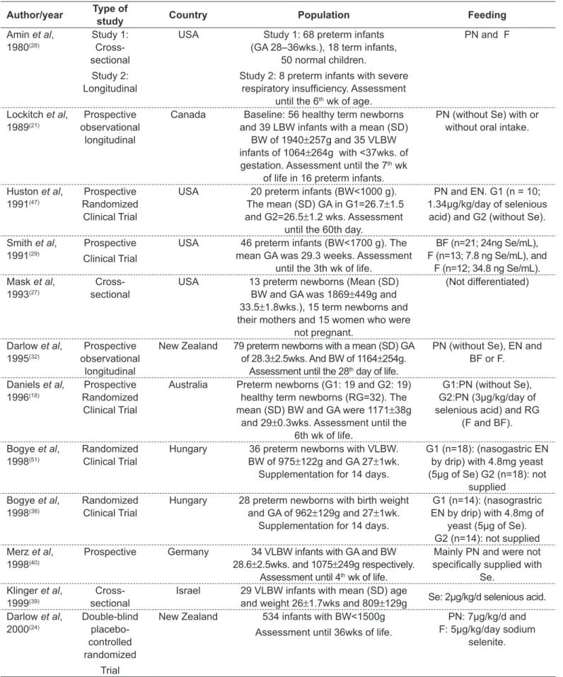
Continue... Table 1 - Type of study, location, case study, feeding form and conclusion in selected studies
Author/year Type of
study Country Population Feeding
Amin et al, 1980(28) Study 1: Cross- sectional Study 2: Longitudinal
USA Study 1: 68 preterm infants (GA 28–36wks.), 18 term infants,
50 normal children.
Study 2: 8 preterm infants with severe respiratory insuficiency. Assessment
until the 6th wk of age.
PN and F
Lockitch et al, 1989(21)
Prospective observational
longitudinal
Canada Baseline: 56 healthy term newborns and 39 LBW infants with a mean (SD)
BW of 1940±257g and 35 VLBW infants of 1064±264g with <37wks. of gestation. Assessment until the 7th wk
of life in 16 preterm infants.
PN (without Se) with or without oral intake.
Huston et al, 1991(47)
Prospective Randomized
Clinical Trial
USA 20 preterm infants (BW<1000 g). The mean (SD) GA in G1=26.7±1.5
and G2=26.5±1.2 wks. Assessment until the 60th day.
PN and EN. G1 (n = 10; 1.34µg/kg/day of selenious
acid) and G2 (without Se).
Smith et al, 1991(29)
Prospective
Clinical Trial
USA 46 preterm infants (BW<1700 g). The mean GA was 29.3 weeks. Assessment
until the 3th wk of life.
BF (n=21; 24ng Se/mL), F (n=13; 7.8 ng Se/mL), and
F (n=12; 34.8 ng Se/mL). Mask et al,
1993(27)
Cross-sectional
USA 13 preterm newborns (Mean (SD) BW and GA was 1869±449g and 33.5±1.8wks.), 15 term newborns and their mothers and 15 women who were
not pregnant.
(Not differentiated)
Darlow et al, 1995(32)
Prospective observational
longitudinal
New Zealand 79 preterm newborns with a mean (SD) GA of 28.3±2.5wks. And BW of 1164±254g.
Assessment until the 28th day of life.
PN (without Se), EN and BF or F.
Daniels et al,
1996(18)
Prospective Randomized
Clinical Trial
Australia Preterm newborns (G1: 19 and G2: 19) healthy term newborns (RG=32). The mean (SD) BW and GA were 1171±38g
and 29±0.3wks. Assessment until the 6th wk of life.
G1:PN (without Se), G2:PN (3µg/kg/day of selenious acid) and RG
(F and BF).
Bogye et al, 1998(51)
Randomized Clinical Trial
Hungary 36 preterm newborns with VLBW. BW of 975±122g and GA 27±1wk.
Supplementation for 14 days.
G1 (n=18): (nasogastric EN by drip) with 4.8mg yeast (5µg of Se) G2 (n=18): not
supplied Bogye et al,
1998(38)
Randomized Clinical Trial
Hungary 28 preterm newborns with birth weight and GA of 962±129g and 27±1wk.
Supplementation for 14 days.
G1 (n=14): (nasograstric EN by drip) with 4.8mg of
yeast (5µg of Se). G2 (n=14): not supplied Merz et al,
1998(40)
Prospective Germany 34 VLBW infants with GA and BW 28.6±2.5wks. and 1075±249g respectively.
Assessment until 4th wk of life.
Mainly PN and were not speciically supplied with
Se. Klinger et al,
1999(39)
Cross-sectional
Israel 29 VLBW infants with mean (SD) age
and weight 26±1.7wks and 809±129g Se: 2µg/kg/d selenious acid. Darlow et al,
2000(24) Double-blind placebo-controlled randomized Trial
New Zealand 534 infants with BW<1500g
Assessment until 36wks of life.
PN: 7µg/kg/d and F: 5µg/kg/day sodium
Table 1 - Continuation
One hundred and eighty-nine (189) articles were found. Of these, 18 were selected and 171 excluded (63 repeated studies, 50 animal studies, 14 review articles, 27 with inad-equate age group, 11 did not address the topic, 6 reported dead children).
Thus, based on titles and abstracts, 18 studies were chosen for this systematic review. After the selection of studies, level of evidence and grades of recommendation were classiied according to Brazilian Medical Association(20).
Results
Eighteen articles analyzing selenium concentrations in preterm infants were selected. Table 1 shows study design, population characteristics and forms of feeding. According to the criteria of the Brazilian Medical Association(20), studies were classiied as A or B. In table 2, a relationship between selenium status and age of the child is observed.
Concerning birth weight, Makhoul et al(8) observed that the lower the weight, the lower the selenium concentration (r=0.237; p=0.002). Lockitch et al(21) found a signiicant
correlation between BW and plasma Se (r=0.47; p<0.001). Plasma GPx levels were more highly correlated with birth weight (r=0.64; p<0.001). In addition, studies have related altera-tions in selenium concentration and clinical status (Table 3). Table 4 shows studies correlating the amount of selenium provided by feeding routes used with selenium concentra-tions observed in studies of children.
Discussion
It is known that the pediatric population, particularly pre-mature infants(8-10), is vulnerable to low Se concentrations due to nutritional changes(22), possible clinical complications(10,23) and low selenium liver stores(8,9,24-26). This occurs because of immature chorionic villi that acts in the transport of this min-eral and also due to inadequate intestinal absorption(8,9,25,26).
During pregnancy, maternal blood selenium levels decrease, relecting a greater amount of selenium trans-ported to the fetus in the 3rd trimester of pregnancy. Mask et al(27), Amin et al(28) and Smith et al(29) suggest that low selenium values found in preterm infants must be associated
Author/year Type of
study Country Population Feeding
Winterbourn
et al, 2000(48)
Randomized controlled
Trial
New Zealand 173 newborns with weight <1500g. Assessment until 36wks of life.
PN: 7µg/kg/d and F: 5µg/kg/day sodium
selenite. Sievers et al,
2001(45)
Prospective observational
longitudinal
Germany 16 preterm newborns (GA 25–32wk and BW 595–1495g), 14 term newborns with F and 17 term newborns with BF.
Assessment until 1st year of life.
BF, F and complementary feeding.
Makhoul et al, 2004(8)
Cross-sectional
Israel 165 preterm newborns and term newborns (24–42wks.) and their
mothers.
(Not differentiated)
Mentro et al, 2004(9)
Prospective observational
longitudinal
14 Caucasians, 3 Hispanics and 1 Asian-American
18 preterm newborns with BW of 1013g (650–1370g) and risk of BPD. Mean GA was 27 weeks. Assessment
until 4th week of life.
BF, PN, EN and F. Mean ingestion of Se in the 1st
wk was 0.82µg/kg/day and 1.7µg/kg/day in the 4th wk.
Galinier et al, 2005(30)
Cross-sectional
France 248 preterm newborns and 262 term newborns. The mean GA and BW (SD) for preterm newborns was
32.4±2.52wks. and 1845±489g.
(Not differentiated)
Nassi et al, 2009(10)
Prospective observational
longitudinal
Italy 30 preterm infants with mean (SD) BW and GA of 1605±122 g and
34.5±0.5 wk. The control group included 30 term infants. Assessment
until the 100th day of life.
BF
with selenium accumulation during gestation. This fact was observed in studies by Makhoul et al(8) and Galinier et al(30), who analyzed umbilical cord selenium concentration and noted a signiicant association with gestational age of newborn infants. Selenium concentration increased after 36 weeks in the former study and from the 26th to the 38th week in the latter study.
Mentro, Smith and Moyer-Mileur(9) suggested that pre-term infant’s small Se stores are used preferentially for GPx
production, occurring in stable or increased GPx and decreased Se concentrations. This could explain the poor correlation between selenium and GPx concentrations observed in the studies(9,18,21,24,31).Another possibility is that the natural defenses antioxidant like enzyme GPx, mature along the gestation. So, in premature animal, the GPx probably are poorly developed(32).
According to Daniels, Gibson and Simmer(18) and Mentro, Smith and Moyer-Mileur(9), GPx concentrations might be
Table 2 - Main results found in publications about the relationship between alterations in selenium status and age
Author Results
Amin et al, 1980(28) The mean serum concentration in term infants (0.098±0.025μg/mL) was slightly higher
than preterm infants (0.032 μg/ml), but there was not difference signiicant. Lockitch et al, 1989(21) The mean concentration decreased from 0.74±0.13 to 0.63±0.15μmol/L at day 7
(p=0.01) and at day 14 decreased to 0.51±0.19μmol/L (p<0.001). Se values decreased in all 16 preterm infants followed over the irst 50 days. In 11 infants, levels dropped to <0.22μmol/L (17μ/L).
Smith et al,1991(29) After 3th week there were no signiicant differences of Se concentration between groups
(preterm infants with F and BF).
Mask et al, 1993(27) The plasma Se was lower in preterm newborns (0.08±0.02μg/mL) than term newborns
(0.10±0.02μg/ml), p=0.052.
Darlow et al, 1995(32) There was no signiicant correlation between gestational period and plasma Se. The
correlations among GPx and plasma Se was weak at birth (0.39) and at 28 days (0.17).
Merz et al, 1998(40) After birth the value of plasma Se was 34.2μg/L and reduced to 16.1μg/L after 4 weeks
(p<0.001).
Klinger et al, 1999(39) No correlation was observed between the plasma Se and gestational age (r=0.27,
p=0.16). There was signiicant correlation between gestational age and the level of T4
(r=0.45; p=0.02).
Winterbourn et al, 2000(48) There was no statistically signiicant difference in GA between the group with and without Se.
Sievers et al, 2001(45) Plasma Se concentrations in preterm newborns were 11.7 (6.5–20.8)μg/L (assessment in
the hospital). At 4 months: preterm newborns = 11.6 (8.8–16.7)μg/L, term newborns fed with IF=31.3 (24.3–47.5)μg/L and term newborns BF=45.6 (27.1–65.1)μg/L.
Makhoul et al, 2004(8) Linear relationship between umbilical cord blood concentrations and GA (r =0.341,
p<0.0001).
Mentro, Smith, Moyer-Mileur, 2004(9)
Se concentrations decreased from the 1st to 4th wk of life. Plasma Se (SD)=0.97±0.21μmol/L
in the 1st wk and 0.72±0.27 μmol/L in the 4th wk (p=0.001). There was not change in plasma
GPx among week 1 and 4. The erythrocyte GPx increased along the time period (t =-3.38;
p=0.004) and was associated to the GA.
Galinier et al, 2005(30) Se concentration increased with GA from 0.4±0.1μmol/L (26th to 33rd week) to
0.5±0.1μmol/L (from the 33rd to the 37th week) and 0.6±0.1μmol/L (>37th weeks) (p<0.001,
r=0.593).
Nassi et al, 2009(10) Up until 20 days postnatal, the GPx was lower in the preterm infants than in the
term infants.
confounded by supplemental oxygen and steroids which are common practice for preterm infants. Thus according to au-thors, GPx activity may be a poor functional indicator of seleni-um status in infants(10,31-33), especially in preterm infants(8,9,24,32). But GPx, seems to be a good marker for adults(8,18,32).
Prematurity also affects birth weight(34). Birth weight is the anthropometric indicator that has the greatest inluence on health and newborn survival(33,35-37). Makhoul et al(8) and Lockitch et al(21) observed that the lower the weight, the lower the concentration of selenium in newborns. Bogye, Alfthan and Machay(38) stated that very low birth weight premature infants are obviously susceptible to selenium deiciency.
Selenium deiciency has also been associated with a greater number of diseases and clinical complications. Klinger
et al(39) found selenium deiciency in most premature infants however, there was not a signiicant correlation between selenium levels and thyroid hormones.
Merz et al(40) found no relationship between the incidence of bronchopulmonary dysplasia and selenium status. Darlow
et al(24) suggested that low Se concentrations may be associ-ated an increase in risk to lung injury.
Darlow et al(32) were the irst to demonstrate in humans an association between low plasma selenium levels and a greater risk of lung disease, evidenced by oxygen require-ment and dependency in the 28th day of life of the affected patients. Mentro, Smith and Moyer-Mileur(9) showed that, despite a reduction in selenium plasma levels, increased selenium ingestion was associated with a reduction in oxy-gen dependency. In fact, selenium supplementation would act against oxidative stress caused by early exposure to an oxygen-rich environment, in addition to supplemental oxygen provided in some cases.
Daniels, Gibson and Simmerb(18) found no signiicant difference in the incidence of retinopathy of prematurity
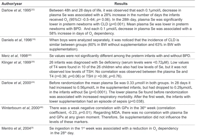
Table 3 - Main results found in publications about alterations in selenium concentration and clinical status
Author/year Results
Darlow et al, 1995(32) Between 48h and 28 days of life, it was observed that each 0.1µmol/L decrease in
plasma Se was associated with a 28% increase in the number of days the infants received O2 (95%CI -0.5–64; p= 0.06). In the 28th day, plasma Se was signiicantly
lower in preterm newborns with CLD (p<0.001). Mean plasma Se was lower in preterm newborns with BPD. And each 0.1 µmol/L decrease in plasma Se was associated with a 58% increase in days of O2 dependency.
Daniels et al, 1996(18) When boys were analyzed separately, it was noticed that the incidence of CLD is
similar between groups (60% in BW without supplementation and 63% in BW with supplementation).
Merz et al, 1998(40) Se values were not signiicantly different among the preterm infants with and without BPD.
Klinger et al, 1999(39) 26 infants was diagnosed with Se deiciency (serum levels were <0.72µM). Low values
of T4 were found in 10 of the 26 children who also had low levels of Se, but it was not observed low levels of TSH. No correlation was observed between the plasma Se and T4 (r=0.36; p=0.06) or TSH (r =0.06; p=0.76).
Darlow et al, 2000(24) Before randomization the mean plasma Se was 0.33 μmol/l in both groups. In 28 days it
had increased to 0.56μmol/L in the supplemented infants, but had dropped to 0.29μmol/L in the infants without Se (p<0.0001). The lower plasma Se found before randomization was associated with increased respiratory morbidity. After the irst week, the infants with lower supplementation had an episode of sepsis (p<0.038).
Winterbourn et al, 2000(48) There was a weak negative correlation with GPx in the 36th week (correlation
coeficient, -0.23, p=0.01). Regarding MDA, there was no correlation with plasma Se and GPx at any given moment. Therefore, Se supplementation did not inluence the levels of these markers.
Mentro et al, 2004(9) Se ingestion in the 1st week was associated with a reduction in O
2 dependency
in the 28th day.
and intraventricular hemorrhage, but observed a higher in-cidence of sepsis among premature infants without selenium supplementation.
Thus, feeding newborns with an adequate amount of sele-nium is important to restore and to maintain selesele-nium liver stores(9), preventing a number of disorders and complications,
as well as to support the appropriate growth and develop-ment of newborn infants.
Daniels et al(31) suggest that supplementation should be at least equivalent to the amount of selenium in breast milk of women from the same geographical region; after all, there are some regions where soils are low in selenium,
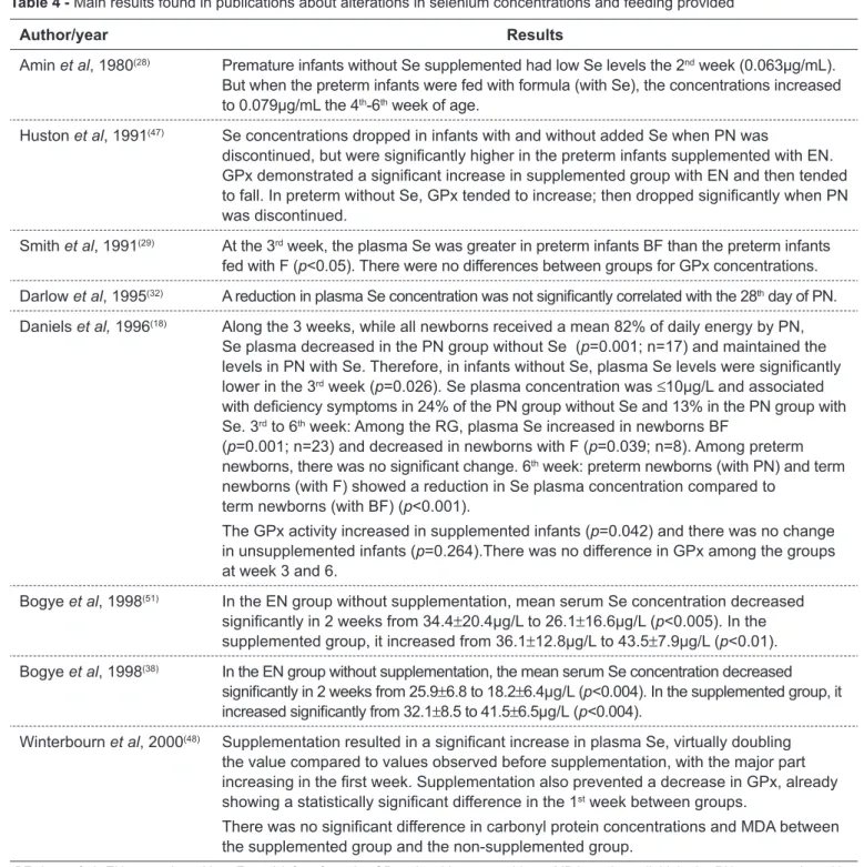
Table 4 - Main results found in publications about alterations in selenium concentrations and feeding provided
Author/year Results
Amin et al, 1980(28) Premature infants without Se supplemented had low Se levels the 2nd week (0.063μg/mL).
But when the preterm infants were fed with formula (with Se), the concentrations increased to 0.079μg/mL the 4th-6th week of age.
Huston et al, 1991(47) Se concentrations dropped in infants with and without added Se when PN was
discontinued, but were signiicantly higher in the preterm infants supplemented with EN. GPx demonstrated a signiicant increase in supplemented group with EN and then tended to fall. In preterm without Se, GPx tended to increase; then dropped signiicantly when PN was discontinued.
Smith et al, 1991(29) At the 3rd week, the plasma Se was greater in preterm infants BF than the preterm infants
fed with F (p<0.05). There were no differences between groups for GPx concentrations. Darlow et al, 1995(32) A reduction in plasma Se concentration was not signiicantly correlated with the 28th day of PN.
Daniels et al, 1996(18) Along the 3 weeks, while all newborns received a mean 82% of daily energy by PN,
Se plasma decreased in the PN group without Se (p=0.001; n=17) and maintained the levels in PN with Se. Therefore, in infants without Se, plasma Se levels were signiicantly lower in the 3rd week (p=0.026). Se plasma concentration was ≤10μg/L and associated
with deiciency symptoms in 24% of the PN group without Se and 13% in the PN group with Se. 3rd to 6th week: Among the RG, plasma Se increased in newborns BF
(p=0.001; n=23) and decreased in newborns with F (p=0.039; n=8). Among preterm newborns, there was no signiicant change. 6th week: preterm newborns (with PN) and term
newborns (with F) showed a reduction in Se plasma concentration compared to term newborns (with BF) (p<0.001).
The GPx activity increased in supplemented infants (p=0.042) and there was no change in unsupplemented infants (p=0.264).There was no difference in GPx among the groups at week 3 and 6.
Bogye et al, 1998(51) In the EN group without supplementation, mean serum Se concentration decreased
signiicantly in 2 weeks from 34.4±20.4μg/L to 26.1±16.6μg/L (p<0.005). In the supplemented group, it increased from 36.1±12.8μg/L to 43.5±7.9μg/L (p<0.01). Bogye et al, 1998(38) In the EN group without supplementation, the mean serum Se concentration decreased
signiicantly in 2 weeks from 25.9±6.8 to 18.2±6.4μg/L (p<0.004). In the supplemented group, it increased signiicantly from 32.1±8.5 to 41.5±6.5μg/L (p<0.004).
Winterbourn et al, 2000(48) Supplementation resulted in a signiicant increase in plasma Se, virtually doubling
the value compared to values observed before supplementation, with the major part increasing in the irst week. Supplementation also prevented a decrease in GPx, already showing a statistically signiicant difference in the 1st week between groups.
There was no signiicant difference in carbonyl protein concentrations and MDA between the supplemented group and the non-supplemented group.
as in New Zealand(32), Switzerland(41), China(42) and some states of Brazil(43).
Makhoul et al(8) stated that infants fed with maternal milk, regardless of being premature or not, do not require selenium supplementation. However, when feeding of these infants is based on infant formula, enteral or parenteral nutrition, supplementation is necessary even in term newborn infants. Most publications studied in this review showed an as-sociation between feeding provided to infants — infant formulas administered by oral, enteral or parenteral route containing little or no addition of selenium — and low selenium concentrations(9,18,23,28,31,32,44).
The current recommendations for selenium supple-ments are based on the ingestion of selenium by infants fed with maternal milk, since it appears to meet newborn requirements(15,31,33).
Currently, ASPEN (2012)(13) recommends 2μg/kg/day selenium in parenteral nutrition for the pediatric popula-tion. There is no differentiation between preterm and term neonates, healthy and sick neonates, and between neonates with appropriate or low birth weight for gestational age.
Surveys claim that selenium concentrations are lower in preterm infants, especially in those with low birth weight (˂1,500 g) and very low birth weight (˂1,000g), when compared with term infants(22,37,38,45,46).
Daniels, Gibson and Simmer(18), studying preterm infants receiving parenteral nutrition, found selenium levels similar to those observed in children with Keshan disease. Huston, Jelen and Vidgoff(47) concluded that adding 1.34 µg/kg/day of selenium in PN is not adequate for LBW. Those authors ad-ditionally suggested that supplementation with 3µg/kg/day of selenious acid was incapable of preventing signiicant decreases in plasma selenium concentration when compared with term newborn infants fed breast milk(18). This fact is concerning, since, according to the literature, supplementa-tion may revert several clinical complicasupplementa-tions, although it is not eficient for reverting Keshan disease.
Klinger et al(39) reported that supplementation of 2µg/kg/day of selenium has not been able to prevent or reverse selenium deiciency. Thus, the authors support the recommendation to review premature infants guidelines. Makhoul et al(8) suggested that measurement of selenium levels recommended in parenteral nutrition should increase twofold (up to 7µg/kg/day).
In a research conducted by Darlow(24), supplementa-tion prevented the fall and achieved levels similar to those
reported in term infants fed human milk. So, the authors suggest that VLBW infants should receive suficient supple-mentation to achieve levels observed in term infants fed human milk, despite the minimal beneits in the clinical picture found in research.
In a study by Winterbourn et al(48), supplementation (7µg/kg/day and 5µg/kg/day of sodium selenite in parenteral and oral nutrition, respectively) did not have an effect on oxidative stress, although selenium levels almost doubled and GPx showed a signiicant difference between groups with and without supplementation. This fact may be explained by the inadequate dose of selenium, the late supplementa-tion, and also due to the scant evidence of oxidative stress among premature infants.
Despite the discussion about the optimal dose and length of selenium supplementation, several studies have shown that the addition of selenium may prevent diseases and their complications(6,17,49,51), including a shortened hospital stay and, consequently, lower inancial costs.
Conclusion
Nutritional assessment of selenium status in the body to analyze biochemical indicators and clinical manifestations should be performed, especially in premature newborns who were not breastfed. Blood selenium concentrations are reduced in neonates, especially in those with lower gestational age and birth weight. Furthermore, newborn infants who are not breastfed and supplemented show the lowest selenium levels, including newborns, without any underlying disease. Therefore, supplementation is impor-tant in preterm infants who were not breastfed in order to minimize the risks of diseases and complications associated with selenium deiciency, contributing to a healthy growth and development of the child.
References
1. Porras IC, Muriel AC, Morales BO, Pozo JF, Aranda JG, Pérez L. Evaluación de nutrición parenteral estandarizada en niños. Nutr Hosp 2010;25:449-55. 2. Uslu N, Saltik-Temizel IN, Demir H, Gürakan F, Özen H, Yüce A. Serum
selenium concentrations in cirrhotic children. Turk J Gastroenterol 2010;21:153-5.
3. Cominetti C, de Bortoli MC, Purgatto E, Ong TP, Moreno FS, Garrido Jr AB et al. Associations between glutathione peroxidase-1 Pro198Leu polymorphism, selenium status, and DNA damage levels in obese women after consumption of Brazil nuts. Nutrition 2011;27:891-6.
4. Ashton K, Hooper L, Harvey LJ, Hurst R, Casgrain A, Fairweather-Tait SJ. Methods of assessment of selenium status in humans: a systematic review. Am J Clin Nutr 2009;89:2025S-39S.
5. Harrison I, Littlejohn D, Fell GS. Distribution of selenium in human blood plasma and serum. Analyst 1996;121:189-94.
6. Nogueira RJ, Lima AE, Prado CC, Ribeiro AF. Nutrição em pediatria oral, enteral e parenteral. São Paulo: Sarvier; 2011.
7. Keshan Disease Research Group. Observations on effect of sodium selenite in prevention of Keshan disease. Chin Med J (Engl) 1979;92:471-6. 8. Makhoul IR, Sammour RN, Diamond E, Shohat I, Tamir A, Shamir R.
Selenium concentrations in maternal and umbilical cord blood at 24-42 weeks of gestation: basis for optimization of selenium supplementation to premature infants. Clin Nutr 2004;23:373-81.
9. Mentro AM, Smith AM, Moyer-Mileur L. Plasma and erythrocyte selenium and glutathione peroxidase activity in preterm infants at risk for bronchopulmonary dysplasia. Biol Trace Elem Res 2005;106:97-106.
10. Nassi N, Ponziani V, Becatti M, Galvan P, Donzelli G. Anti-oxidant enzymes and related elements in term and preterm newborns. Pediatr Int 2009;51:183-7.
11. National Health and Medical Research Council. Nutrient Reference Values for Australia and New Zealand. Canberra: Commonwealth of Australia; 2006.
12. Koletzko B, Goulet O, Hunt J, Krohn K, Shamir R. Guidelines on Paediatric Parenteral Nutrition of the European Society of Paediatric Gastroenterology, Hepatology and Nutrition (ESPGHAN) and the European Society for Clinical Nutrition and Metabolism (ESPEN), Supported by the European Society of Paediatric Research (ESPR). J Pediatr Gastroenterol Nutr 2005;41 (Suppl 2):S1-87.
13. Vanek VW, Borum P, Buchman A, Fessler TA, Howard L, Jeejeebhoy K et al. A.S.P.E.N. position paper: recommendations for changes in commercially available parenteral multivitamin and multi-trace element products. Nutr Clin Pract 2012;27:440-91.
14. Canada T, Crill C, Guenter P. A.S.P.E.N. Parenteral nutrition handbook. Silver Spring: American Society Parenteral for Parenteral and Enteral Nutrition; 2009.
15. Forchielli ML. Pediatric nutrition in your pocket. Silver Spring: American Society for Parenteral and Enteral Nutrition; 2002.
16. Rayman JM. The importance of selenium to human health. Lancet 2000;356:233-41.
17. Dylewski ML, Bender JC, Smith AM, Prelack K, Lydon M, Weber J et al. The selenium status of pediatric patients with burn injuries. J Trauma 2010;69:584-88.
18. Daniels L, Gibson R, Simmer K. Randomised clinical trial of parenteral selenium supplementation in preterm infants. Arch Dis Child Fetal Neonatal Ed 1996;74:F158-64.
19. Kien CL, Ganther HE. Manifestations of chronic selenium deiciency in a child receiving total parenteral nutrition. Am J Clin Nutr 1983;37:319-28. 20. Associação Médica Brasileira, Conselho Federal de Medicina [homepage
on the Internet]. Projeto Diretrizes - Associação Médica Brasileira e Conselho Federal de Medicina [cited 2013 Jul 10]. Available from: http:// www.portalmedico.org.br/diretrizes/100_diretrizes/Texto_Introdutorio.pdf 21. Lockitch G, Jacobson B, Quigley G, Dison P, Pendray M. Selenium
deiciency in low birth weight neonates: an unrecognized problem. J Pediatr
1989;114:865-70.
22. Trindade CE. Importância dos minerais na alimentação do pré-termo extremo. J Pediatr (Rio J) 2005;81 (Suppl 1):s43-51.
23. Muntau AC, Streiter M, Kappler M, Röschinger W, Schmid I, Rehnert A et al. Age-related reference values for serum selenium concentrations in infants and children. Clin Chem 2002;48:555-60.
24. Darlow BA, Winterbourn CC, Inder TE, Graham PJ, Harding JE, Weston PJ
et al. The effect of selenium supplementation on outcome in very low birth weight infants: a randomized controlled trial. J Pediatr 2000;136:473-80. 25. Friel JK, Andrews WL, Long DR, L’Abbe MR. Selenium status of very low
birth weight infants. Pediatr Res 1993;34:293-6.
26. Hambidge KM. Trace element requirements in the premature infants. In: Lebenthal E, editor. Textbook of gastroenterology and nutrition in infancy. 2nd ed. New York: Raven Press; 1992. p. 393-401.
27. Mask GR, Lane HW. Selected measures of selenium status in full-term and preterm neonates, their mothers and nonpregnant women. Nutr Res 1993;13:901-11.
28. Amin S, Chen SY, Collipp PJ, Castro-Magana M, Maddaiah VT, Klein SW. Selenium in premature infants. Nutr Metab 1980;24:331-40.
29. Smith AM, Chan GM, Moyer-Mileur LJ, Johnson CE, Gardner BR. Selenium status of preterm infants fed human milk, preterm formula, or selenium-supplemented preterm formula. J Pediatr 1991;119:429-33.
30. Galinier A, Périquet B, Lambert W, Garcia J, Assouline C, Rolland M et al. Reference range for micronutrients and nutritional marker proteins in cord blood of neonates appropriated for gestational ages. Early Hum Dev 2005;81:583-93.
31. Daniels L, Gibson RA, Simmer K, Van Dael P, Makrides M. Selenium status of term infants fed selenium-supplemented formula in a randomized dose-response trial. Am J Clin Nutr 2008;88:70-6.
32. Darlow BA, Inder TE, Graham PJ, Sluis KB, Malpas TJ, Taylor BJ et al. The relationship of selenium status to respiratory outcome in the very low birth weight infant. Pediatrics 1995;96:314-9.
33. Food and Nutrition Board, Institute of Medicine. Dietary reference intakes for vitamin C, vitamin E, selenium and carotenoids. Washington, DC: National Academy Press; 2002.
34. Monteiro CA, Benicio MH, Ortiz LP. Tendência secular do peso ao nascer na cidade de São Paulo (1984-1996). Rev Saude Publica 2000;34 (Suppl 6):26-40.
35. Kilsztajn S, Rossbach A, Nunes do Carmo MS, Sugahara GT. Assistência pré-natal, baixo peso e prematuridade no Estado de São Paulo, 2000. Rev Saude Publica 2003;37:303-10.
36. Nascimento LF, Gotlieb SL. Fatores de risco para o baixo peso ao nascer, com base em informações da declaração de nascido vivo em Guaratinguetá, SP, 1998. Inf Epidemiol SUS 2001;10:113-20.
37. Cormick MC. The contribution of low birth weight to infant mortality and childhood morbidity. N Engl J Med 1985;312:82-90.
38. Bogye G, Alfthan G, Machay T. Randomized clinical trial of enteral yeast-selenium supplementation in preterm infants. Biofactors 1998;8:139-42. 39. Klinger G, Shamir R, Singer P, Diamond EM, Josefsberg Z, Sirota L.
Parenteral selenium supplementation in extremely low birth weight infants: inadequate dosage but no correlation with hypothyroidism. J Perinatol 1999;19:568-72.
40. Merz U, Peschgens T, Dott W, Hörnchen H. Selenium status and bronchopulmonary dysplasia in premature infants <1,500 g. Z Geburtshilfe Neonatol 1998;202:203-6.
41. Eichholzer M. Micronutrient deficiencies in Switzerland: causes and consequences. J Food Eng 2003;56:171-9.
42. Xia Y, Hill KE, Burk RF. Biochemical studies of a selenium-deficient population in China: measurement of selenium, glutathione peroxidase and other oxidant defense indices in blood. J Nutr 1989;119:1318-26. 43. Moraes MF, Welch RM, Nutti MR, Carvalho JL, Watanabe E. Evidences
of selenium deiciency in Brazil: from soil to human nutrition. Proceedings
44. Andrews PJ, Avenell A, Noble DW, Campbell MK, Battison CG, Croal BL et al. Randomised trial of glutamine and selenium supplemented parenteral nutrition for critically ill patients. Protocol Version 9, 19 February 2007 known as SIGNET (Scottish Intensive care Glutamine or seleNium Evaluation Trial). Trials 2007;8:25.
45. Sievers E, Arpe T, Schleyerbach U, Garbe-Schönberg D, Schaub J. Plasma
selenium in preterm and term infants during the irst 12 months of life. J
Trace Elements Med Biol 2001;14:218-22.
46. Klein CJ. Nutrient requirements for preterm infant formulas. Minerals: calcium and phosphorus. J Nutr 2002;132:1395S-577S.
47. Huston RK, Jelen BJ, Vidgoff J. Selenium supplementation in low-birthweight premature infants: relationship to trace metals and antioxidant enzymes. JPEN J Parenter Enteral Nutr 1991;15:556-9.
48. Winterbourn CC, Chan T, Buss IH, Inder TE, Mogridge N, Darlow BA. Protein carbonyls and lipid peroxidation products as oxidation markers in preterm infant plasma: associations with chronic lung disease and retinopathy and effects of selenium supplementation. Pediatr Res 2000;48:84-90. 49. Forceville LX. Selenium and the “free” electron. Selenium – a trace to
be followed in septic or inlammatory ICU patients? Intensive Care Med
2001;27:16-8.
50. Heyland DK, Dhaliwal R, Suchner U, Berger MM. Antioxidant nutrients: a systematic review of trace elements and vitamins in the critically ill patient. Intensive Care Med 2005;31:327-37.