ABSTRACT
BACKGROUND AND OBJECTIVES: Craniomandibular
and cervical symptoms interfere with mandibular stability. De-termining whether these disorders inluence muscle activation when chewing, it is possible to improve interventions for this population. he objective of this study was to verify the efect of the temporomandibular joint dysfunction, craniomandibular and cervical pain on the electromyographic activity of the masti-catory muscles during rest and mandibular isometry.
METHODS: Fifty-ive women aged between 18 and 30 years were divided into two groups: with temporomandibular dysfunction (n=28) and without temporomandibular dysfunction (n=27). he diagnosis of temporomandibular dysfunction was established using the Research Diagnostic Criteria for Temporomandibular Disorder (RDC/TMD). he RDC was also used to determine the presence of craniomandibular pain. Cervical pain was deined by physical examination. he electromyographic activity of masseter and tem-poralis muscles was evaluated in the rest position and mandibular isometry. he amplitude of muscle activation was represented by the root mean square values (RMS%) and normalized by maximum voluntary contraction. he Mann-Whitney U test was used to de-tect diferences between the groups with and without temporoman-dibular dysfunction; with and without myofascial craniomantemporoman-dibular pain; and with and without cervical myofascial pain.
RESULTS: It was observed greater amplitude in the activation of masseter and right temporalis muscles in the rest position in individuals with myofascial craniomandibular pain compared to asymptomatic (p<0.05). here was no diference among indi-viduals with and without cervical myofascial pain, and with and without temporomandibular dysfunction.
CONCLUSION: he presence of myofascial craniomandibular pain did not afect the masticatory activity, with greater muscle activation in mandibular rest.
Temporomandibular dysfunction, myofascial, craniomandibular and cervical
pain: effect on masticatory activity during rest and mandibular isometry
Disfunção temporomandibular, dor miofascial crâniomandibular e cervical: efeito na atividade
mastigatória durante o repouso e isometria mandibular
Carlos Eduardo Fassicollo1, Maylli Daiani Graciosa1, Barbara Flissak Graeling1, Lilian Gerdi Kittel Ries1
1. Universidade do Estado de Santa Catarina, Florianópolis, SC, Brasil.
Submitted in March 09, 2017.
Accepted for publication in August 04, 2017. Conlict of interests: none – Sponsoring sources: none.
Correspondence to:
Rua Paschoal Simone, 385, Coqueiros 88080-350 Florianópolis, SC, Brasil. E-mail: maygraciosa@gmail.com
© Sociedade Brasileira para o Estudo da Dor
Keywords: Chewing, Electromyography, Myofascial pain, Tem-poromandibular dysfunction.
RESUMO
JUSTIFICATIVA E OBJETIVOS: Sintomas crâniomandibu-lares e cervicais interferem na estabilidade mandibular. Ao de-terminar se essas desordens inluenciam na ativação muscular durante a mastigação é possível aprimorar intervenções para essa população. O objetivo deste estudo foi veriicar o efeito da dis-função temporomandibular, dores crâniomandibular e cervical na atividade eletromiográica dos músculos mastigatórios, du-rante o repouso e a isometria mandibular.
MÉTODOS: Cinquenta e cinco mulheres com idade entre 18 e 30 anos, foram divididas em grupo com disfunção temporoman-dibular (n=28) e sem disfunção temporomantemporoman-dibular (n=27). O diagnóstico de disfunção temporomandibular foi estabelecido por meio do Research Diagnostic Criteria for Temporomandibular Disorder (RDC/TMD). O RDC também foi utilizado para de-terminar a presença de dor crâniomandibular. A dor cervical foi deinida por meio de um exame clínico. A atividade eletromiográ-ica dos músculos temporal e masseter foi avaliada durante o re-pouso e a isometria mandibular. A amplitude de ativação muscular foi representada por valores de raiz quadrada da média (RMS%) e normalizada pela contração voluntária máxima. O teste U de Mann-Whitney foi utilizado para detectar diferenças entre os gru-pos, com e sem disfunção temporomandibular; com e sem dor miofascial crâniomandibular; e com e sem dor miofascial cervical.
RESULTADOS: Observou-se maior amplitude de ativação dos músculos temporais e masseter direito durante o repouso para indivíduos com dor miofascial crâniomandibular em relação a assintomáticos (p<0,05). Não houve diferença entre indivíduos com e sem dor miofascial cervical e com e sem disfunção tem-poromandibular.
CONCLUSÃO: A presença de dor miofascial crâniomandibular exerceu efeito sobre a atividade mastigatória, com uma maior ativação muscular no repouso mandibular.
Descritores: Disfunção temporomandibular, Dor miofascial, Eletromiograia, Mastigação.
INTRODUCTION
Temporomandibular disorder (TMD) is characterized by a group of clinical conditions associated with noises and block-ages in the temporomandibular joint (TMJ)1. he Research
Diagnostic Criteria (RDC/TMD) is composed of a set of cri-teria, determining the TMD’s diagnosis through a variety of signs and symptoms2. his evaluation method can diagnose
an individual with TMD through the disc and articular symp-toms, or due to muscular pain’s presence, and also in a mixed way, including the alteration types3.
Masticatory muscles pain and in the TMJ region is the most prevalent symptom in TMD-individuals4. Besides these
symp-toms, 60% of TMD-individuals have pain in other regions such as head and cervical5. he pain presence in this region is due
to the connection between the cervical structures and the TMJ, which through muscles and ligaments form the complex called the craniocervical-mandibular system6. hese structural
connec-tions have encouraged studies aiming to understand the relation-ship between TMD and cervical symptoms.
It is already known that individuals with craniomandibular symptoms have more frequently cervical pain than do the as-ymptomatics1,7. Another study observed the relationship between
cervical postural changes and increased activation of the masseter muscle8. In addition, a relationship was observed between the
cervical pain presence and the increased muscle sensitivity in the skull-mandibular system9.
Factors such as increased sensitivity and pain presence are associ-ated with proprioceptive deicits and interfere with the muscle activation pattern10,11. hus, mandibular movements’
dysfunc-tions may also be inluenced by cervical symptoms in TMD-patients. hese symptoms’ frequency in this population suggests the presence of compensatory strategies, aiming at promoting stability for mandibular movements and maintaining the mus-culoskeletal system functional efectiveness12.
his way, it is important to consider cervical and cranioman-dibular symptoms during masticatory muscle evaluation. hese muscles’ electromyographic analysis will enable to determine if these disorders inluence the muscular activation pattern of symptomatic subjects, enhancing assessments and therapeutic interventions for this population.
he purpose of this study was to verify the efect of TMD, myo-fascial, craniomandibular and cervical pain on the electromyo-graphic activity of the masticatory muscles during rest and man-dibular isometry.
METHODS
he probabilistic and intentional sample was recruited through the research project’s dissemination in universities and health centers in Florianopolis.
Volunteers were clariied about the research’s objectives and signed the Free and Informed Consent Form (FICT).
Inclusion criteria were: age between 18 and 30 years old and fe-male. Exclusion criteria were: use of functional orthodontic/ orthopedic appliances, use of analgesic and anti-inlammatory drugs, systemic diseases such as arthritis and arthrosis, classiied as Angle’s13 classes II and III, vestibular system’s alterations, dental
failures, cervical trauma history, shoulder girdle, face, and TMJ. Sixty women were evaluated. Fifty-ive participated in the study, and ive were excluded due to data processing problems.
Volunteers evaluated by the present study were classiied accord-ing to the presence or absence of three conditions: TMD, myo-fascial pain in the craniomandibular region and cervical pain.
Clinical instruments
All volunteers were assessed by RDC/TMD14 to determine
TMD-presence. Volunteers who had one or more TMD diag-noses were included in the TMD group, based on the history and presence of clinical signs according to the RDC/TMD. In the group without TMD, volunteers who did not present TMD diagnoses according to RDC/TMD were included.
RDC/TMD14 is an instrument that considers physical (axis
I) and psychosocial aspects (axis II) and determines the TMD presence or absence, classifying individuals into three groups: I) Muscular diagnoses (myofascial pain with or without limited opening); II) Disc displacement (with or without reduction and with limited opening or without reduction, and without limited opening); III) Arthralgia, osteoarthritis, osteoarthrosis of TMJ. To be classiied as TMD, the individual must present at least one diagnosis and may have a maximum of ive diagnoses14.
he clinical examination by muscle palpation of the RDC/TMD was also used to determine the myofascial pain presence in the craniomandibular region, regardless of the TMD diagnosis. Vol-unteers were classiied as “myofascial craniomandibular pain present” when reporting pain in at least one muscle area during evaluation by palpation.
he cervical pain presence was detected by a clinical examina-tion15, consisting of the evaluation of active and passive
move-ments, tests (dynamic-static) and cervical muscles palpation. hose volunteers who presented pain during muscle palpation and head movement were classiied as “Myofascial cranioman-dibular pain present”, according to this examination.
Based on the anteroposterior relationship’s visual inspection between the mandible and the maxillary, Angle’s malocclusion classiication was used to evaluate the morphological aspects of dental occlusion13.
Electromyography
Electromyography (EMG) was used to evaluate the electrical activity of the masseter (MA) and temporal (TA) muscles bilat-erally, during isometry and mandibular rest. he Miotool USB (Miotec) electromyography was used with 14-bit resolution ana-log-to-digital converter board for an acquisition rate of 2000 Hz, minimum Common Mode Rejection Ratio of 110 dB. In order to capture the electromyographic signal, the surface electrodes of Meditrace Kendall-LTP brand, model Chicopee MA 01022 were adopted.
For this evaluation, individuals remained seated on a chair with back support, knees at 90° and head in the Frankfurt position (parallel plane to the ground). he skin was cleaned on the elec-trode ixing place with 70% alcohol, and trichotomy was done, as necessary.
Electrodes’ ixing on the skin surrounding the MA and TA mus-cles followed the SENIAM recommendations (Surface Electro-MyGraphy for the Non-Invasive Assessment of Muscles)16. he
con-traction reference, through dental tightening. Electrodes were bilaterally ixed on the MA (2 cm above the mandible angle), and TA (vertically, from the muscle’s anterior margin)17,18. he
reference electrode was ixed on the sternal manubrium.
A pre-protocol evaluation training was done for the participants to understand the activities execution. he electromyographic signal acquisition occurred during the following activities: Rest: lips touching lightly with teeth out of the occlusion for three 10-second repetitions;
Isometry: with an M Parailm bar (Neenah, Wisconsin, USA), 15 times-folded (1.5 cm x 3.5 cm), positioned bilaterally be-tween the last dental contacts; was requested a maximum vol-untary contraction, maintained for ive seconds. hree attempts were made with a one-minute interval between them.
Data analysis
he MATLAB R2009a software was used for data processing. he amplitude analysis was calculated by RMS, root-mean square, in micro volts (μv). Two thousand one-second data (the second most central of each muscle) were selected. To reduce ex-ternal noise, the 20Hz high-pass ilter and 500Hz low-pass ilter were used. he amplitude normalization of masticatory muscu-lar activity (RMS%) was made by the RMS value percentage during one second of each muscle by isometry.
his study was approved by the Ethics and Research Committee on Human Beings of Santa Catarina›s State University-UDESC, under Report Nr. 149,333.
Statistical analysis
Descriptive statistics were used by average and standard de-viation with a 95% interval. he data normality was tested by Kolmogorov-Smirnov test. he Mann-Whitney U test was used to detect diferences between the groups’ averages: a) with and without TMD; B) with and without myofascial craniomandibu-lar pain; C) with and without cervical myofascial pain.
For this, the Statistical Package for the Social Science (SPSS) version 20.0 was used with a signiicance level of 5% (p<0.05) and two-tailed distribution.
RESULTS
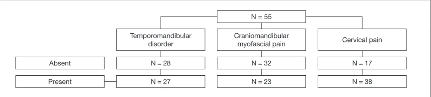
Figure 1 shows the volunteers distribution (number of individu-als) regarding the presence or absence of the three conditions: TMD, craniomandibular myofascial pain, and cervical pain. Individuals with craniomandibular myofascial pain exhibited greater activation amplitudes during the rest of the RT, LT and RM muscles than asymptomatic individuals (p<0.05). he cra-niomandibular pain did not afect isometry (p>0.05). For cer-vical myofascial pain, the electrical activity of the masticatory muscles during rest and isometry did not present statistical dif-ferences between groups (p>0.05) (Table 1).
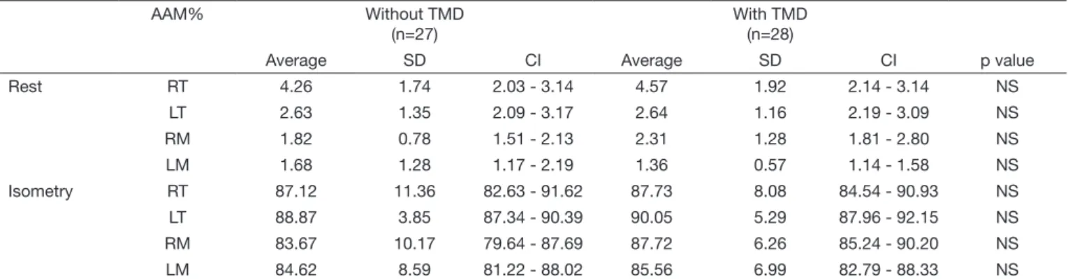
Comparison of the average of amplitude surface EMG index-es (RMS%) during rindex-est and isometry did not prindex-esent statisti-cal diferences (p>0.05) between subjects with and without TMD (Table 2).
Figure 1. Volunteers’ distribution regarding the presence or absence of temporomandibular disorder, craniomandibular myofascial pain and cervical pain
N = number of volunteers.
Absent
Present
Temporomandibular disorder
N = 28
N = 27
N = 55
Craniomandibular myofascial pain
N = 32
N = 23
Cervical pain
N = 17
N = 38
Table 1. Comparison between the averages of activation amplitude for right and left temporal muscles, right and left masseter muscles, during rest and isometry according to the myofascial pain presence in the craniomandibular region and the cervical region
AAM% Average SD CI Average SD CI p value
Absent (n=23) Present (n=32)
Craniomandibular myofascial pain
Rest RT 2.39 1.30 1.98 - 2.81 3.20 1.28 2.49 - 3.91 0.02
LT 2.44 1.19 2.06 - 2.82 3.15 1.29 2.44 - 3.86 0.03
RM 1.82 0.72 1.59 - 2.05 2.73 1.55 1.87 - 3.59 0.03
LM 1.52 1.10 1.17 - 1.87 1.50 0.63 1.16 - 1.86 NS
Isometry RT 88.00 9.71 84.85 - 91.14 85.95 10.12 80.35 - 91.55 NS
LT 89.73 4.13 88.39 - 91.07 88.76 5.82 85.54 - 91.98 NS
RM 85.55 9.33 82.52 - 88.57 86.08 6.65 82.39 - 89.77 NS
LM 85.36 7.79 82.83 - 87.88 84.40 7.95 79.99 - 88.80 NS
DISCUSSION
his study evaluated the electric activity behavior of the masticatory musculature during rest and mandibular isometry in women with and without TMD, seeking to understand the efect of the cranio-mandibular myofascial pain and cranio-mandibular pain on the activity of these individuals’ masticatory muscles. he main result showed that the activation of RT, LT and RM muscles at rest is greater in the presence of craniomandibular myofascial pain than in the absence of this symptom, independent of the TMD diagnosis.
As during rest the mandible is in a position of involuntary sus-pension resulting from the masticatory muscles’ relaxation, the electromyographic activity expected is minimal19,20. However, in
this study, individuals with craniomandibular myofascial pain presented greater electrical activation amplitude of the mastica-tory muscles during rest than those asymptomatic ones, a result found by Bodéré et al.21. As to the TMD presence, there was no
diference between the groups. It was considered that the greater activation amplitude of the masticatory muscles during rest in subjects of the TMD group, compared to the control group found in other studies22,23 was due to methodological diferences.
he severity degree of TMD and the inclusion in the non-TMD group of individuals who presented craniomandibular myofascial pain may have inluenced the results. Individuals with moderate to
severe intensity of TMD showed greater activation during rest com-pared to those with mild pain or from control group23. he present
study included subjects with mild, moderate and severe TMD in the same group. he inclusion of individuals with mild TMD may have reduced the diferences between this group and the control. Despite the presence of TMD, the presence of craniomandibu-lar myofascial pain in the control group may have increased the activation of the masticatory muscles during rest. he exclusion in the control group of any dysfunction sign or symptom was considered in another study22. When some disturbance interferes
with the stomatognathic system functioning, the organism uses several adaptive processes to maintain the eiciency of the func-tions that involve its structures24. It is likely that by means of
sensory and motor interactions, the presence of myofascial pain in this region modiies the generation of a resting action poten-tial, changing the activation pattern of the craniomandibular muscles22. In TMD’s study, it is necessary that the control group
does not present any dysfunction sign and symptom.
he muscle activation amplitude did not difer in the isometry between the groups with and without myofascial pain and with and without TMD. he same result was observed by Rodrigues-Bigaton et al.22 and Lauriti et al.23, indicating that both the
pres-ence and intensity of pain in TMD subjects did not inlupres-ence the electrical activity of the masticatory muscles.
Table 2. Comparison between the averages of activation amplitude for right and left temporal muscles, right and left masseter muscles (AAM%), during rest and isometry according to the temporomandibular disorder presence
AAM% Without TMD
(n=27)
With TMD (n=28)
Average SD CI Average SD CI p value
Rest RT 4.26 1.74 2.03 - 3.14 4.57 1.92 2.14 - 3.14 NS
LT 2.63 1.35 2.09 - 3.17 2.64 1.16 2.19 - 3.09 NS
RM 1.82 0.78 1.51 - 2.13 2.31 1.28 1.81 - 2.80 NS
LM 1.68 1.28 1.17 - 2.19 1.36 0.57 1.14 - 1.58 NS
Isometry RT 87.12 11.36 82.63 - 91.62 87.73 8.08 84.54 - 90.93 NS
LT 88.87 3.85 87.34 - 90.39 90.05 5.29 87.96 - 92.15 NS
RM 83.67 10.17 79.64 - 87.69 87.72 6.26 85.24 - 90.20 NS
LM 84.62 8.59 81.22 - 88.02 85.56 6.99 82.79 - 88.33 NS
AAM = activation amplitude; RT = right temporal muscle; LT = left temporal; RM = right masseter; LM = left masseter; NS = not signiicant; SD = standard deviation; CI = conidence interval; Mann-Whitney U test.
Table 1. Comparison between the averages of activation amplitude for right and left temporal muscles, right and left masseter muscles, during rest and isometry according to the myofascial pain presence in the craniomandibular region and the cervical region – continuation
AAM% Average SD CI Average SD CI p value
Absent (n=23) Present (n=32)
Cervical myofascial pain
Rest RT 2.64 1.49 2.15 - 3.13 2.55 0.91 2.08 - 3.02 NS
LT 2.61 1.27 2.19 - 3.03 2.69 1.21 2.07 - 3.31 NS
RM 2.05 1.14 1.67 - 2.42 2.12 0.95 1.63 - 2.61 NS
LM 1.55 1.12 1.18 - 1.92 1.45 0.61 1.14 - 1.76 NS
Isometry RT 88.08 7.84 85.47 - 90.69 86.01 13.22 79.21 - 92.81 NS
LT 89.72 4.63 88.17 - 91.26 88.90 4.71 86.48 - 91.32 NS
RM 85.84 9.34 82.73 - 88.95 85.38 7.02 81.77 - 88.99 NS
LM 85.30 8.52 82.46 - 88.14 84.64 6.04 81.53 - 87.75 NS
However, other studies have found less electrical activity during the maximal voluntary contraction of the masticatory muscles in subjects with TMJ disorders25, or with arthrogenetic and psychogenic TMD,
but not myogenic TMJ26. In this study, the DTM group consisted of
individuals with one or more RDC/TMD diagnosis. Most individuals with TMD presented pain only due to muscle palpation14.
Spontane-ous pain (active pain) at rest, which is characteristic in myofascial pain because of muscle tension and contracture27, was not frequent among
subjects in the TMD group. And some of the individuals with TMD classiied according to RDC/TMD as group II (disc displacement) showed no signs of joint and muscular pain. As pain is an important modiier of muscle function26,28,29, it is probable that the absence of
the symptom or its location may also have contributed to the similar-ity in the amplitude parameters during mandibular isometry between the groups with and without TMD.
Due to the anatomical and neurophysiological connection be-tween the craniomandibular structures and the cervical6, the
ini-tial hypothesis was that pain in this region could inluence the masticatory muscles’ activity. However, no signiicant diference was observed in the isometry or at rest of the masticatory mus-cles between the groups. Corroborating these results, Svensson et al.30 veriied through an experimental study that cervical pain
is not associated with changes in the electromyographic activ-ity of the mandibular muscles. Nevertheless, it has been shown that masticatory dysfunction seems to be more associated with chronic cervical pain, lasting at least three months31.
As the presence of pain may alter the functional balance and the masticatory action efectiveness32, the results of this study
rein-force the importance of considering craniomandibular myofas-cial pain during the evaluation and treatment of TMD-patients. he information obtained through this research provides subsi-dies for research protocols that identify more speciic aspects of the pain inluence on the masticatory muscles’ activity, contrib-uting to the clinical intervention of these subjects.
CONCLUSION
Cervical pain and TMD did not afect masticatory electrical activity at rest or in isometry. At rest, there was a greater mas-ticatory muscles activation amplitude in the presence of cranio-mandibular myofascial pain. hus, craniocranio-mandibular myofas-cial pain seems to interfere in the physiological behavior of the masticatory muscles when the mandible is at rest and should be investigated in the evaluation and intervention of TMD-indi-viduals. he importance of the symptoms absence in the TMD study control groups is emphasized.
REFERENCES
1. Visscher CM, Lobbezoo F, de Boer W, van der Zaag J, Naeije M. Prevalence of cervical spinal pain in craniomandibular pain patients. Eur J Oral Sci. 2001;109(2):76-80. 2. Piccin CF, Corrêa EC, Pasinato F, Bouleus J, Chiodelli L, Pozzebon D. Aspectos
clínicos e psicossociais avaliados por critérios de diagnóstico para disfunção temporo-mandibular. Rev CEFAC. 2016;18(1):113-9.
3. Manfredini D, Guarda-Nardini L, Winocur E, Piccotti F, Ahlberg J, Lobbezoo F. Research diagnostic criteria for temporomandibular disorders: a systematic review of axis I epidemiolo-gic indings. Oral Surg Oral Med Oral Pathol Oral Radiol Endod. 2011;112(4):453-62. 4. Ardizone I, Celemin A, Aneiros F, del Rio J, Sanchez T, Moreno I. Electromyographic
study of activity of the masseter and anterior temporalis muscles in patients with
tem-poromandibular joint (TMJ) dysfunction: comparison with the clinical dysfunction index. Med Oral Patol Oral Cir Bucal. 2010;15(1):e14-9.
5. Ozkan F, Cakır Özkan N, Erkorkmaz U. Trigger point injection therapy in the mana-gement of myofascial temporomandibular pain. Agri. 2011;23(3):119-25. 6. Armijo-Olivo S, Fuentes JP, da Costa BR, Major PW, Warren S, hie NM, et al.
Reduced endurance of the cervical lexor muscles in patients with concurrent tempo-romandibular disorders and neck disability. Man her. 2010;15(6):586-92. 7. Weber P, Corrêa EC, Ferreira Fdos S, Soares JC, Bolzan Gde P, Silva AM. Cervical
spi-ne dysfunction signs and symptoms in individuals with temporomandibular disorder. J Soc Bras Fonoaudiol. 2012;24(2):134-9. English, Portuguese.
8. McLean L. he efect of postural correction on muscle activation amplitudes recorded from the cervicobrachial region. J Electromyogr Kinesiol. 2005;(15):527-35. 9. Stiesch-Scholz M, Fink M, Tschernitschek H. Comorbidity of internal derangement
of the temporomandibular joint and silent dysfunction of the cervical spine. J Oral Rehabil. 2003;30(4):386-91.
10. Silveira A, Gadotti IC, Armijo-Olivo S, Biasotto-Gonzalez DA, Magee D. Jaw dys-function is associated with neck disability and muscle tenderness in subjects with and without chronic temporomandibular disorders. Biomed Res Int. 2015;2015:512792. 11. Cheng CH, Wang JL, Lin JJ, Wang SF, Lin KH. Position accuracy and electromyo-graphic responses during head reposition in young adults with chronic neck pain. J Electromyogr Kinesiol. 2010;20(5):1014-20.
12. Ries LG, Bérzin F. Cervical pain in individuals with and without temporomandibular disorders. Braz J Oral Sci. 2007;6(20):1301-7.
13. Winter K, Baccaglini L, Tomar S. A review of malocclusion among individuals with mental and physical disabilities. Spec Care Dentist. 2008;28(1):19-26.
14. Dworkin SF, LeResche L. Research diagnostic criteria for temporomandibular disor-ders: review, criteria, examinations and speciications, critique. J Craniomandib Dis-ord. 1992;6(4):301-55.
15. Visscher CM, Lobbezoo F, de Boer W, van der Zaag J, Verheij JG, Naeije M. Clinical tests in distinguishing between persons with or without craniomandibular or cervical spinal pain complaints. Eur J Oral Sci. 2000;108(6):475-83.
16. Hermens HJ, Freriks B, Disselhorst-Klug C, Rau G. Development of recommenda-tions for SEMG sensors and sensor placement procedures. J Electromyogr Kinesiol. 2000;10(5):361-74.
17. Ries LG, Alves MC, Bérzin F. Asymmetric activation of temporalis, masseter, and sternocleidomastoid muscles in temporomandibular disorder patients. Cranio. 2008;26(1):59-64.
18. Briesemeister M, Schmidt KC, Ries LG. Changes in masticatory muscle activity in children with cerebral palsy. J Electromyogr Kinesiol. 2013;23(1):260-6.
19. Carr AB, Christensen LV, Donegan SJ, Ziebert GJ. Postural contractile activities of hu-man jaw muscles following use of an occlusal splint. J Oral Rehabil. 1991;18(2):185-91. 20. Galo R, Vitti M, Santos CM, Hallak JE, Regalo SC. he efect of age on the
func-tion of the masticatory system--an electromyographical analysis. Gerodontology. 2006;23(3):177-82.
21. Bodéré C, Téa SH, Giroux-Metges MA, Woda A. Activity of masticatory muscles in subjects with diferent orofacial pain conditions. Pain. 2005;116(1-2):33-41. 22. Rodrigues-Bigaton D, Berto R, Oliveira AM, Bérzin F. Does masticatory muscle
hy-peractivity occur in individuals presenting temporomandibular disorders? Braz J Oral Sci. 2008;24(7):1497-501.
23. Lauriti L, Motta LJ, de Godoy CH, Biasotto-Gonzalez DA, Politti F, Mesquita-Ferrari RA, et al. Inluence of temporomandibular disorder on temporal and masseter muscles and occlusal contacts in adolescents: an electromyographic study. BMC Musculoskelet Disord. 2014;15:123.
24. Douglas CR, Avoglio JL, de Oliveira H. Stomatognathic adaptive motor syn-drome is the correct diagnosis for temporomandibular disorders. Med Hypotheses. 2010;74(4):710-8.
25. Ferrario VF, Tartaglia GM, Luraghi FE, Sforza C. he use of surface electromyography as a tool in diferentiating temporomandibular disorders from neck disorders. Man her. 2007;12(4):372-9.
26. Tartaglia GM, Moreira Rodrigues da Silva MA, Bottini S, Sforza C, Ferrario VF. Masticatory muscle activity during maximum voluntary clench in diferent research diagnostic criteria for temporomandibular disorders (RDC/TMD) groups. Man her. 2008;13(5):434-40.
27. Partanen JV, Ojala TA, Arokoski JP. Myofascial syndrome and pain: A neurophysi-ological approach. Pathophysiology. 2010;17(1):19-28.
28. Ries LG, Graciosa MD, Medeiros DL, Pacheco SC, Fassicolo CE, Graeling BC, et al. Inluence of craniomandibular and cervical pain on the activity of masticatory muscles in individuals with temporomandibular disorder. Codas. 2014;26(5):389-94. English, Portuguese.
29. Santana-Mora U, Cudeiro J, Mora-Bermúdez MJ, Rilo-Pousa B, Ferreira-Pinho JC, Otero-Cepeda JL, et al. Changes in EMG activity during clenching in chronic pain patients with unilateral temporomandibular disorders. J Electromyogr Kinesiol. 2009;19(6):e543-9.
30. Svensson P, Wang K, Sessle BJ, Arendt-Nielsen L. Associations between pain and neu-romuscular activity in the human jaw and neck muscles. Pain. 2004;109(3):225-32. 31. Catanzariti JF, Debuse T, Duquesnoy B. Chronic neck pain and masticatory
dysfunc-tion. Joint Bone Spine. 2005;72(6):515-9.