88
VASTUS LATERALIS OBLIQUE ACTIVITY DURING GAIT OF
SUBJECTS WITH PATELLOFEMORAL PAIN
ATIVIDADE DO VASTO LATERAL OBLÍQUO NA MARCHA DE SUJEITOS COM DOR FEMOROPATELAR
ACTIVIDAD DEL VASTO LATERAL OBLICUO EN LA MARCHA DE SUJETOS CON DOLOR FEMOROPATELAR
Gilmar Moraes Santos1
(Fisioterapeuta)
Karina Gramani Say2 (Fisioterapeuta)
Flávio Pulzatto3 (Fisioterapeuta)
Thiele de Cássia Libardoni1
(Fisioterapeuta)
Larissa Milani Brognoli Sinhorim1
(Fisioterapeuta) Tamiris Beppler Martins1
(Fisioterapeuta) Vanessa Monteiro Pedro2
(Fisioterapeuta)
1. Universidade do Estado de Santa Catarina, Florianópolis, SC, Brasil. 2. Universidade Federal de São Carlos, São Carlos, SP, Brasil. 3. Centro Universitário Toledo, Araçatuba, SP, Brasil.
Correspondência:
Rua Professor Bayer Filho, 125, Coqueiros, Florianópolis, SC, Brasil. 88080-300. gilmar.santos@udesc.br
ORIGINAL ARTICLE
ARTIGO ORIGINAL
ARTÍCULO ORIGINAL
ABSTRACT
Introduction: So far, little is known about the behavior of electromyographic activity of vastus lateralis oblique muscle during treadmill gait in subjects with and without patellofemoral pain syndrome. Objective: The purpose of this study was to investigate the electromyographic activity of the patellar stabilizers muscles and the angle of the knee joint flexion in subjects with and without patellofemoral pain syndrome. Method: Fifteen subjects without (21 ± 3 years) and 12 with patellofemoral pain syndrome (20 ± 2 years) were eval-uated. The electromyographic activity and flexion angle of the knee joint were obtained during gait on the treadmill with a 5 degree inclination. Results: The knee flexion angle was significantly lower in the subjects with patellofemoral pain syndrome when compared with the healthy controls. The electromyographic activity of vastus lateralis longus was significantly greater during gait on the treadmill with inclination in subjects with patellofemoral pain syndrome. The results also showed that the electromyographic activity of vastus lateralis oblique and vastus medialis oblique were similar in both groups, regardless of the condition (with/without inclination). Conclusion: We have shown that knee kinematics during gait differs among patients with and without patellofemoral pain syndrome and healthy controls and that a different motor strategy persists even when the pain is no longer present. In addition, the findings suggested that the vastus lateralis oblique has a minor role in patellar stability during gait.
Keywords: gait; knee joint; quadriceps muscle.
RESUMO
Introdução: Até o presente, pouco é conhecido sobre o comportamento da atividade eletromiográfica do músculo vasto lateral oblíquo durante a marcha em esteira em sujeitos com e sem síndrome da dor femoropatelar. Objetivo: A finalidade deste estudo foi investigar a atividade eletromiográfica dos músculos estabilizadores patelares e o ângulo de flexão do joelho em sujeitos com e sem síndrome da dor femoropatelar. Método: Quinze sujeitos sem (21 ± 3 anos) e 12 com síndrome da dor femoropatelar (20 ± 2 anos) foram avaliados. A atividade eletromiográfica e o ângulo de flexão da articulação do joelho foram obtidos durante a marcha em esteira com inclinação de 5 graus. Resultados: O ângulo de flexão do joelho foi significativamente menor nos sujeitos com síndrome da dor femoropatelar quando comparados aos controles saudáveis. A atividade eletromiográfica do vasto lateral longo foi significativamente maior durante a marcha na esteira com inclinação nos sujeitos com síndrome da dor femoropatelar. Os resultados também mostraram que a atividade eletromiográfica dos músculos vasto lateral oblíquo e vasto medial oblíquo foram simi-lares em ambos os grupos, independentemente da condição (com e sem inclinação). Conclusão: Mostramos que a cinemática do joelho durante a marcha é diferente entre sujeitos com e sem síndrome da dor femoropatelar e que a estratégia motora diferente persiste mesmo quando a dor não está mais presente. Adicionalmente, os achados sugerem que o vasto lateral oblíquo tem papel secundário na estabilidade patelar durante a marcha.
Descritores: marcha; articulação do joelho; músculo quadríceps.
RESUMEN
89
INTRODUCTION
Patellofemoral Pain Syndrome (PFPS) is the most commonly diagnosed knee condition seen in patients under the age of 501, with women having a higher incidence than men2. The etiology of PFPS has its origin in many factors, with various theories and clinical tests being proposed3. However, the etiological factor most widely thought to cause PFPS is abnormal patellar tracking4.
A number of studies can be found in the literature concerning the balance between vastus medialis oblique and vastus lateralis muscles in different exercises, like functional activities5,6, isometric exercises in an open kinetic chain7, a closed kinetic chain8 and rehabilitation9. However, this previous researches evaluating the patellofemoral pain syndrome has not included the oblique portion of the vastus lateralis muscle, which is separated from the longus component by fascial planes and has different functions10,11.
The most oblique portions of vastus medialis and of vastus lateralis, would be the portions most prepared anatomically to control the position of the patella with respect to the trochlear surface of the femur7, play an important function in patellar orientation within the femoral trochlea10 and would be responsible for the control and stability of the patella7.
The eccentric quadriceps contraction, in the stance phase of the gait cycle, is considered primary dynamic shock absorbing mechanism during the weight-acceptance7. However, for patients with PFPS, the increase at the knee joint flexion during the loading response phase of gait and the contraction of the quadriceps muscle would increase the patellofemoral joint reaction force, inducing biomechanical alterations in patellofemoral joint, influencing as such the function of the lower limb, especially locomotion12. In addition, based on clinical experience of authors, the condition showed change in walking patterns that is exacerbated by activities such as stair climbing and walking along slopes. The discomfort associated with these activities generally results in modifications of gait patterns, in a possible strategy to reduce muscular demands and subsequently the pain5.
Although studies exist about the analysis of gait parameters, only six evaluated subjects with patellofemoral pain syndrome. These studies utilized different system for gait evaluation, including movement analysis systems13-18 , force plates13,14,16,17 and electromyography15. In addition, although reduced loading response phase knee flexion is a finding in subjects with patellofemoral pain syndrome13,16, others investigators have not found such finding17,18.
Therefore, the objective of this study was to evaluate kinematic of the knee joint and behavior of the electromyographic activity of the vastus medialis oblique (VMO), vastus lateralis oblique (VLO) and vastus lateralis longus (VLL) muscles during graded treadmill walking of subjects with and without patellofemoral pain syndrome. Thus, according to the above considerations, we hypothesized that during gait the vastus lateralis oblique muscle could also be a primary dynamic lateral stabilizer of the patella, being one of the force vectors responsible for patellar stability.
METHODS
Two groups of participants were evaluated during treadmill walk-ing. Twelve women diagnosed with patellofemoral pain syndrome (PFPS) based on clinical examination by an experienced musculoskeletal
physiotherapist, and 15 asymptomatic women were recruited for the study. It was used treadmill for its convenience in inclination of walk way and simplicity in measurement techniques, i.e., no need for long cabled or telemetry recording system.
The mean ± standard deviation age, weight, height and Q angle of the patellofemoral pain participants were 21 ± 2 yr, 52.81 ± 7.73Kg, 1.63 ± 0.07m and 19.5° ± 3.3 respectively. The mean ± standard deviation age, weight, height and Q angle of the control participants were 22 ± 3 yr. 57.76 ± 5.96Kg, 1.64 ± 0.07m and 17.9° ± 3.6 respectively. The Research Ethics Committee at Federal University of São Carlos approved this study (n° 101/2005) and subjects were included in the study after signing a free informed consent form.
The inclusion criteria were based on those used in other PFPS studies19-21:
• pain on patella palpation of 3 cm or more on a 10 cm visual analogue scale (VAS), while going down a step of 25 cm and squatting at 90°, both with 30 sec duration;
• have had an average pain level of 3 or more on a 10 visual analogue scale (VAS) in the patellofemoral joint in the previous week, and; • have had an insidious onset of pain not related to traumatic
incident.
The asymptomatic control participants were recruited from the Federal University of São Carlos. Subjects were examined by an experi-enced musculoskeletal physiotherapist and were excluded if they had any history of lower limb pathology or other disorder that might interfere with treadmill walking.
Active differential surface bipolar electrodes (EMG System (EMG System - BrazilTM) detected electromyography activity (EMG) of the VMO, VLO and VLL muscles.
The electrodes are constituted by two bars of Ag/AgCl, 10 mm long, 1 mm wide each and 2 mm high each, positioned parallelly and separated from each other by 10 mm. These electrodes enable a differential amplification of 50x, with input impedance of 10GΩ and common mode rejection ratio (CMRR) of 130dB at 60Hz.
The electrodes were connected to the signal conditioner module (EMG System - BrazilTM) to be amplified and filtered by the Butterworth band pass filter of low-pass filtering of 500 Hz and high-pass filtering of 20 Hz. A dynamic resolution band 12-bit A/D card digitized the electro-myographic signals of the VMO, VLO and VLL muscles and the sampling frequency was adjusted to 2 KHz, with an input band of ± 10V.
Angular movement of the knee was obtained by a digital electrogo-niometer (EMG System - BrazilTM) strapped to the articular rotation axis of the knee joint, which enabled the angular register in a continuous, automatic and synchronized way to the electromyographic recordings. For the participants with bilateral PFPS, only the side with more pain was tested, whilst for participants from the control group, the dominant lower limb was chosen. The dominant leg was defined as the leg that the participant would choose to kick a ball.
Prior to placing the electrodes, the skin was shaved, cleaned with alcohol and lightly rubbed to reduce impedance and eventual interference. The electrodes for the VMO were placed approximately 4 cm above the superomedial edge of the patella and orientated to 55° in relation to the femur axis21. For the VLL muscle, the electrodes were positioned
Article received on 02/27/2015 accepted on 11/07/2016 DOI: http://dx.doi.org/10.1590/1517-869220172302146524
dolor ya no está presente. Además, los hallazgos sugieren que el músculo vasto lateral oblicuo tiene un papel menor en la estabilidad de la rótula durante la marcha.
90
approximately 11 cm above the superolateral edge of the patella with an inclination of 15° to the femur axis20. For the placing of the VLO electrodes the lateral epicondyle of the femur and the beginning and middle of the muscle belly were located with an inclination of 50° in relation the femur axis20. The reference electrode was attached right above the lateral malleolus. Electrode placement was confirmed by voluntary muscle contraction
A footswitch (EMG System - BrazilTM) was taped to the underside of each participant’s heel prior to the walking trials. Both the footswitch and the EMG recording system were connected to an interface unit (EMG System Brazil), which enabled the start of each stride to be recorded together with the EMGs by the acquisition software thereby synchronizing the two (Figure 1). From heel strike on one leg to the next heel strike on the same leg was defined as a stride, being considered 100% of the stride time21.
For the electromyography and electrogoniometer data collection the participants walked on an electric treadmill (Pro-action Fitness) in bare feet, with and without an inclination of 5°, at preferred speed (characterized by the average speed during walking 5 meters in 3 trials) about seven minutes. The inclination of 5° was chosen because would be comfortable for all patients during treadmill walking, around 15 minutes.
After application of the electrodes and recording equipment, but prior to recording data, the subjects exercised on the treadmill for familiarization.
The participants’ self-selected walking speeds do not showed signif-icant differences between PFPS (3.8 ± 0.4 Km/h) and CG (3.9 ± 0.3 Km/h) groups. Pain assessment was made on one visual analogue scale (VAS) before (PFPS = 0.2 cm; CG = 0 cm) and after (PFPS = 0.4 cm; CG = 0 cm) to walk on a treadmill. The pain of PFPS participants was minimum, do not showing significant differences in healthy participants’ comparison.
Data analysis
The data from the goniometer were calculated for the three trials. The values recorded by the electrogoniometer were compared using the coefficient of variation (CV=2%), in order to analyze the reproduc-ibility of the tool.
The myoelectric signals were full-wave rectified, low-pass filtered, smoothing using a Butterworth filter in a cutoff frequency of 5Hz and then A/D converted together with the footswitch signal. Next, the
linear envelopes were also temporally normalized to the stride period, adjusted for 100% (gait cycle), using as reference the heel strike, and a mathematical routine elaborated in the program MatLab (Santa Catarina State University). In this study, our option was to analyze the heel strike, i.e., stage in gait cycle at which the heel of the foot or shoe first makes contact with the walking surface.
The final EMG pre-processing stage was amplitude normalization. For this study, the EMG signal was normalized using the mean value during the gait cycles from each specific muscle during treadmill gait conditions. Normalized EMG was ensemble averaged over the full strides recorded during a 10 seconds trial then a group ensemble average was determined by pooling participant data. The average rectified value was computing in 10s because this time allowed collecting enough number of strides (8) to be able to compute EMG variables of interests.
The mean normalization method, mainly in dynamic activities, it is based on the studies of Winter22 and Benoit et al.23 that verified larger efficiency and applicability of that normalization type during dynamic activities, as well as lower degree of variation from one data series to another when compared the other methods.
Statistical analysis
The characteristics of the participants (age, height, and weight) were compared between the groups using test t for independent samples. In order to verify if the amplitude of the knee joint movement (dependent factor) varied between the groups and the walking conditions (independent factors), an ANOVA 2x2 was calculated (groups x walking conditions). Repeated measures ANOVA with “muscles” and “walking conditions” as within-subjects factors and “groups” as between-subjects factor was carried out to determine if normalized EMG activity was different between the groups and muscles. All the variance analyses used the Newman-Keuls Post Hoc test, to identify the significant comparisons. All the significance levels were determined as p ≤0.05 with sequential Bonferroni adjustment. The statistical analyses were carried out with SPSS 17.0 software (Santa Catarina State University).
RESULTS
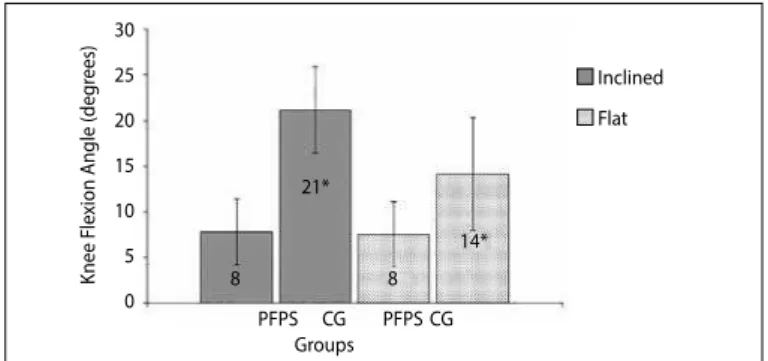
The results of this study showed that there was a significant difference in the knee joint flexion angle in the heel strike (p=0.02) between the healthy control and participants with PFPS (Figure 2).
The analysis of the participants with PFPS, showed that the results of the normalized EMG activity during walking on an inclined surface were significantly greater when compared with walking on a flat sur-face in all muscles (vastus medialis oblique - p=0.03; vastus lateralis oblique - p=0.05; vastus lateralis longus - p=0.04). However, significant differences in the control group were not found (Table 1). In addition a repeated-measures analysis of variance revealed no interactionbetween group and walking conditions (F=1.271; p=0.607).
Figure 1. EMG, footswitch and eletrogoniometer signals synchronized.
Figure 2. Knee joint flexion angle in the heel strike (KJF), in degree, in the CG (n= 15) and PFPS (n= 12) during walking on the flat and inclined surface.
*KFJ, significantly greater in the CG regardless condition.
Inclined
Flat
Groups
K
nee F
le
xion A
ngle (deg
rees)
30
25
20
15
10
5
0
PFPS
8 8
21*
14*
PFPS CG CG
Footswitch 6
4 2 0 -2
80 60 40 20 0 150 100 50 0 -50 -100 150 100 50 0 -50 -100 150 100 50 0 -50 -100
Eletrognoniometer
VMO muscle
VLL muscle
VLO muscle
91 The results, in each group, showed that there is a significant difference
between the three muscles studied. The Newman-Keuls test in the CG showed that during walking on a flat and inclined surface, EMG activity of the VMO (p=0.04) and VLL (p=0.04) muscles was significantly greater when compared with the EMG activity of the VLO muscle. For the PFPS participants, EMG activity of the VMO (p=0.04) and VLL (p=0.03) muscles during gait on a flat surface also showed to be greater when compared the EMG activity of the VLO muscle. However, during gait on an inclined surface, no difference in the EMG activity was found (Table 1). In addi-tion arepeated-measures analysis of variance revealed no interaction between group and muscles conditions (F=1.271; p=0.607).
A significant increase (p=0.04) was observed for EMG activity of the VLL muscle in PFPS participants during gait on an inclined surface when compared with healthy controls (Table 1). However, significant differences were not observed either between the groups or in the interaction (groups x muscles) during gait on a flat surface.
DISCUSSION
The results of this study showed a large knee flexion angle in heel strike phase of gait of control group, when compared with patellofemoral pain group, both during walking on a flat surface and on an inclined surface. According Dillon et al.13 and Nadeau et al.16, the small knee flexion angle would be a strategy utilized during gait to avoid the contraction of the quadriceps and as such reducing the reaction force in the patellofem-oral joint and consequently the pain. We believe that the reduction in angle of the knee joint flexion, found in this study in participants with patellofemoral pain syndrome, can be a compensatory mechanism utilized during gait and could perhaps explain the absence of pain seen when carrying out the task (patellofemoral pain participants did not experience pain while walking on a treadmill). In addition, Perry24 reported that individuals with weak or painful femoral quadriceps avoid knee joint flexion in the loading response phase, since it is at this point in the gait cycle that muscular demands and joint reaction forces are greater. Some authors examined factors for the characterization of patellofem-oral pain syndrome, like valgus knee, pronation and Q angles14, hyper-mobile patella25, among others. We believe that the reduction in the angle of the knee joint flexion could also be a factor to characterize these participants and is as such important data in the evaluation and functional rehabilitation of this pathology and not only a strategy utilized during gait by individuals with patellofemoral pain syndrome.
Patellofemoral pain participants presented greater vastus lateralis longus EMG activity than healthy controls during gait on an inclined surface. This result conflicts with previously published studies. Powers et al.5,15, when analyzing functional activities, verifiedthat patellofem-oral pain participants showed less activity in the quadriceps muscle, when compared with healthy participants whilst they walked on a flat and inclined surface. It is possible that methodological differences can have generated these contradictory findings. To calculate the
EMG activity in each group, these authors utilized the percentage of a maximum voluntary isometric contraction test, whilst in this study the EMG activity was normalized by using the mean value during the
gait cycles. Moreover, the inclination utilized in this study was less than that in previously mentioned studies.
In addition, the EMG activity of both VMO and VLL muscles was significantly greater than EMG activity of the vastus lateralis oblique during two conditions tested on healthy participants and during gait on flat surface in the patellofemoral pain participants. These results suggested a lower activation of the vastus lateralis oblique, refuting the hypothesis of the participation of this muscle in the patellar sta-bility suggested by some authors7. It is possible that this controversial finding is because we are analyzing in this study, a dynamic activity, gait, where biomechanical, anatomical and functional characteristics are very different to those found when analyzing corpses as well as isometric contraction in an open kinetic chain. Evidence exists that muscular activity is specific to the type of exercise carried out; in as much that muscular activation cannot necessarily be the same in dynamic movements as in isometric contractions. In addition, there are several possible explanations for this finding. Merchant and Mercer26 described the fibers of VLO muscle attached to the lateral border of patella with point of the insertion of the terminal fibers proximal or distal to the most superior point of the patella, which might affect its angle of insertion. Women have a predominance of insertion of VLO muscle proximal to the most superior point of the patella. This less obtuse angle of insertion the VLO muscle in women should be result a lower lateral drift of the patella, which produce a lower lateral deforming force on the patella, especially when there are similar factors pertaining to static alignment (Q angle) between GC (17.9°) and patellofemoral pain group (19.5°) and a dynamic task. In addition, Salsich and Perman27 showed that it is possible to have subgroups of patients in this syndrome, which would show different characteristics from each other.
The EMG activity in the control group was similar for all muscles during walking on an inclined surface when compared with walking on a flat surface. However, our results showed that the patellofemoral pain participants presented greater EMG activity, in all muscles, during gait on an inclined surface when compared with a flat one. Our findings also showed that an inclination of 5° was sufficient to modify the EMG activity in all muscles in patellofemoral pain participants.
The fact of the knee joint presenting a smaller flexion angle in the loading response phase with the objective of reducing the activity of the quadriceps16 is not corroborated by our findings, during gait on an inclined surface. It is possible that, with the inclination and decrease in the knee flexion, a reduction occurs of the congruence of the contact surfaces of the patellofemoral joint, which would cause more instability and consequently the necessity of more activation of the primary stabilizers, to compensate the reduction in the anatomical stability. According to Hungerford and Barry28, the patella articulates with the trochlea in approximately 10° of the knee joint flexion, which would permit the beginning of the congruence of the articulate surfaces and consequently more stability. The participants with patellofemoral pain syndrome in this study, presented values (flat-8.35°; inclined-8.76°) in the knee joint flexion angle, which did not favor the functional stability of the patella with the trochlea.
Another possible explanation for greater EMG activity during gait on an inclined surface, in patellofemoral pain group, is related to a mechanical factor. According to Smidt29, the quadriceps moment arm increases with greater knee joint flexion. As the patellofemoral pain participants presented a smaller knee joint flexion angle when compared with the participants of control group, it is possible that a greater activation of the quadriceps muscle is necessary with the objective of compensating the
Table 1. Mean and standard devition of normalized EMG activity between the VMO, VLO and VLL muscles during the gait of the CG (n= 15) and PFPS (n= 12) groups.
Groups Muscles
VMOA VLOB VLLC
CG
Flat 247.8# (SD 56.7) 177.6 (SD 16.0) 239.0# (SD 40.7) Inclined 259.8# (SD 39.3) 183.8 (SD 15.2) 237.4# (SD 28.6)
PFPS
Flat 221.5# (SD 22.8) 181.8 (SD 27.2) 236.5# (SD 76.5) Inclined 258.9* (SD 28.0) 203.0* (SD 21.0) 299.0*ab (SD 98.8)
92
reduction in the lever arm and consequently the knee extensor moment. In addition, we believe that a possible explanation for reduced knee flexion and increased level of activation of the VLL in patellofemoral pain participants compared with controls is related to the imbalance of the medial and lateral forces acting on the patella, with the net force on the patella is being directed too laterally.
Moreover, these alterations are peculiar as the patellofemoral pain and healthy participants had similar level of pain, that is, changes persisted even without pain. According to Grenholm et al.30 strategies may become habitual and remain also when the pain disappeared, in order to protect the patellofemoral joint and avoid the pain. It´s important to report that findings of this study only applied to subjects with patellofemoral pain syndrome with minimal or no pain during walking
CONCLUSION
The results of this presente study showed that knee kinematics in walking differs between participants with patellofemoral pain
syndrome and healthy controls and that a different motor strate-gy persists also when the pain is no longer present. In participants with patellofemoral pain syndrome a reduced angle of the knee joint flexion, produced more muscle activity in all the knee muscles while walking on a surface with an inclination of 5° and might not be safe for the treatment or training exercises of these patients. In addition it was observed, during gait on an inclined surface, that the vastus lateralis longus presented more activation in patellofemoral pain participants than control subjects, while vastus lateralis oblique muscle has minor role in patellar stability and control during gait in both conditions. Our findings also suggest that the alteration in the angle of the knee joint flexion could be a factor of discrimination of patellofemoral pain participants and thus an important factor in the evaluation of this pathology.
All authors declare no potential conflict of interest related to this article.
REFERENCES
1. Lankhorst NE, Bierma-Zeinstra SM, van Middelkoop M. Risk factors for patellofemoral pain syndrome: a systematic review. J Orthop Sports Phys Ther. 2012;42(2):81-94.
2. Boling M, Padua D, Marshall S, Guskiewicz K, Pyne S, Beutler A. Gender differences in the incidence and prevalence of patellofemoral pain syndrome. Scand J Med Sci Sports. 2010;20(5):725-30.
3. Nunes GS, Stapait EL, Kirsten MH, de Noronha M, Santos GM. Clinical test for diagnosis of patellofemoral pain syndrome: Systematic review with meta-analysis. Phys Ther Sport. 2013;14(1):54-9.
4. Insall J. “Chondromalacia patellae”: patellar malalignment syndrome. Orthop Clin North Am. 1979;10(1):117-27.
5. Powers CM, Perry J, Hsu A, Hislop HJ. Are patellofemoral pain and quadriceps femoris muscle torque associated with locomotor function? Phys Ther. 1997;77(10):1063-75.
6. Morrish GM, Woledge RC, Haddad FS. Activity in three parts of the quadriceps recorded isometrically at two different knee angles and during a functional exercise. Electromyogr Clin Neurophysiol. 2003;43(5):259-65. 7. Morrish GM, Woledge RC. A comparison of the activation of muscles moving the patella in normal subjects and in patients with chronic patellofemoral problems. Scand J Rehabil Med. 1997;29(1):43-8. 8. Coqueiro KR, Bevilaqua-Grossi D, Bérzin F, Soares AB, Candolo C, Monteiro-Pedro V. Analysis on the activation of the VMO and VLL muscles during semisquat exercises with and without hip adduction in individuals with patellofemoral pain syndrome. J Electromyogr Kinesiol. 2005;15(6):596-603. 9. Mostamand J, Bader DL, Hudson Z. The effect of patellar taping on EMG activity of vasti muscles during
squatting in individuals with patellofemoral pain syndrome. J Sports Sci. 2011;29(2):197-205. 10. Weinstabl R, Scharf W, Firbas W. The extensor apparatus of the knee joint and its peripheral vasti: anatomic
investigation and clinical relevance. Surg Radiol Anat. 1989;11(1):17-22.
11. Bennett WF, Doherty N, Hallisey MJ, Fulkerson, JP. Insertion orientation of terminal vastus lateralis obliquus and vastus medialis obliquus muscle fibers in human knees. Clin Anat. 1993:6(3);129-34. 12. Nyland JA, Ullery LR, Caborn DN. Medial patellar taping changes the peak plantar force location and
timing of female basketball players. Gait Posture. 2002;15(2):146-52.
13. Dillon PZ, Updyke WF, Allen WC. Gait analysis with reference to chondromalacia patellae. J Orthop Sports Phys Ther. 1983;5(3):127-31.
14. Callaghan MJ, Baltzopoulos V. Gait analysis in patients with anterior knee pain. Clin Biomech (Bristol, Avon). 1994;9(2):79-84.
15. Powers CM, Landel R, Perry J. Timing and intensity of vastus muscle activity during functional activities in subjects with and without patellofemoral pain. Phys Ther. 1996;76(9):946-55.
16. Nadeau, S, Gravel, D, Hébert, LJ, Arsenault, AB, Lepage, Y. Gait study of patients with patellofemoral pain syndrome. Gait Posture. 1997;5(1):21-7.
17. Powers CM, Heino JG, Rao S, Perry J. The influence of patellofemoral pain on lower limb loading during gait. Clin Biomech (Bristol, Avon). 1999;14(10):722-8.
18. Brechter JH, Powers CM. Patellofemoral joint stress during stair ascent and descent in persons with and without patellofemoral pain. Gait Posture. 2002;16(2):115-23.
19. Cowan SM, Hodges PW, Bennell KL, Crossley KM. Altered vastii recruitment when people with patellofe-moral pain syndrome complete a postural task. Arch Phys Med Rehabil. 2002;83(7):989-95. 20. Santos GM, Ries LGK, Sperandio FF, Say KG, Pulzatto F, Monteiro-Pedro V. Tempo de início da atividade
elétrica dos estabilizadores patelares na marcha em sujeitos com e sem síndrome de dor femoropatelar. Fisioter Mov. 2011:24(1);125-32.
21. Yang JF, Winter DA. Electromyographic amplitude normalization methods: improving their sensitivity as diagnostic tools in gait analysis. Arch Phys Med Rehabil. 1984;65(9):517-21.
22. Winter DA. Pathologic gait diagnosis with computer-averaged electromyographic profiles. Arch Phys Med Rehabil. 1984;65(7):393-8.
23. Benoit DL, Lamontagne M, Cerulli G, Liti A. The clinical significance of electromyography normalisation techniques in subjects with anterior cruciate ligament injury during treadmill walking. Gait Posture. 2003;18(2):56-63.
24. Perry J. Gait analysis: normal and pathological function. Thorofare, New Jersey: Slack Inc; 1992 25. Witvrouw E, Sneyers C, Lysens R, Victor J, Bellemans J. Reflex response times of vastus medialis oblique
and vastus lateralis in normal subjects and in subjects with patellofemoral pain syndrome. J Orthop Sports Phys Ther. 1996;24(3):160-5.
26. Merchant AC, Mercer RL. Lateral release of the patella. A preliminary report. Clin Orthop Relat Res. 1974;(103):40-5.
27. Salsich GB, Perman WH. Tibiofemoral and patellofemoral mechanics are altered at small knee flexion angles in people with patellofemoral pain. J Sci Med Sport. 2013;16(1):13-7.
28. Hungerford DS, Barry M. Biomechanics of the patellofemoral joint. Clin Orthop Relat Res. 1979;(144):9-15. 29. Smidt GL. Biomechanical analysis of knee flexion and extension. J Biomech. 1973;6(1):79-92. 30. Grenholm A, Stensdotter AK, Häger-Ross C. Kinematic analyses during stair descent in young women
with patellofemoral pain. Clin Biomech (Bristol, Avon). 2009;24(1):88-94.