Randomized controlled trial comparing nasal intermittent positive
pressure ventilation and nasal continuous positive airway pressure
in premature infants after tracheal extubation
DANIELA FRANCO RIZZO KOMATSU1, EDNA MARIADE ALBUQUERQUE DINIZ2*, ALEXANDRE ARCHANJO FERRARO3,
MARIA ESTHER JURVEST RIVERO CECCON4, FLÁVIO ADOLFO COSTA VAZ5
1MSc in Pediatrics from Faculdade de Medicina, Universidade de São Paulo (FMUSP), São Paulo, SP, Brazil
2Associate Professor, Habilitation (BR: Livre-docência) in Neonatology, Department of Pediatrics, FMUSP, São Paulo, SP, Brazil 3PhD Professor, Division of Preventive and Social Pediatrics, Department of Pediatrics, FMUSP, São Paulo, SP, Brazil 4Professor, Habilitation (BR: Livre-docência) in Neonatology, Department of Pediatrics, FMUSP, São Paulo, SP, Brazil 5Full Professor, Department of Pediatrics, FMUSP, São Paulo, SP, Brazil
S
UMMARYStudy conducted at the Department of
Pediatrics, Faculdade de Medicina, Universidade São Paulo (FMUSP),
São Paulo, SP, Brazil
Article received: 6/29/2016 Accepted for publication: 7/26/2016
*Correspondence:
Departamento de Pediatria Address: Av. Dr. Enéas de Carvalho
Aguiar, 647 São Paulo, SP – Brazil
Postal code: 05403-900 [email protected]
http://dx.doi.org/10.1590/1806-9282.62.06.568
Objective: To analyze the frequency of extubation failure in premature infants using conventional mechanical ventilation (MV) after extubation in groups subjected to nasal intermittent positive pressure ventilation (nIPPV) and con-tinuous positive airway pressure (nCPAP).
Method: Seventy-two premature infants with respiratory failure were studied, with a gestational age (GA) ≤ 36 weeks and birth weight (BW) > 750 g, who required tracheal intubation and mechanical ventilation. The study was controlled and ran-domized in order to ensure that the members of the groups used in the research were chosen at random. Randomization was performed at the time of extubation using sealed envelopes. Extubation failure was deined as the need for re-intuba-tion and mechanical ventilare-intuba-tion during the irst 72 hours after extubare-intuba-tion.
Results: Among the 36 premature infants randomized to nIPPV, six (16.6%) present-ed extubation failure in comparison to 11 (30.5%) of the 36 premature infants ran-domized to nCPAP. There was no statistical difference between the two study groups regarding BW, GA, classiication of the premature infant, and MV time. The main cause of extubation failure was the occurrence of apnea. Gastrointestinal and neuro-logical complications did not occur in the premature infants participating in the study.
Conclusion: We found that, despite the extubation failure of the group of pre-mature infants submitted to nIPPV being numerically smaller than in prepre-mature infants submitted to nCPAP, there was no statistically signiicant difference be-tween the two modes of ventilatory support after extubation.
Keywords: infant respiratory distress syndrome, continuous positive airway
pressure, intermittent positive pressure ventilation, premature, controlled and random clinical trials.
I
NTRODUCTIONGiven the advances in neonatology, particularly in venti-latory assistance, premature infants have increasingly sur-vived, especially those with very low birth weight (VLBW).1,2 Respiratory distress is one of the most common diseases that affect these premature infants, and they most often require respiratory support through the use of mechani-cal ventilation (MV).3,4
Medical conduct from the respiratory perspective has been directed to minimize the need for prolonged MV
concern-ing because in addition to prolongconcern-ing the ventilatory support time, it also increases the likelihood of airway trauma and nosocomial infection upon re-intubation.
Some meta-analysis studies have shown the use of various interventions to reduce atelectasis or respiratory failure after extubation of premature infants. These in-terventions include the use of nasal continuous positive airway pressure (nCPAP),8,9 which has also demonstrat-ed extubation failure at around 28% in newborns, which justiies the development and/or improvement of meth-ods to increase its eficacy.9
A new non-invasive, nasal method known as nasal in-termittent positive pressure ventilation (nIPPV) has been used in extubated premature infants in order to decrease the incidence of extubation failure. This ventilation meth-od seems to ensure better respiratory support, particu-larly in premature infants with VLBW, decreasing the need for tracheal re-intubation.1,10,11 Although there are few studies on the use of nIPPV, most research consists of a small number of newborns and/or observation for a short period after extubation.1,7,12
M
ETHODSample
Seventy-two newborns were studied, all with a GA ≤ 36 weeks and 6 days and BW > 750 g, and respiratory failure requiring orotracheal intubation and mechanical venti-lation at the Unidade de Cuidados Intensivos Neonatal, Instituto da Criança, Hospital das Clínicas, Faculdade de Medicina, Universidade de São Paulo (HC-FMUSP) and the Unidade de Terapia Intensiva Neonatal, Hospital Es-tadual Mário Covas de Santo André (UTIN HESA).
The study excluded premature infants with congen-ital anomalies that could compromise the cardiorespira-tory system.
The Research Project was approved by the Ethics Com-mittee of both services (HC-FMUSP and UTIN HESA).
Informed consent was obtained from the guardians of all of the premature infants studied.
Deinitions
• Respiratory failure: Respiratory distress character-ized by the presence of acute clinical symptoms (in-tercostal and/or subcostal retraction, nasal laring, grunting and cyanosis) and laboratory indings [the need to maintain the fraction of inspired oxygen (FiO2) over 65% to maintain the saturation of O2 between 90 and 92%, the partial pressure of CO2 (PaCO2) > 65 mmHg and pH < 7.25] with the need for tracheal in-tubation and mechanical ventilation.
• Extubation failure: The occurrence of clinical and lab-oratory signs of acute respiratory failure during the 72 hours under study, which could evolve into respirato-ry fatigue and recurrent severe apnea, with the need for tracheal intubation and mechanical ventilation.
• Neurological complications: The presence of
peri--intraventricular or intraventricular hemorrhage (IVH) according to Papile’s classiication.13
• Gastrointestinal complications: The occurrence of
food intolerance characterized by the presence of gas-tric residue and/or abdominal distension.
• Sepsis: Diagnosed by the presence of a positive blood culture and/or clinical and laboratory signs, result-ing in a clinical decision of treatment with antibiot-ics for at least 10 days.
• Bronchopulmonary dysplasia (BPD): Deined as
oxygen dependence with a corrected GA of 36 weeks and/or presence of characteristic radiographic paren-chymal abnormalities.8
• Patent ductus arteriosus (PDA): Characterized
through clinical examination and echocardiogram.
Study design
Premature infants who fulilled the inclusion criteria of the study received treatment for respiratory failure accord-ing to the routine of the services, includaccord-ing the use of an exogenous surfactant.
Premature infants with a GA < 34 weeks and/or BW < 1500 g received treatment with aminophylline.
In order to obtain two homogeneous samples for the study, the distribution of premature infants was per-formed by randomization, which was carried out at the time of extubation using sealed envelopes: 36 cards were identiied as nIPPV and 36 as nCPAP, placed in dark en-velopes and distributed randomly in order to conduct the draw, and subsequently discarded.
When the premature infant reached the criteria for ex-tubation from mechanical ventilation, that is, the ventila-tory parameters: PIP (peak inspiraventila-tory pressure) ≤ 16 cmH20; PEEP (positive end-expiratory pressure) ≤ 4 cmH20; RF (re-spiratory frequency) = 12 bpm; FiO2 ≤ 40%, MAP (mean airway pressure) ≤ 7 cmH20; and laboratory parameters: pH ≥ 7.25; PaO2 ≥ 50 mmHg; PaCO2 ≤ 60 mmHg; hemato-crit ≥ 40%, extubation was performed followed by installa-tion of nIPPV or nCPAP, according to the randomizainstalla-tion result. Newport® – model E100 or Inter III® fans were used, as well as nasal tubes by Argyle® or Hudson RCI® marks.
The initial parameters used for the premature in-fants randomized to nCPAP were: P (pressure) = 6 cmH20; FiO2 ≤ 40%.
The blood gases were monitored during the irst 24 hours after extubation and/or when necessary.
During the study, the premature infants were fed via enteral route through orogastric tubes every 3 hours or by continuous infusion, if necessary. Gastric residue was measured before the next feeding.
The study was concluded after an extubation time of 72 hours or when the premature infant fulilled the cri-teria for extubation failure.
Statistical analysis
The study groups were compared using Student’s t-test and chi-square test. The cutoff point for rejecting the null hypothesis was p≤0.05.
The univariate analysis was conducted using the Mantel-Haenszel test. A multivariate logistic analysis was conducted.
The association and univariate analyses were under-taken using EPI Info™ software version 6.0b. The multi-variate logistic regression was carried out using Stata soft-ware version 8.0.
R
ESULTSThirty-six of the 72 premature infants studied were ran-domized to receive nIPPV, and 36 to receive nCPAP. The average birth weight was between 1,271 and 1,425 g (p=0.10), respectively; and the average GA was between 30.2 and 31.3 weeks (p=0.043), respectively. As shown in Table 1, the distribution of the demographic and birth variables were similar, with the exception of gestational age, which had a signiicant difference between the two study groups (p=0.04).
To avoid the interference of potential confounding variables we compared the rates of common complica-tions of prematurity prior to extubation.
The incidence of PDA in the nIPPV group was great-er than the pgreat-ercentage in the nCPAP group (19.4% vs.
5.5%), without any statistical difference (p=0.15). Like-wise, the two study groups were similar regarding the in-cidence of sepsis, BPD and IVH, with no statistically sig-niicant difference. We emphasize that among the two study groups there was no premature infant with a diag-nosis of necrotizing enterocolitis.
Ventilatory and clinical evolution prior to extubation was similar in both groups (Table 2). Aminophylline was used in 91.6% of the children who were randomized to
TABLE 1 Demographic and initial clinical data of 72 premature infants with respiratory failure undergoing nCPAP or nIPPV ventilation.
nCPAP n=36
nIPPV n=36
p
BW (g) Mean (SD) 1425.0 (±431) 1271.0 (±356) 0.10
Male 18.0 (50.0) 17.0 (47.2) 1.00
GA (weeks) Mean (SD) 31.3 (±2.5) 30.2 (±2) 0.04
Vaginal delivery n (%) 11.0 (30.6) 14.0 (38.9) 0.62
1 minute Apgar Mean (SD) 5.4 (±2.4) 4.9 (±2.6) 0.40
5 minute Apgar Mean (SD) 7.7 (±1.6) 8.0 (±1.1) 0.36
SD: standard deviation; BW: birth weight; GA: gestational age.
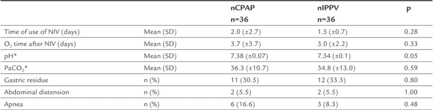
TABLE 2 Respiratory support time and incidence of complications after extubation in 72 infants undergoing nCPAP or nIPPV ventilation.
nCPAP n=36
nIPPV n=36
p
Time of use of NIV (days) Mean (SD) 2.0 (±2.7) 1.5 (±0.7) 0.28
O2 time after NIV (days) Mean (SD) 3.7 (±3.7) 3.0 (±2.2) 0.33
pH* Mean (SD) 7.38 (±0.07) 7.34 (±0.1) 0.05
PaCO2* Mean (SD) 36.3 (±10.7) 34.8 (±13.0) 0.59
Gastric residue n (%) 11 (30.5) 12 (33.3) 0.80
Abdominal distension n (%) 2 (5.5) 2 (5.5) 1.00
Apnea n (%) 6 (16.6) 3 (8.3) 0.48
nCPAP, and in 86.1% of those randomized to the nIPPV group, with no statistical difference. Most of the prema-ture infants received at least one dose of exogenous sur-factant (80.5% in the nIPPV group and 83.3% in the nC-PAP group, p=1.00). The average time on mechanical ventilation (6.2 and 7.3 days respectively, p=0.59) and the average FiO2 prior to extubation (29.9 and 28.8 respec-tively, p=0.71) were similar between the two groups.
The average time of use of the ventilatory methods was two days for nCPAP and one day and a half for the nIPPV group (p=0.28). Also regarding use of the respira-tory assistance methods, inhaled oxygen support time af-ter the suspension of the ventilatory methods was also similar. As for blood gas analysis, pH values in the nIPPV group were lower than in the nCPAP group (7.34 vs. 7.38).
Although this difference was statistically significant (p=0.05), there was no clinical signiicance because both results are in the normal range, and therefore could not interfere in the occurrence of extubation failure. The val-ues of PaCO2 on the irst day after extubation were also similar in both groups.
In our study the occurrence of gastric residue or ab-dominal distension that would prohibit feeding was sim-ilar between the two groups, and none of the patients pre-sented a diagnosis of gastric perforation.
The evaluation of extubation failure among the pre-mature infants who were randomized to the nIPPV group and the nCPAP group showed no statistically signiicant difference. Among the 36 premature infants randomized to receive nIPPV, six (16.6%) presented extubation failure
in comparison to 11 (30.5%) of the 36 premature infants randomized to nCPAP (Chart 1).
We sought to verify if there was a relationship between extubation failure of the two methods and BW, GA, clas-siication of the premature infant, and the MV time. We found no statistically signiicant difference between the association of the characteristics described above and the methods under study, and all adjusted relative risk esti-mates (nIPPV versus nCPAP) were not greatly different
from the gross value of 0.55.
In relation to the factors determining failures, that is, recurrent episodes of apnea (54.5% vs. 16.6%), frequent
decreases in oxygen saturation (27.2% vs. 50%) and
clini-cal signs of respiratory distress (18.1% vs. 33.3%), we did
not ind a statistically signiicant difference between the nCPAP and nIPPV groups. Although the occurrence of apnea in the nCPAP group was numerically higher in re-lation to the nIPPV group (6 vs. 1), this difference was not
signiicant (p=0.30).
Most premature infants participating in the study underwent a cranial ultrasound after extubation, and the results showed no statistically signiicant difference be-tween the two study groups.
The occurrence of side effects was not observed dur-ing the 72 hour period of the study.
D
ISCUSSIONIn recent decades the number of premature infants with a VLBW requiring prolonged mechanical ventilation has increased, and currently the challenge for neonatologists
CHART 1 Extubation failure among the 72 premature infants with respiratory failure submitted to ventilation by nCPAP or nIPPV. nCPAP: nasal continuous positive airway pressure; nIPPV: nasal intermittent positive pressure ventilation.
Total nIPPV
nCPAP
30.5
16.6
23.6
%
35
30
25
20
15
10
5
is no longer treating newborns with low birth weight but those with extremely low birth weight (PN < 1,000 g).14 Efforts to limit the duration of MV have been conduct-ed with the purpose of rconduct-educing not only the incidence of mortality but also morbidity, which is high in this pop-ulation of premature infants. However, early extubation has been accompanied by various dificulties, particular-ly due to instability of the ribcage, the presence of alveo-lar atelectasis and residual pulmonary damage.9 Despite the non-invasive ventilation method used, the extubation failure rate was 23.6%, a similar result to those found in the literature. Barrington et al., in their controlled and randomized study, compared the extubation failure rate of 54 premature infants with BW < 1,250 g and GA of 26.1 weeks, verifying a failure rate of 29.6%.1 Among the 64 pre-mature infants studied by Khalaf et al., around 21.8% showed extubation failure during the use of non-invasive ventilation.6 When assessing extubation in premature in-fants with a BW < 1,000 g, Stefanescu et al. reported fail-ure in 38.2% of their patients.15
In our study, even though the distribution of the fail-ure frequencies showed a numerically lower failfail-ure rate in premature infants treated with nIPPV compared to those undergoing nCPAP, there was no statistically sig-niicant difference between the two methods of ventila-tory support after extubation.
The consensus in the literature is that the nIPPV meth-od can cause gastrointestinal complications due to the possibility of gastric distension leading to suspension of feeding, higher frequency of gastric residue, and even gas-tric perforation. Garland et al. conducted a retrospective evaluation of 20 premature infants that presented gas-tric perforation in order to determine whether the type of ventilatory support used was associated with the oc-currence of gastric perforation.16 The authors found that ventilation through the use of a facial mask or nasal prong favored an increased risk of gastric perforation at around 30% compared to the use of a tracheal tube. Nevertheless, the authors themselves call attention to possible bias in the study, which is the underdiagnosis of necrotizing en-terocolitis. They also point out that most premature in-fants participating in the study received both types of ventilation (via the nasal prong and endotracheal tube), therefore gastric perforation could have occurred during the ventilation period with the tracheal tube, and the symptoms may have manifested during the use of the na-sal prong or the facial mask.16
In our study the occurrence of gastric residue or ab-dominal distension that could prohibit feeding was sim-ilar between the two study groups, and none of the
pa-tients participating in the research presented gastric perforation. Kugelman et al. also did not observe the oc-currence of gastrointestinal complications in their study.17 Similar facts have been described by other authors.9,18,19 Barrington et al. did not demonstrate an increased inci-dence of abdominal distention or food intolerance in the 54 premature infants subject to two non-invasive venti-lation methods, and also did not ind any cases of gastric perforation.1 A similar fact was described by Kiciman et al. among the 14 premature infants studied.10
In some studies we veriied variable rates in relation to extubation failure, sometimes associated with lower birth weight and gestational age.1,15,20,21 As such, we sought to verify if there was a relationship between extubation failure in the two methods and the BW and GA, classii-cation of the premature infant, and the MV time. We found that there was no signiicant difference between the asso-ciation of the characteristics described and the methods under study.
The mechanism explaining the fact that nIPPV is more beneicial or effective than nCPAP has not yet been fully elucidated. Kishore et al. believe that with nIPPV there is better alveolar recruitment, with improved residual func-tional capacity.11 Moretti et al. consider that nIPPV may increase upper airway patency by creating high pressure in the pharynx and promoting intermittent inlation of the pharynx, thereby activating the respiratory rhythm.22 In their study, the authors performed pulmonary func-tion tests and found that during nIPPV the pulmonary volumes (tidal volume and minute volume) were signii-cantly higher compared to nCPAP, and the values of the transcutaneous PaCO2 were also signiicantly lower com-pared to those in the nCPAP group.22 Lin et al. also believe that the intermittent pressure created in the pharynx is higher when using nIPPV, and that the low of air passing through the pharynx can activate the pharyngeal dilator muscles, stimulating breathing and reducing episodes of apnea. According to the authors, increases in chest wall in-cursions and respiratory impulses were observed when the premature infant underwent nIPPV. These impulses were not noted when the premature infant received nCPAP.7 There is evidence that these impulses are induced by nIPPV, and could be responsible for recruiting more alveoli and reversing areas of pulmonary microatelectasis, which ex-plains its success.17
by Stefanescu et al., the occurrence of apnea and brady-cardia was responsible for failure in 58% of the prema-ture infants participating in the study.15 Likewise, Khalaf et al. reported episodes of apnea as being responsible for 41% of failures.6 When evaluating the incidence of apnea in relation to the study group, the authors observed that, in the nCPAP group, the occurrence of apnea was respon-sible for 46.7% of failures, and 32.3% in the nIPPV group, with no signiicant difference, similar to our indings. With respect to the number of episodes of apnea, the pre-mature infants submitted to nIPPV showed a lower num-ber in relation to the group submitted to nCPAP, although this difference was not signiicant. Among the three stud-ies evaluated by Davis et al. in their meta-analysis, only one investigated the occurrence of apnea, and the results of which showed a tendency to reduce the episodes of ap-nea in premature infants randomized to nIPPV, although not statistically signiicant.9
During the study period, whether in the nIPPV or nCPAP method, the occurrence of side effects was not observed, similar to that noted by other authors.1,6,7,11,19
Barrington et al. and Davis et al. were unanimous in afirming that nIPPV is a useful and effective method to increase the beneits of nCPAP in the prevention of extu-bation failure of premature infants in the irst hours af-ter extubation.1,8
Likewise, Khalaf et al. consider nIPPV as more effec-tive than nCPAP in the weaning of premature infants with respiratory distress syndrome (RDS) using mechanical ventilation, and recommend that nIPPV should be used as the primary extubation method even in premature in-fants with impaired pulmonary function.6
Kahramaner et al. also concluded that nIPPV has a better effect than nCPAP after extubation of premature infants, as it reduced the occurrence of atelectasis, the need for re-intubation and even the number of deaths.21
C
ONCLUSIONAlthough the distribution of the failure frequencies showed a numerically lower failure rate in premature infants treat-ed with nIPPV compartreat-ed to those undergoing nCPAP, there was no statistically signiicant difference between the two methods of ventilatory support after extubation. We believe that, to the extent in which mechanical ventilation can be avoided, through the use of non-inva-sive ventilation, particularly in the nIPPV method, there will be a decrease in the incidence of sequelae such as bronchopulmonary dysplasia and cerebral hemorrhage, improving not only survival, but also the quality of life of these patients.
R
ESUMOEstudo controlado e randomizado entre uso de ventila-ção positiva intermitente e pressão positiva contínua em vias aéreas em recém-nascidos prematuros após a extu-bação traqueal
Objetivo: analisar a frequência de falha da extubação em recém-nascidos pré-termo (RNPT) em uso de ventilação mecânica (VM) convencional após a extubação traqueal nos grupos submetidos à ventilação por pressão positiva intermitente por via nasal (nIPPV) e pressão positiva con-tínua em vias aéreas (nCPAP).
Método: foram estudados 72 RNPT portadores de
insu-iciência respiratória, com idade gestacional (IG) ≤ 36 se-manas e peso de nascimento (PN) > 750 g, que necessita-ram de entubação traqueal e ventilação mecânica. O estudo foi controlado e randomizado a im de garantir a aleatoriedade na escolha dos integrantes dos grupos. A randomização foi realizada no momento da extubação por meio de envelopes selados. Falha da extubação foi de-inida como necessidade de reentubação e ventilação me-cânica durante as primeiras 72 horas após a extubação.
Resultados: entre os 36 RN randomizados para nIPPV,
seis (16,6%) apresentaram falha de extubação em com-paração a 11 (30,5%) dos 36 RN randomizados para nC-PAP. Não houve diferença estatística entre os dois gru-pos de estudo em relação a PN, IG, classiicação do RN e tempo de VM. A principal causa de falha da extubação foi a ocorrência de apneia. Complicações gastrointesti-nais e neurológicas não ocorreram nos RNPT partici-pantes do estudo.
Conclusão: constatamos que no grupo dos RNPT
sub-metidos à nIPPV, apesar da falha da extubação ser nume-ricamente menor que nos RNPT submetidos à nCPAP, não houve diferença estatisticamente signiicante entre os dois modos de suporte ventilatório após a extubação.
Palavras-chave: síndrome do desconforto respiratório
do recém-nascido, pressão positiva contínua nas vias aé-reas, ventilação com pressão positiva intermitente, pre-maturo, ensaios clínicos controlados e aleatórios.
R
EFERENCES1. Barrington KJ, Bull D, Finer NN. Randomized trial of nasal synchronized intermittent mandatory ventilation compared with continuous positive airway pressure after extubation of very low birth weight infants. Pediatrics. 2001; 107(4):638-41.
3. Malik RK, Gupta RK. A two year experience in continuous positive pressure ventilation using nasal prongs and pulse oximetry. Med J Armed Forces India. 2003; 59(1):36-9.
4. Ramanathan R, Sekar KC, Rasmussen M, Bhatia J, Soll RF. Nasal intermittent positive pressure ventilation after surfactant treatment for respiratory distress syndrome in preterm infants < 30 weeks’ gestation: a randomized, controlled trial. J Perinatol. 2012; 32(5):336-43.
5. Jobe AH, Bancalari E. Bronchopulmonary dysplasia. Am J Respir Crit Care Med. 2001; 163(7):1723-9.
6. Khalaf MN, Brodsky N, Hurley J, Bhandari V. A prospective randomized, controlled trial comparing synchronized nasal intermittent positive pressure ventilation versus nasal continuous positive airway pressure as modes of extubation. Pediatrics. 2001; 108(1):13-7.
7. Lin CH, Tsay WH, Lin YJ, Wang ST, Yeh TF. Eficacy of nasal intermittent positive pressure ventilation in treating apnea of prematurity. Pediatr Pulmonol. 1998; 26(5):349-53.
8. Davis PG, Lemyre B, De Paoli AG. Nasal intermittent positive pressure ventilation (NIPPV) versus nasal continuous positive airway pressure (NCPAP) for preterm neonates after extubation. Cochrane Database Syst Rev. 2001(3):CD003212. 9. Davis PG, Hendderson-Smart DJ. Nasal continuous positive airways pressure
immediately after extubation for preventing morbidity in preterm infants. Cochrane Database Syst Rev. 2003;(2):CD000143.
10. Kiciman NM, Andreasson B, Bernstein G, Mannino FL, Rich W, Henderson C, et al. Thoracoabdominal motion in newborns during ventilation delivered by endotracheal tube or nasal prongs. Pediatr Pulmonol. 1998; 25(3):175-81. 11. Sai Sunil Kishore M, Dutta S, Kumar P. Early nasal intermittent positive pressure ventilation versus continuous positive airway pressure for respiratory distress syndrome. Acta Paediatr 2009; 98(8):1412-5.
12. Ryan CA, Finer NN, Peters KL. Nasal intermittent positive-pressure ventilation offers no advantages over nasal continuous positive airway pressure in apnea of prematurity. Am J Dis Child. 1989; 143(10):1196-8. 13. Papile LA, Burstein J, Burstein R, Kofler H. Incidence and evolution of
subependymal and intraventricular hemorrhage: a study of infants with weight less than 1500 grams. J Pediatr. 1978; 92(4):529-34.
14. Darlow BA, Cust AE, Donoghue DA. Improved outcomes for very low birthweight infants: evidence from New Zealand national population based data. Arch Dis Child Fetal Neonatal. 2003; 88:F23-8.
15. Stefanescu BM, Murphy WP, Hansell BJ, Fuloria M, Morgan TM, Aschner JL. A randomised, controlled trial comparing two different continuous positive airway pressure systems for successful extubation of extremely low birth weight infants. Pediatrics. 2003; 112(5):1031-8.
16. Garland JS, Nelson DB, Rice T, Neu J. Increased risk of gastrointestinal perforation in neonates mechanically ventilated with either face mask or nasal prongs. Pediatrics. 1985; 76(3):406-10.
17. Kugelman A, Feferkorn I, Riskin A, Chistyakov I, Kaufman B, Bader D. Nasal intermittent mandatory ventilation versus nasal continuous positive airway pressure for respiratory distress syndrome: a randomized, controlled, prospective study. J Pediatr. 2007; 150(5):521-6.
18. De Paoli AG, Davis PG, Lemyre B. Nasal continuous pressure versus nasal intermittent positive pressure ventilation for preterm neonates: a systematic review and meta-analysis. Acta Paediatr. 2003; 92(1):70-5.
19. Khorana M, Paradeevisut H, Sangtawesin V, Kanjanapatanakul W, Chotigeat U, Ayutthaya JK. A randomized trial of non-synchronized nasopharyngeal intermittent mandatory ventilation (nsNIMV) vs. nasal continuous positive airway pressure (NCPAP) in the prevention of extubation failure in pre-term < 1,500 grams. J Med Assoc Thai. 2008; 91 Suppl 3:136-42.
20. Dimitrou G, Greenough A, Endo A, Cherian S, Rafferty GF. Prediction of extubation failure in preterm infants. Arch dis Child Fetal Neonatal Ed. 2002; 86(1):F32-5.
21. Kahramaner Z, Erdemir A, Turkoglu E, Cosar H, Suctuoglu S, Ozer EA. Unsyncronized nasal intermitente positive pressure versus nasal continuous positive airway pressure in preterm infants after extubation. J Matern Fetal Neonatal Med. 2014; 27(9):926-9.