1984-1462/© 2015 Sociedade de Pediatria de São Paulo. Published by Elsevier Editora Ltda. All rights reserved.
REVISTA PAULISTA
DE PEDIATRIA
www.rpped.com.brCASE REPORT
Early manifestations of cystic ibrosis in a premature
patient with complex meconium ileus at birth
Ieda Regina Lopes del Ciampo*, Tainara Queiroz Oliveira, Luiz Antonio del Ciampo,
Regina Sawamura, Lidia Alice Gomes Monteiro Marin Torres, Albin Eugenio Augustin,
Maria Inez Machado Fernandes
Escola de Medicina de Ribeirão Preto, Universidade de São Paulo (USP), Ribeirão Preto, SP, Brazil
Received 3 October 2014; accepted 25 December 2014
KEYWORDS
Cystic ibrosis;
Meconium ileus; Prematurity
Abstract
Objective: To report a case of a preterm infant with complex meconium ileus at birth
and cystic ibrosis.
Case description: A male infant was born by vaginal delivery at 33 weeks and 5 days of gestational age with respiratory distress and severe abdominal distension. The
explora-tory laparotomy in the irst day of life identiied meconium ileus and secondary perito -nitis. Ileal resection and ileostomy were performed, followed by reconstruction of the
bowel transit at 20 days of life. At 11 days of life, the irst immunoreactive trypsinogen
(IRT) was 154 ng/mL (reference value = 70), and oral pancreatic enzymes replacement therapy was started. After 23 days, the second IRT was 172ng/mL (reference value = 70). At 35 days of age he was discharged with referrals to primary care and to a special clinic for CF for the determination of sweat chloride. He was received in the outpatient clinic for neonatal screening for CF at 65 days of life presenting malnutrition and respiratory distress. The sweat chloride test was performed, with a positive result (126mEq/L).
Comments: This case illustrates the rapid evolution of CF in a premature patient with
complex meconium ileus as the irst clinical manifestation.
© 2015 Sociedade de Pediatria de São Paulo. Published by Elsevier Editora Ltda. All rights reserved.
DOI of original article: http://dx.doi.org/10.1016/j.rpped.2014.12.004 *Corresponding author.
Manifestações precoces da ibrose cística em paciente prematuro com íleo meconial complexo ao nascimento
Resumo
Objetivo: Relatar o caso de um recém-nascido prematuro com íleo meconial complexo e
ibrose cística.
Descrição do caso: Recém-nascido do sexo masculino nasceu de parto vaginal com 33 se-manas e cinco dias de idade gestacional e apresentou desconforto respiratório e disten-são abdominal grave. Foi submetido à laparotomia exploratória no primeiro dia de vida
e identiicado íleo meconial com peritonite secundária. Foram feitas ressecção ileal e ileostomia, com reconstrução do trânsito intestinal aos 20 dias de vida. Com 11 dias de
idade, a primeira dosagem sérica de tripsina imunorreativa (TIR) foi 154ng/mL (valor
de referência = 70) e optou-se pelo início da terapia de reposição oral de enzimas pan
-creáticas. Após 23 dias, a segunda TIR foi 172ng/mL (valor de referência = 70). Recebeu alta com 35 dias de vida com encaminhamentos à rede básica de saúde e ao serviço de referência para a detecção de ibrose cística. Foi atendido no ambulatório de triagem neonatal para ibrose cística aos 65 dias de vida e apresentava desnutrição e desconforto
respiratório. O resultado do teste do cloro no suor foi positivo (126 mEq/L).
Comentários: O caso ilustra a rápida evolução da ibrose cística em um paciente prema
-turo com íleo meconial complexo como primeira manifestação clínica.
© 2015 Sociedade de Pediatria de São Paulo. Publicado por Elsevier Editora Ltda. Todos os direitos reservados.
PALAVRAS-CHAVE
Fibrose cística; Íleo meconial; Prematuridade
Introduction
Cystic Fibrosis (CF) is the most prevalent lethal autosomal recessive disorder, affecting 1:2,000 caucasians. It is caused by the alteration of a gene located on the long arm of chro-mosome 7 that encodes a protein of 1,480 amino acids, the cystic fibrosis transmembrane conductance regulator (CFTR), which functions as a chloride channel on the apical membrane of epithelial cells.1 This alteration results in a
change of the viscosity of secretions, with the production of thick mucus, leading mainly to malabsorption, loss of electrolytes in sweat and alteration of pulmonary secre-tions. There are more than 1,900 known genetic mutations, as well as disease modifying genes.2 This phenotypic
het-erogeneity involves different clinical presentations, rang-ing from mild to severe, which can determine a lethal out-come. The classic presentation of CF is chronic lung disease (recurrent pulmonary infections), exocrine pancreatic insufficiency (diarrhea and malnutrition), loss of salt, and obstructive azoospermia syndrome.3 In cases without
clini-cal manifestations suggestive of CF in the first month of life, neonatal screening may lead to early detection and allows immediate treatment of pancreatic insufficiency, nutritional deficiencies and pulmonary involvement, improving survival and facilitating the design of treatment strategies.4
Less frequently, meconium ileus (MI) may be the first manifestation of CF in the neonatal period, occurring in approximately 20% of patients with pancreatic insufficien-cy. This clinical picture is caused by obstruction of the ter-minal ileus with thick meconium containing high amounts of protein. Complex MI is a severe condition, significantly more frequent in patients without CF of lower gestational age and birth weight than in patients with CF. MI is
classi-fied as complex when associated with ileal perforation.5,6
About 80% of cases of MI are due to CF, and it would be ideal to perform an early sweat chloride test before 48 hours of life, although this is not always feasible.7,8 Children
with MI appear to have normal pulmonary function at CF diagnosis, with slower progression of lung disease than those diagnosed due to respiratory symptoms.9,10 However,
it is currently believed that lung inflammation may occur early, and may even precede the onset of infection in infants with newly diagnosed cystic fibrosis.11
The objective of the present communication is to report the case of a child with complex MI who had a poor early evolution despite the clinical suspicion of CF.
Case description
Parenteral nutrition was gradually transitioned to infant formula up to a volume of 160mL/kg/day. At 11 days of life the result of the first immunoreactive trypsin (IRT) was 154ng/mL, and oral pancreatic enzymes replacement ther-apy was started (1,000U/kg/dose every 3 hours before feeding). After 23 days, the second IRT was 172ng/mL. At 35 days of age he was weighing 2,040 g and was discharged with referrals to primary care and to a special clinic for CF for the determination of sweat chloride.
After 65 days of life, the sweat chloride test was per-formed in the reference service with a positive result (126mEq/L). That same day, the patient was attended at the follow-up clinic for neonatal screening for CF. According to his mother, the same guidelines were maintained regard-ing the volume of infant formula and the dosage of pancre-atic enzymes, which she was instructed to offer mashed in water. He weighed 2,610 g and his length was 48cm. He had gained 19g/day after discharge. He had tachypnea (62 respi-ratory cycles/minute), pallor (2/4+) and persistent dry cough. His chest X-ray revealed pulmonary hyperinflation and condensation compatible with pneumonic disease. The patient was hospitalized and the laboratory tests per-formed during hospitalization are shown in Table 1. The C-reactive protein (CRP) test performed at admission was
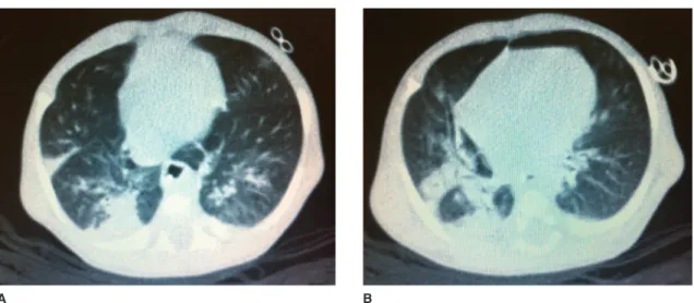
18.8mg/dL (Reference Value <0.5mg/dL by turbidimetry). Laboratory tests for respiratory syncytial virus, adenovirus and influenza, as well as blood cultures were negative. A pulmonary CT scan was performed on the 6th day of hos-pital stay (Fig. 1).
An enteral diet was prescribed, with a volume of 150mL/ kg/day of semi-elemental formula at 1:25 dilution. The dose of pancreatic enzymes was adjusted to 5,000 U before feed-ing, every 3 hours. A transfusion of red blood cells was per-formed. Enteral sodium (4mEq/kg/day) and potassium (1mEq/kg/day) were replaced. Multivitamins (24 drops/day) and zinc sulfate (1mg/kg/day) were prescribed. After cul-ture, and based on the microorganism isolated from the upper respiratory tract secretion (Table 1), ceftazi-dime (200mg/kg/day), gentamicin (5mg/kg/day), and oxa-cillin (200mg/kg/day) were prescribed. Due to ileal resec-tion, vitamin B12 (100mcg) was administered intramuscular-ly. On the seventh day of hospital stay, due to progressive worsening of the respiratory pattern, noninvasive ventilation (CPAP) was applied for two weeks. After stabilization of pul-monary disease he progressed with satisfactory weight gain. He was discharged after 40 days, weighing 3,490 g, without antibiotics, with sodium replacement (adjusted to 2mEq/kg/ day) and ferrous sulfate (4mg/kg/day).
Table 1 Exams performed at the beginning and at the end of hospitalization of the patient at the referral service for CF management.
Laboratory test Reference values Day of hospitalization
1 2 6 35
Serum
Hb (g/dL) 10-14 6.6 9.4 8.8
Ht (%) 28-42 21 30 26
WBC (/µL) 5,000-15,000 15,000 7,300
Differential (%) (7B/5Me/2My) No immature forms
Platelets (/µL) 150-300.103 662,000 532,000 391,000
Na+ (mEq/L) 135-145 125 121 137 135
K+ (mEq/L) 3.5-5.0 3.1 3.1 4.5 4.3
Cl– (mmol/L) 98-1,107 94 92 101 105
Total Protein (g/dL) 6.0 5.2 5.7
Albumin (g/dL) 3.5 3.2 4.0
B12 Vitamin (pg/mL) 175-878 >1,000
Zinc (mcg/%) 50-120 74.5
Arterial Blood Gas
pH (mmHg) 7.35-7.45 7.45 7.43 7.35
pO2 (mmHg) 75-100 61.0 72.7 78.4
pCO2 (mmHg) 35-45 37.6 40.0 40.3
HCO3 (mmHg) 21-28 25.6 25.9 21.7
BE (mmol/L) 0- ±2 +2 +1.9 –3.0
SaO2 (%) 95-98 94.1 95 96
URT Secretion
Culture Staphylococcus
aureus
Pseudomonas aeruginosa, Burkholderia cepaccia
Negative
Faecal
Steatrocrit (%) <10 — — 50 35
Discussion
It is known that the confirmation of CF may be difficult during the first days of life, but in the presence of meconi-um ileus this differential diagnosis should be compulsorily considered. The sweat chloride test can be performed after 48 hours of life, but more reliable results are obtained after the second week of life, with the patient weighing more than 2 kg, showing adequate hydration status and without significant systemic disease.12 The sweat chloride
test is the gold standard for the diagnosis of CF; however, some situations may alter the results, such as dehydration, low weight, skin rash or a bad general condition.13,14 Despite
the importance of an early sweat chloride test, the clinical status of the present patient was not appropriate for test-ing him durtest-ing the first days of life.
Cystic fibrosis transmembrane conductance regulator (CFTR) genetic analysis also helps the diagnosis of CF when it detects two known mutations.15 This exam would have
been useful for the present patient if it had detected the two characteristic mutations for CF, however, the exam is not routinely available in our center due to its high cost. The nationwide Brazilian Newborn Screening Program start-ed in 2011, although it was first implementstart-ed in 2010 in the state of São Paulo.16 The method of two IRT samples was
adopted, with the first sample being collected between 3 and 7 days of life and the second up to 30 days of life. If
both (IRT) samples are positive (Reference Value ≥70ng/mL),
CF is confirmed by two positive sweat chloride tests
(Reference Value ≥60mEq/L). A sweat chloride test between
30 and 60mEq/L is “borderline” for neonates and does not immediately exclude or confirm the disease.17
Although the importance of two IRTs neonatal screenings for CF detection, the benefit seems smaller for infants with meconium ileus because neonates with CF and MI may have low initial IRT values.18 Even if IRT levels remain elevated,
this would simply alert to the possible presence of CF with-out confirming the disease, because several factors increase
the possibility of false-positive IRT values, including perina-tal stress.19 Although low IRT values were expected for the
present patient, the levels detected were high, supporting the suspicion of CF and probably also the decision of empir-ically starting the treatment of pancreatic insufficiency due to CF.
Neonates with MI should receive specific treatment for pancreatic insufficiency while pending confirmation of CF by the sweat chloride test. Weight gain during hospital stay indicates a good response.20 Current reports state that MI is
no longer regarded as a poor prognostic factor in patients with effective treatment for CF.21 The present patient was
discharged using pancreatic enzymes, with the mother dis-solving them in water. This procedure illustrates the impor-tance of an early and frequent monitoring of children with a suspected chronic, rare and severe disease by the refer-ence service for CF treatment, both regarding support to the family and the refinement of the guidelines offered, so that they will be closely followed. It should be emphasized that this type of care involves, but does not replace patient monitoring by a general pediatrician. The management of pancreatic insufficiency involves the oral ingestion of intact microspheres of pancreatic enzymes immediately before breastfeeding, ranging from 2,000 to 4,000IU lipase for each 120mL of formula or breastfeeding. Although less physiological, the calculation can also be performed using a dose of 1,000IU lipase/kg/meal for children younger than 4 years, avoiding doses higher than 2,500IU/kg/meal and 10,000IU/kg/day, which could trigger fibrosing colonopa-thy.22
The lungs may be affected since the period of CF neona-tal screening, with 81% of cases showing structural abnor-malities, 45% bronchial wall thickening, and 21% lung infec-tion.23 Cough and dyspnea in neonates and infants indicate
the need to include CF in the list of differential diagnoses. Pulmonary inflammation or infection may already be pres-ent, with Staphylococcus aureus being the microorganism most frequently detected, followed by Pseudomonas
aeru-A B
Figure 1 Pulmonary computer scanner images. A. Reveals several bands of atelectasis with bronchiectasis and some bronchi
with thickened walls. B. Reveals other bronchial wall thickening, peribronchial inlammation, and extensive area of
ginosa, with significant respiratory symptoms. Researchers observed that infants with cystic fibrosis detected by new-born screening may have lung disease with bacterial infec-tion since the first days of life.24,25 This is a source of
con-cern, because it is linked to early onset of bronchiectasis and more severe lung inflammation.
The present case report illustrates the rapid multisystem evolution of CF in a premature patient with complex MI as the first clinical manifestation. It also shows the presence of paradoxical results of neonatal screening (IRT/IRT) in patients with MI, and the difficulties of the early chloride sweat testing and of genotyping for CF in MI cases. Furthermore, it emphasizes the importance of a rapid referral of patients with MI and suspected CF to a team of experts.
Funding
This study did not receive funding.
Conlicts of interest
The authors declare no conflicts of interest.
References
1. Raskin S, Phillips JA 3rd, Krishnamani MR, Vnencak-Jones C,
Parker RA, Rozov T, et al. DNA analysis of cystic ibrosis in Brazil by direct PCR ampliication from Guthrie cards. Am J Med Genet. 1993;46:665-9.
2. Stanke F, Becker T, Kumar V, Hedtfeld S, Becker C, Cuppens H,
et al. Genes that determine immunology and inlammation
modify the basic defect of impaired ion conductance in cystic
ibrosis epithelia. J Med Genet Jan. 2011;48:24-31.
3. Sing CF, Risser DR, Howatt WF, Erickson RP. Phenotypic
heterogeneity in cystic ibrosis. Am J Med Genet.
1982;13:179-95.
4. Comeau AM, Accurso FJ, White TB, Campbell PW, Hoffman G, Parad RB, et al. Guidelines for implementation of cystic ibrosis
newborn screening programs: Cystic Fibrosis Foundation workshop report. Pediatrics. 2007;119:e495-518.
5. Gorter RR, Karimi A, Sleeboom C, Kneepkens CM, Heij HA.
Clinical and genetic characteristics of meconium ileus in
newborns with and without cystic ibrosis. J Pediatr
Gastroenterol Nutr. 2010;50:569-72.
6. Ziegler MM. Meconium ileus. Curr Probl Surg. 1994;31:731-77. 7. Fakhoury K, Durie PR, Levison H, Canny GJ. Meconium ileus in
the absence of cystic ibrosis. Arch Dis Child. 1992;67:1204-6.
8. AACB, Sweat Testing Working Party, Australian Guidelines for
the Performance of the Sweat Test for the Diagnosis of Cystic Fibrosis. Clin Biochem. 2006;27 Suppl i:S1-9.
9. Tepper RS, Hiatt P, Eigen H, Scott P, Grosfeld J, Cohen M. Infants with cystic ibrosis: pulmonary function at diagnosis.
Pediatr Pulmonol. 1988;5:15-8.
10. Hudson I, Phelan PD. Are sex, age at diagnosis, or mode of
presentation prognostic factors for cystic ibrosis? Pediatr
Pulmonol. 1987;3:288-97.
11. Armstrong DS, Grimwood K, Carzino R, Carlin JB, Olinsky A, Phelan PD. Lower respiratory infection and inlammation in infants with newly diagnosed cystic ibrosis. BMJ. 1995;310:1571-2.
12. Farrell PM, Rosenstein BJ, White TB, Accurso FJ, Castellani C,
Cutting GR, et al. Guidelines for diagnosis of cystic ibrosis in
newborns through older adults: Cystic Fibrosis Foundation consensus report. J Pediatr. 2008;153:S4-14.
13. Littlewood JM. The sweat test. Arch Dis Child. 1986;61:1041-3. 14. Di Sant’Agnese PA, Darling RC, Perera GA, Shea E. Abnormal
electrolyte composition of sweat in cystic ibrosis of the pancreas; clinical signiicance and relationship to the disease.
Pediatrics. 1953;12:549-63.
15. Castellani C, Cuppens H, Macek M Jr, Cassiman JJ, Kerem E, Durie P, et al. Consensus on the use and interpretation of cystic
ibrosis mutation analysis in clinical practice. J Cyst Fibros.
2008;7:179-96.
16. Kerem E, Conway S, Elborn S, Heijerman H. Consensus
Committee. Standards of care for patients with cystic ibrosis:
a European Consensus. J Cyst Fibros. 2005;4:7-26.
17. São Paulo. Justiça Federal [Internet page]. Ação Pública n. (0021921-14.2009.403.6100. Exame para diagnosticar ibrose cística em recém-nascidos é obrigatório [accessed 30 June 2014]. Available from:
http://www.jfsp.jus.br/20110329-ibrosecistica/
18. Heeley AF, Bangert SK. The neonatal detection of cystic ibrosis
by measurement of immunoreactive trypsin in blood. Ann Clin Biochem. 1992;29:361-76.
19. Rock MJ, Mischler EH, Farrell PM, Bruns WT, Hassemer DJ, Laessig RH. Immunoreactive trypsinogen screening for cystic
ibrosis: characterisation of infants with a false-positive
screening test. Pediatr Pulmonol. 1989;6:42-8.
20. Ribeiro JD, Ribeiro MA, Ribeiro AF. Controversies in cystic
ibrosis – From pediatrician to specialist. J Pediatr (Rio J).
2002;78Suppl 2:S171-86.
21. Corey M, Farewell V. Determinants of mortality from cystic ibrosis
in Canada, 1970-1989. Am J Epidemiol. 1996;143:1007-17. 22. Sinaasappel M, Stern M, Littlewood J, Wolfe S, Steinkamp G,
Heijerman HG, et al. Nutrition in patients with cystic ibrosis:
a European Consensus. J Cyst Fibros. 2002;1:51-75.
23. Mott LS, Park J, Murray CP, Gangell CL, De Klerk NH, Robinson
PJ, et al. Progression of early structural lung disease in young
children with cystic ibrosis assessed using CT. Thorax.
2012;67:509-16.
24. Sly PD, Brennan S, Gangell C, De Klerk N, Murray C, Mott L, et al. Lung disease at diagnosis in infants with cystic ibro-sis
detected by newborn screening. Am J Resp Crit Care Med. 2009;180:146-52.
25. Stick SM, Brennan S, Murray C, Douglas T, Von Ungern-Sternberg
BS, Garratt LW, et al. Bronchiectasis in infants and preschool children diagnosed with cystic ibrosis after newborn screening.