CASE REPORT
93
Bras J Rheumatol 2010;50(1):90-5
Received on 10/02/2009. Approved on 11/14/2009. We declare no conflict of interest.
Rheumatology Department of the University Hospital of the Medical School of Universidade Federal de Mato Grosso do Sul (FAMED – UFMS)
1. Professor of the Internal Medicine Department of the Medical School (FAMED)/ UFMS; chief of the Rheumatology Department of the University Hospital of UFMS 2. Rheumatology resident (fourth year) of the Rheumatology Department of the University Hospital of UFMS
3. Rheumatology resident (third year) of the Rheumatology Department of the University Hospital of UFMS
Correspondence to: Izaias Pereira da Costa. Av Senador Filinto Muller, s/n. Hospital Universitário / UFMS. Serviço de Reumatologia. Campo Grande, MS – CEP: 79080-190. Tel: 55 67 3349-3203. E-mail: [email protected]
Polymyositis associated with lymphocytic
arteritis of the central nervous system
Izaias Pereira da Costa1, Elisangela Possebon Pradebon2, Julia Villegas Campos2,
Fabrícia Santos Melo3, Flávia Midori Arakaki Ayres Tavares3
ABSTRACT
Central Nervous System (CNS) complications in idiopathic inlammatory myopathies are seldom reported. The authors describe the case of a 48-year old female with polymyositis and positive anti-Jo-1 autoantibody who, after ive years of
evolution, developed extensive CNS demyelinating injury associated with lymphocytic arteritis.
Keywords: polymyositis, central nervous system vasculitis, myositis-speciic autoantibodies, anti-Jo-1.
INTRODUCTION
Idiopathic inflammatory myopathies represent a group of diseases characterized by proximal weakness,
non-suppurative inlammation of skeletal muscles, and production
of autoantibodies, without sensorial changes and deep tendon
relexes abnormalities.1,2
Polymyositis is included in this group of diseases, and an interaction between environmental and genetic factors seems to be responsible for the onset of this disorder, clinical
manifestations, and autoantibody proile.3 It has characteristic
histologic changes.1 Women are affected more often than men,
at a proportion of 2:1.1
The classiication criteria of Bohan and Peter, published in 1975 and modiied by Targoff and Miller in 1997, are still used.4 In
case of diagnostic uncertainties, the criteria proposed by Dalakas (1991), revised in 2003, which uses immunohistochemistry in the differential diagnosis, can be used.4,5
Anti-synthetase antibodies, deemed specific for inflammatory myopathies, are directed against different aminoacyl-tRNA synthetase enzymes found in the cytoplasm: anti-PL-7, anti-PL-12. Anti-EJ, anti-OJ, anti-Ks, anti-Zo,
anti-tyrosyl-tRNA synthetase, and anti-Jo1; the latter is more prevalent in patients with polymyositis.3,6,7
Recent studies indicate a role for anti-Jo1 antibodies in the induction and maintenance of the so-called “anti-synthetase syndrome”,6,8,9 and its serum levels correlate with disease
activity.6,7
Among the extramuscular manifestations of polymyositis, pulmonary involvement is the most common.3,10,11 On the other
hand, complications of the central nervous system (CNS) are rarely reported.12
CASE REPORT
This is a 48 years old female admitted to our service in 2002 with proximal muscular weakness of the lower limbs for “a few months”, which became more severe three months before admission, with involvement of the scapular girdle and dysphagia for solid foods. She also had type II diabetes mellitus with adequate control of blood glucose levels with metformin.
Physical exam showed grade III proximal muscular
strength in all four limbs, and preserved deep tendon relexes
Costa etal.
94 Bras J Rheumatol 2010;50(1):90-5
Except for alanine aminotransferase (ALT) levels, which were three times higher, and ESR of 43 mm/1st hour, all other
routine laboratorial parameters and muscle enzymes, including CK and aldolase, were normal. Electroneuromyography of the limbs showed myopathic pattern without changes in the peripheral nervous system. Biopsy of the right deltoid muscle
showed scarce muscle ibers permeated by globules of adipose
tissue, suggesting adipose substitution.
The patient was treated with pulse therapy with methylprednisolone (1 g) and cyclophosphamide (20 mg/kg), with complete remission of her symptoms; she was maintained on prednisone (1 mg/kg/d), programmed for gradual dose reduction.
After five months, still asymptomatic, the patient discontinued her treatment.
In 2007, the patient had recurrence of the muscular weakness, psychomotor agitation, hallucinations, signs of dementia, and left spastic hypertonia. She was using prednisone (10 mg/d) irregularly, glibenclamide, and diazepam.
Laboratorial exams, including serology for the main infectious diseases, were unchanged. Among all antibodies investigated, only anti-Jo-1 was positive. The cerebral spinal
luid had increased of protein levels (74 mg%) and glucose
(94 mg%).
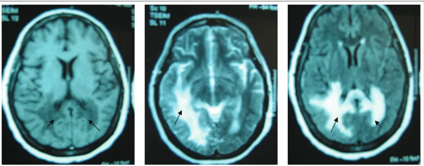
Magnetic resonance imaging of the head showed extensive bilateral signal changes, right temporo-parieto-occipital and left parieto-occipital, with involvement of the periventricular white matter (Figure 1).
Cerebral biopsy showed diffuse hystiocytic iniltration of
demyelinated areas associated with non-necrotizing chronic lymphocytic angiitis with reduction in the lumen of the vessels (Figure 2). Investigation for acid-fast bacilli, fungi, and neoplastic cells was negative.
After six cycles of monthly pulse therapy with methylprednisolone (1 g) and cyclophosphamide (20 mg/kg), and prednisone (0.5 mg/kg) between cycles, improvement of hallucinations and psychomotor agitation was observed.
Four months after the discontinuation of pulse therapy, and while on 20 mg/d of prednisone, the level of protein in the CSF increased even more (468 mg%). After new cycles of pulse therapy with cyclophosphamide (20 mg/kg) and methylprednisolone (1 g), the levels of protein in the CSF reduced to 78 mg%. Objective proximal motor strength and left spastic hypertonia improved; however, the patient is not able to walk.
DISCUSSION
In the present case, the patient presented inlammatory myo -pathy.
The initial fast response to corticosteroids and the absence of distal muscular involvement of the limbs ruled out the diagnosis of inclusion body myositis. Dermatomyositis was excluded due to the absence of cutaneous manifestations.
Electrolyte abnormalities, infections, and drug toxicity were excluded as the cause of CNS damage.
Figure 1. MRI of the head showing right temporal-parietal-occipital and left parietal-occipital lesions, affecting especially the periventricular white
Polymyositis associated with lymphocytic arteritis of the central nervous system
95
Bras J Rheumatol 2010;50(1):90-5
The fact that the majority of the muscle enzymes of the patient were normal has been reported in the literature.2,6 Besides,
adipose substitution predominated over the inflammatory process, explaining the moderate elevation of ALT.
Therefore, the case fulilled the criteria of Bohan and Peter
for polymyositis, with positive anti-Jo-1.
Very few cases of CNS involvement in idiopathic
inlammatory myopathy have been reported.12,13 Reports on
complications of polymyositis with CNS damage, similar to the case presented here, were not found.
In the studies reviewed, several etiological hypotheses for the CNS involvement were raised. Among them, vascular involvement, which could be due to: vasculopathy, as part of the clinical manifestations of dermatomyositis, hypoxic ischemic encephalopathy secondary to cerebral hypoperfusion, hypertensive encephalopathy, and as a consequence of corticosteroid use.12
The use of glucocorticoids by the patient does not justify the myopathic presentation, since it affects almost exclusively patients treated with more than 30 mg/day of prednisone or the equivalent, and muscular electromyographic and histological changes are different.14
The presence of lymphocytic iniltrate in the arterial wall,
with reduction in vascular lumen, does not only characterize
inlammatory arteritis, but it also excludes the possibility that
the cerebral lesion was secondary to hypoxia or hypertensive encephalopathy. Besides, lymphocytic arteritis is not compatible with diabetic arteriopathy.
Studies have demonstrated that the production of myositis specific auto-antibodies correlates with the pathogenic mechanisms of idiopathic inflammatory myopathies and
extra-muscular manifestations.6,7,8,9 Anti-Jo-1 antibodies would
have a pathogenic role in the organic lesion, triggering a local
inlammatory process and systemic immune response.6,8,9
In the present case, we raise the hypothesis of the possible role of anti-Jo-1 antibodies in the pathogenesis of lymphocytic arteritis, which would have caused the CNS damage, indicated by elevated protein levels in the CSF, as well as the favorable clinical response after treatment with immunosuppressors.
REFERÊNCIAS REFERENCE
1. Wortmann RL. Inflammatory diseases of muscle and other myopathies. In: Harris Jr ED, Budd RC, Firestein GS, Genovese MC, Sergent JS, Ruddy S et al. (eds.). Kelley’s Textbook of Rheumatology. 7 ed. Philadelphia: Elsevier Saunders, 2005, pp. 1309-35.
2. Sultan SM. Clinical assessment in adult onset idiopathic
inlammatory myopathy. Curr Opin Rheumatol 2004; 16:668-72.
3. Hassan AB, Fathi M, Dastmalchi M, Lundberg IE, Padyukov L. Genetically determined imbalance between serum levels of tumour factor (TNF) and interleukin (IL)-10 is associated with anti-Jo-1 and anti-Ro52 autoantibodies in patients with poly- and dermatomyositis. J Autoimmunity 2006; 27:62-8.
4. Baer AN. Advances in the therapy of idiopathic inlammatory
myopathies. Curr Opin Rheumatol 2006; 18:236-41.
5. Dalakas MC, Hohlfeld R. Polymyositis and dermatomyositis. Lancet 2003; 362:971-82.
6. Stone KB, Oddis CV, Fertig N, Katsumata Y, Lucas M, Vogt M et al. Anti-Jo-1 antibody levels correlate with disease activity in idiopathic
inlammatory myopathy. Arthritis Rheum 2007; 56 (9):3125-31.
7. Casciola-Rosen L. Autoimmune myositis: new concepts for disease initiation and propagation. Curr Opin Rheumatol 2005; 17:699-700. 8. Mimori T, Imura Y, Nakashima R, Yoshifuji H. Autoantibodies
in idiopathic inlammatory myopathy: an update on clinical and
pathophysiological significance. Curr Opin Rheumatol 2007; 19:523-9.
9. Sordet C, Goetz J, Sibilia J. Contribution of autoantibodies to the
diagnosis and nosology of inlammatory muscle disease. Joint Bone
Spine 2006; 73:646-54.
10. Songcharoen S, Raju SF, Pennebaker JB. Interstitial lung disease in polymyositis and dermatomyositis. J Rheumatol 1980; 7:353-60. 11. Lakhanpal S, Lie JT, Conn DL, Martin WJ. Pulmonary disease in
plymyositis/dermatomyositis: a clinicopathological analysis of 65 autopsy cases. Ann Rheum Dis 1987; 46:23-9.
12. Ramanan AV, Sawhney S, Murray KJ. Central nervous system complications in two cases of juvenile onset dermatomyositis. Rheumatology 2001; 40:1293-8.
13. Jimenez C, Rowe PC, Keene D. Cardiac and central nervous system vasculitis in a child with dermatomyositis. J Child Neurol 1994; 9:297-300.
14. Jacobs JWG, Bijlsma JWJ. Glucocorticoid therapy. In: Harris Jr ED, Budd RC, Firestein GS, Genovese MC, Sergent JS, Ruddy S et
al. (eds.). Kelley’s Textbook of Rheumatology. 7 ed. Philadelphia:
Elsevier Saunders, 2005, pp. 859-76.
Figure 2. Brain tissue showing diffuse lymphocytic iniltrate and
non-necrotizing chronic angiitis, with reduction in the lumen of the blood