artigo 320
CASE REPORT
Declaramos inexistência de conflito de interesses neste artigo
1 – Doctor of the Shoulder and Elbow Group of the Sports Traumatology Center of the Department of Orthopedics and Traumatology, Universidade Federal de São Paulo (CETE/Unifesp).
2 – Postgraduate Student, Assistant Doctor of the Traumatology Center of the Department of Orthopedics and Traumatology, Federal University of São Paulo/EPM. 3 – Doctor of the Shoulder and Elbow Group of the Sports Traumatology Center of the Department of Orthopedics and Traumatology, Universidade Federal de São Paulo
(CETE/Unifesp).
4 – Doctor and Head of the Traumatology Sector of the Department of Orthopedics and Traumatology, Federal University of São Paulo, Brazil. 5 – Doctorate Professor of the Department of Orthopedics and Traumatology, Federal University of São Paulo, Brazil.
Work carried out by the Department of Orthopedics and Traumatology of the Federal University of São Paulo, SP – Brazil (DOT-Unifesp/EPM). Correspondence: Rua Borges Lagoa, 783 – 5o andar – Vila Clementino – 04038-032 – São Paulo, SP. E-mail: [email protected] Work received for publication: March 30, 2010; Accepted for publication: May11, 2010.
subclaVian Vein ThroMbosis folloWing fracTure
of The claVicle: case reporT
Bernardo Barcellos Terra1, Luiz Fernando Cocco2, Benno Ejnisman3, Hélio Jorge Alvachian Fernandes4,
Fernando Baldy dos Reis5
InTRODUCTIOn
Fractures of the clavicles are common in ortho-pedic practice, representing 5-12% of all fractures and 44% of injuries involving the upper cingulate.
Neurovascular injuries associated with closed
fractures of the clavicle are rare(1-3) and are
associated mainly with penetrating traumas(4-7). The
most common injury mechanism is falling onto the palm of the hand, or onto a point on the shoulder, or direct or indirect traumas associated with contact
sports(1,3,8). Deep vein thrombosis of the upper
limbs involving the subclavian and/or axillary vein is rare compared with deep vein thrombosis of the lower limbs, constituting 1-4% of deep vein
thromboses of the limbs(9,10). Few cases are cited in
the orthopedic literature(11,12). The objective of this
case report is to report on this rare condition, which if not diagnosed and conducted correctly, can lead to fatal complications.
CASE REPORT
Female patient, aged 70, reported that two days ago she suffered onset of a condition of diffuse edema throughout the upper right limb associated with hyperemia, without a history of recent trauma.
She reports that on 05/20/09 (two months and 15 days ago) she fractured her right clavicle after falling onto the ground (Figure 1). She sought medical attendance on that occasion and was prescribed the use of a figure 8 shoulder brace for three months. She reports that two days ago she removed the brace due to strong pain in the upper thoracic spine, when she noted the appearance of edema and hyperemia on the upper right limb. She therefore visited the first aid department of this service, where her clinical history was taken, and she was submitted to physical examination, in which the following alterations to the upper right cingulate were observed:
Rev Bras Ortop. 2011;46(2):215-8 ABSTRACT
Deep vein thrombosis in the upper limbs is uncommon in the orthopedic literature. We report on a case of subclavian vein thrombosis that occurred during conservative treatment of a fracture in the middle third of the clavicle.
This is difficult to diagnose and requires a high degree of suspicion. Treating it may prevent fatal thromboembolism. In some rare cases, it has been described in association with fractures of the clavicle.
216
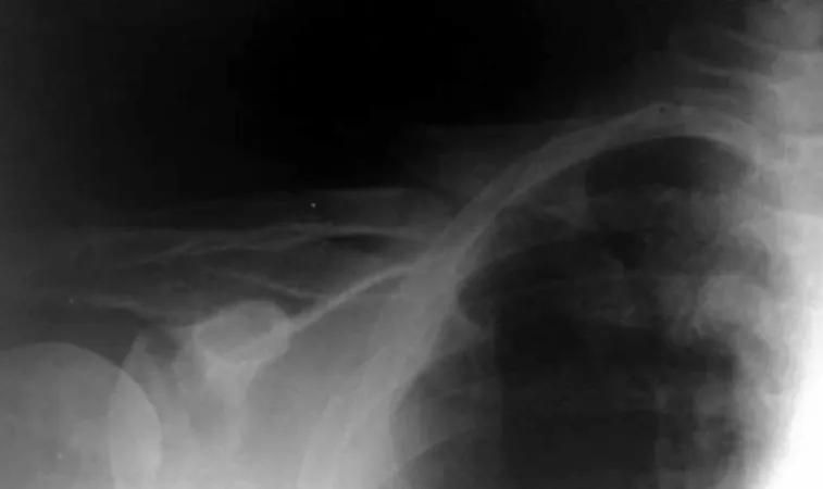
Figure 1 – Fracture of the clavicle following trauma.
Figure 2 – Delayed consolidation of the clavicular fracture. – Deformity in the middle third region of the
cla-vicle associated with pain and crepitation on local palpation.
– Diffuse edema ++/4+ associated with increased temperature and non-fixed cyanosis of the entire upper right limb.
– Brachial and radial pulses 4+/4+, symmetrical. The patient also reported the use of antihyperten-sives (enalapril, hydrochlorothiazide), calcium car-bonate and medications for asthma.
In view of the history and examination, a hypothesis of deep vein thrombosis of the upper right limb (DVT of the URL) was proposed. Radiographs of the shoul-der region and venous doppler ultrasound of the upper right limb were requested, with the following results:
Radiograph: Delayed consolidation/atrophic pseudo-arthrosis of the fracture of the middle third of the clavicle, without evidence of formation of local calli (Figure 2).
Figure 3 – Postoperative view showing decrease in edema of the upper right limb.
vENOuS COlOr duPlEX ulTrASOuNd OF THE url
Subclavian, axillary and branchial veins analyzed.
Proximal subclavian vein presenting thrombi with mixed appearance leading to partial flow.
Compressibility preserved, without signs suggestive of recent and/or old thrombi in the other segments analyzed.
They present phasic flow, without reflux.
CONCluSiON: – Partial segmental thrombosis of the subclavian vein.
and local graft, and by the vascular surgery team which began low-molecular-weight heparin
(Enoxa-parin – Clexane®) at a dose of 40mg subcutaneous
twice a day with clinical support and investigation of coagulopathies.
During hospitalization, the patient developed pul-sating hemicranial migraine on the right side, associa-ted with episodes of presyncope (visual cloudiness, pale skin, sudoresis), therefore computed tomography of the cranium, doppler ultrasound of the carotids and hemogram with biochemistry were requested, ruling out hypotheses of vascular accident, hypoglycemia or other alterations.
– Hemogram on 08/11/09: no alterations.
– Doppler ultrasound of the carotid and vertebral arteries: small calcified atheromatous plaques in the carotid bulb and origin of the internal carotids bila-terally, which did not cause a significant increase in flow. Patent vertebral arteries and ascending flow.
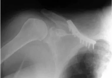
On 08/19/09 (day eight of hospitalization), the surgical procedure was carried out as scheduled (Figure 4). Immediately after surgery, the patient evolved without complications.
On day one after surgery, a decrease in the edema and temperature of the affected limb was observed, becoming equal to the contralateral limb.
On the third day, the patient reported a major im-provement in the clinical condition and in the phy-sical examination total disappearance of the edema was observed (Figure 5).
Rev Bras Ortop. 2011;46(2):215-8
Ultrasound (08/14/09): Partial segmental thrombo-sis of the subclavian vein (Figure 3).
Figure 4 – Radiograph showing osteosynthesis of the clavicle, with plate and screws as well as cortical-spongy autologous graft of the iliac.
Figure 5 – Postoperative view showing decrease in edema of the upper right limb.
Figure 6 – Radiograph two months after surgery.
The patient is currently in follow-up in the ou-tpatient department, with total improvement of the clinical condition and no complaints (Figure 6).
DISCUSSIOn
Generally, vascular lesions following fracture of the clavicle are uncommon, but they are recognized as an immediate complication due to transection of the
vein by a deviated fracture(6,8,13) or by a later
compli-cation, secondary to the compression resulting from a hypertrophic callus.
In 1875, James Paget postulated that a spontaneous thrombosis of the axillary and subclavian veins could cause pain and edema of the upper limb. Effort Syn-drome (Paget-Schroetter) was described at that time. This syndrome occurs as a result of repetitive effort in which demand is placed on the upper limb in positions of extreme abduction and lateral rotation, resulting in endothelial damage with the formation of a blood clot(9,10,14). Deep vein thrombosis of the upper limb can be of idiopathic cause, and is generally associated with neoplasms.
The most common causes associated with deep vein thrombosis of the upper limb are central vein catheter (72%), infection (28%), and renal
insuffi-ciency (21%)(15). Pain and edema are symptoms that
are generally present; pain is infrequent, unlike ede-ma. Other signs and symptoms include decoloration, prominent veins and increased temperature; however,
many may be asymptomatic(10,16).
A high level of suspicion is needed to form an objective diagnosis using invasive methods (veno-gram, angiography) and similar non-invasive
metho-ds (doppler ultrasound)(10,16). Pulmonary embolism
is an important complication, and is sometimes fatal. Recent studies show complications like pulmonary thromboembolism (8-36%), post-anticoagulation re-currence (2-15%) and post-thrombotic syndrome (36-50%)(10,17,18). Acute treatment of deep vein thrombosis of the upper limb includes raising the limb, analge-sia and therapeutic doses of anticoagulant medication with low-molecular-weight or unfractionated heparin,
followed by a period of three months of warfarin(10,17).
Oral or systemic thrombolysis with urokinase and streptokinase has been used with good results. Throm-bolytic therapy with catheter, anticoagulant for three months and surgical decompression have been
218
commended in cases of effort syndrome. Filter of the upper vena cava can be used to prevent pulmonary thromboembolism if anticoagulant therapy is con-traindicated(10).
In the present case, the patient had 75 days of evolution of the fracture, with radiographs showing signs of delayed consolidation, but without signs of hypertrophic calli, either in the radiographic exam or in the physical exam. Treatment was carried out in conjunction with the vascular surgical team, opting for internal fixation with low contact locking plate and locked screws, associated with anticoagulant
the-rapy with low-molecular-weight heparin (Clexane®)
and oral warfarin for three months. Two days after surgery, the edema and hyperemia of the upper limb had practically disappeared.
Mortality rates due to deep vein thrombosis of the
upper limb can reach 12% (ranging from 10-50%), and the majority of patients who die have a central venous catheter or an infection(15,16).
Adla et al(19) reported a case of deep vein
throm-bosis of the upper limb four days after fracture, two days after starting the use of a figure 8 shoulder bra-ce, regressing after removal of the brace and start of anticoagulant therapy.
We did not observe any case in the literature that reports deep vein thrombosis of the upper limb asso-ciated with delayed consolidation of the clavicular fracture with that time. There must be a high level of suspicion to diagnose this uncommon entity, to avoid
fatal complications. Although this entity is rare(11,12),
the possibility of deep vein thrombosis of the subcla-vian veins should always be considered in patients with fracture of the clavicle.
Rev Bras Ortop. 2011;46(2):215-8 REFEREnCES
1. Hill JM, McGuire MH, Crosby LA. Closed treatment of displaced middle-third fractures of the clavicle gives poor results. J Bone Joint Surg Br. 1997;79(4):537-9. 2. Hutchinson MR, Ahuja GS. Diagnosing and treating clavicle injuries. Phys
Sportsmed. 1996;24(3):26-36.
3. Rockwood CA Jr, Williams GR, Young DC. Injuries to the acromioclavicular joint. In: Rockwood CA Jr, Green DP, Bucholz RW, Heckman JD, edtors. Fractures in adults. 3rd ed. Philadelphia: Lippincott; 1991.p. 1181-239.
4. Costa MC, Robbs JV. Nonpenetrating subclavian artery trauma. J Vasc Surg. 1988;8(1):71-5.
5. Katras T, Baltazar U, Rush DS, Davis D, Bell TD, Browder IW, Compton RP,Stanton PE Jr. Subclavian arterial injury associated with blunt trauma. Vasc Surg. 2001;35(1):43-50.
6. Kendall KM, Burton JH, Cushing B. Fatal subclavian artery transection from isolated clavicle fracture. J Trauma. 2000;48(2):316-8.
7. Natali J, Maraval M, Kieffer E, Petrovic P. Fractures of the clavicle and inju-ries of the sub-clavian artery. Report of 10 cases. J Cardiovasc Surg(Torino. 1975;16(5):541-7.
8. Guillemin A. Chirure de la veine sous-claviere par fracture fermee de la clavicule. Bull Meme Soc Nat Chir. 1930;56:302-4.
9. Adams JT, DeWeese JA. “Effort” thrombosis of the axillary and subclavian veins. J Trauma. 1971;11(11):923-30.
10. Kommareddy A, Zaroukian MH, Hassouna HI. Upper extremity deep venous thrombosis. Semin Thromb Hemost. 2002;28(1):89-99.
11. Lim EV, Day LJ. Subclavian vein thrombosis following fracture of the clavicle. A case report. Orthopedics. 1987;10(2):349-51
12. Kanbar MS. Subclavian vein thrombosis following fracture of the clavicle: a case report. Orthopedics. 1988;11(10):1372.
13. Matry C. Fracture de la clavicule gauch au tiers interne. Blessure de la veine sous claviere. Osteosynthese. Bull Meme Soc Nat Chir. 1932;58:75-8. 14. Ellis MH, Manor Y, Witz M. Risk factors and management of patients with upper
limb deep vein thrombosis. Chest. 2000;117(1):43-6.
15. Marinella MA, Kathula SK, Markert RJ. Spectrum of upper-extremity deep venous thrombosis in a community teaching hospital. Heart Lung. 2000;29(2):113-7.
16. Prandoni P, Bernardi E. Upper extremity deep vein thrombosis. Curr Opin Pulm Med. 1999 Jul;5(4):222-6. Review. PubMed PMID: 10407691.
17. Prandoni P, Polistena P, Bernardi E, Cogo A, Casara D, Verlato F, Angelini F,Simioni P, Signorini GP, Benedetti L, Girolami A. Upper-extremity deep vein thrombosis. Risk factors, diagnosis, and complications. Arch Intern Med. 1997;157(1):57-62.
18. Leebeek FW, Kappers-Klunne MC, Gómez-García EB. [Deep venous throm-bosis of the arm: etiology, diagnosis and treatment]. Ned Tijdschr Geneeskd. 2000;144(8):361-4.
SUBSCRIPTIONS
Revista Brasileira de
Ortopedia
All associates of
theBrazilian Society
of Orthopedics and
Traumatology receive this
Journal automatically.
If this does not apply
to you, ensure that you
receive the Journal by
taking out a subscription.
Use one of the adjacent
subscription slips.
Fill it in and send it to:
Sociedade Brasileira de
Ortopedia e Traumatologia.
Alameda Lorena, 427
14º andar – Jd. Paulista
CEP 01424-000
São Paulo/SP
I would like to have an annual subscription to the Revista Brasileira de Ortopedia, starting from issue number __________________________ (indicate the month). For this, I attach a check for the amount of R$ 180.00 (one hundred and eighty reais), made payable to the “Sociedade Brasileira de Ortopedia e Traumatologia”.
Name ________________________________________________________
Specialty ______________________________________________________
Address _______________________________________________________
Postal code _____________ City ________________ State ______________
Date _______________ ___________________________ (signature)
SUBSCRIPTION
Revista Brasileira de Ortopedia
I would like to have an annual subscription to the Revista Brasileira de Ortopedia, starting from issue number __________________________ (indicate the month). For this, I attach a check for the amount of R$ 180.00 (one hundred and eighty reais), made payable to the “Sociedade Brasileira de Ortopedia e Traumatologia”.
Name ________________________________________________________
Specialty ______________________________________________________
Address _______________________________________________________
Postal code _____________ City ________________ State ______________
Date _______________ ___________________________ (signature)
Revista Brasileira de Ortopedia
I would like to inform you that I have changed address, as follows:
Name _________________________________________________________
New Address ___________________________________________________
Postal code _____________ City ________________ State ______________
(abbreviation)
Previous address
______________________________________________________________
(Avenue, Street or Square)
Postal code _____________ City ________________ State ______________
(abbreviation)