Grasieli de Oliveira Ramos(a)
Juliana Cristina Porto(b)
Daniella Serafim Couto Vieira(c)
Filipe Modolo Siqueira(c)
Elena Riet Correa Rivero(c)
(a) Graduate Program in Dentistry, Health
Sciences Center, Universidade Federal de Santa Catarina - UFSC, Florianópolis, SC, Brazil.
(b) Undergraduate Course in Dentistry, Health
Sciences Center, Universidade Federal de Santa Catarina - UFSC, Florianópolis, SC, Brazil.
(c) Department of Pathology, Health Sciences
Center, Universidade Federal de Santa Catarina - UFSC, Florianópolis, SC, Brazil.
Corresponding Author: Elena Riet Correa Rivero E-mail: riet@ccs.ufsc.br
Odontogenic tumors: a 14-year
retrospective study in Santa Catarina,
Brazil
Abstract: Odontogenic tumors (OTs) are lesions that develop exclusively on maxillary bones, and form a heterogeneous group. They vary from hamartomatous lesions to benign and malign tumors. Although they are rarely observed in dentistry clinics, it is extremely important for the den-tist to be aware of them. The aim of this study was to investigate the inci-dence of odontogenic tumors diagnosed in the population of Florianópo-lis, Santa Catarina, Brazil. Cases of odontogenic tumors were selected from the anatomopathological diagnostic services at Federal University of Santa Catarina from 1998 to 2011. Clinical data on these cases were collected from biopsy reports and patient iles. Seventy-eight cases of odontogenic tumors were surveyed. Of these diagnoses, 51% were kera-tocystic odontogenic tumors (KCOTs); the remaining cases were mainly ameloblastomas and odontomas. The most frequently observed lesion in this retrospective study was KCOT (more than half of cases). Thus, this study shows that modifying the classiication of the OTs altered the fre-quency of the lesions, possibly making KCOT the most common lesion observed in diagnostic services worldwide.
Descriptors: Odontogenic Tumors; Pathology, Oral; Epidemiology.
Introduction
Odontogenic tumors (OTs) develop exclusively on gnathic bones through the proliferation of odontogenic tissues (epithelial, mesenchymal or both). They form a heterogeneous group of hamartomatous lesions, benign and malign tumors. According to their origins, they are divided into epithelial, ectomesenchymal, and mesenchymal. Among the most frequent OTs are keratocystic odontogenic tumors (KCOTs), ameloblas-tomas, and odontomas.1
Over time, OTs have passed through several modiications in their classiication. In 1869, Broca used the term “odontoma” for any lesion arising from tissues that were part of the dental formation.2 Later, sev-eral authors began to use and modify this terminology, and it was only in 1971 that the World Health Organization (WHO) published the irst guide for the classiication of OTs.3 In the most recent classiication, “WHO Classiication of Tumors - Head and Neck Tumours” in 2005,1 there were several modiications. One was the inclusion of the kerato-cystic odontogenic tumor in the category of benign tumors; it was no longer categorized as an odontogenic cyst.2 Information was also added
Declaration of Interests: The authors certify that they have no commercial or associative interest that represents a conflict of interest in connection with the manuscript.
Submitted: May 06, 2013
Accepted for publication: Sep 30, 2013 Last revision: Oct 06, 2013
concerning lesions, including their deinition, epide-miology, etiology, clinical and imaging features, and genetics.4
OTs are rare lesions in dentistry clinics, and their incidence varies based on geographic location. In Brazil, studies report that the incidence of these lesions varies between 2.4%,5 and 6.8%6 of all di-agnosed oral lesions. Findings from other parts of Latin America are similar, such as those by Ledes-ma-Montes et al.,7 whose multicentric study (real-ized in Mexico and Guatemala) observed a 2.16%
incidence of OTs, and in another study performed in Chile in which the authors found an incidence of 1.29% for OTs8 among all the oral lesions diag-nosed in their services. Studies from other parts of the world, such as the one conducted by Jones and Franklin9 in the United Kingdom, found a lower fre-quency of 0.8% for all OTs. However, reports from Nigeria showed a greater prevalence of OTs, such as frequencies of 9.6% and 19% observed in studies by Ladeinde et al.10 and Odukoya,11 respectively.
The aim of the present study was to investigate the incidence and main characteristics of OT cases that were diagnosed at the Universidade Federal de
Santa Catarina in two diagnostic services, which
are references to the Santa Catarina State.
Methodology
Cases for the study were selected from histopath-ological reports of two diagnostic services of the
Universidade Federal de Santa Catarina - UFSC,
the Oral Pathology Laboratory and the Pathologi-cal Anatomy Service, from 2006 to 2011 and from 1998 to 2010, respectively. Slides stained with he-matoxylin and eosin (H&E) were selected from the histopathological diagnosis reports for evaluation under light microscopy (Olympus Corporation, To-kyo, Japan) and classiied according to WHO crite-ria.1 Clinical data were collected from biopsy reports stored by both services, as well as from the hospital records. Patient information included gender, age, ethnic group and, in KCOT cases, association with nevoid basal cell carcinoma syndrome (NBCCS), also known as Gorlin syndrome. Lesion-related data were the location, clinical and radiographic aspects of the lesions. Data were collected and,
af-ter being iled in an initial format, they were laaf-ter iled in a Microsoft Excel (Microsoft Corporation, Redmond, USA) spreadsheet. Descriptive statistical analysis was performed with all collected data us-ing Microsoft Excel software. This study was ap-proved by the Ethics Committee for Research with Human Beings at UFSC under number 1055/10.
Results
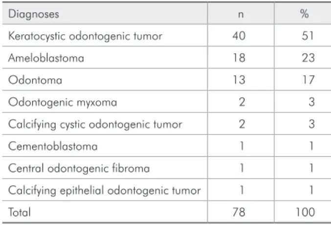
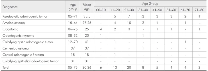
In our study, OTs corresponded to 3% of all oral lesions diagnosed by both services. In Table 1, we can observe the general prevalence of the lesions in these cases. These cases occurred more frequently in male patients (Table 2) and Caucasian (89%). The lesions were most prevalent in the age group be-tween 21 and 30 years (Table 3). The preferential location was the posterior region of the mandible Table 1 - Distribution of the cases diagnosed.
Diagnoses n %
Keratocystic odontogenic tumor 40 51
Ameloblastoma 18 23
Odontoma 13 17
Odontogenic myxoma 2 3
Calcifying cystic odontogenic tumor 2 3
Cementoblastoma 1 1
Central odontogenic fibroma 1 1
Calcifying epithelial odontogenic tumor 1 1
Total 78 100
Table 2 - Cases diagnosed distributed by gender.
Diagnoses Male Female
n % n %
Keratocystic odontogenic tumor 15 62 10 38
Ameloblastoma 9 50 9 50
Odontoma 6 46 7 54
Odontogenic myxoma 1 50 1 50
Calcifying cystic odontogenic tumor - - 2 100
Cementoblastoma - - 1 100
Central odontogenic fibroma 1 100 -
-Calcifying epithelial odontogenic tumor - - 1 100
vices, and it occurred more frequently in male pa-tients with a mean age of 35 years, varying between 5 and 75 years. These KCOT cases occurred in all areas of the maxillary bones, although the most frequent were the posterior mandible. Of all KCOT cases diagnosed in both services, 40% were associat-ed with NBCCS. When we evaluatassociat-ed the KCOT cas-es associated with NBCCS, and the isolated KCOT cases, we veriied that the mean age of the cases as-sociated with the syndrome was 28 years, whereas the mean age for patients without the syndrome was 31 years. The ethnic group, and gender distributions were very similar between the two groups (KCOT cases associated with NBCCS, and isolated KCOT cases). We also checked the number of recurrence, and found seven cases of recurrence, of which two were associated with NBCCS. Occurrence of mul-(Table 4), and the lesions presented as radiolucent
images (74%), mixed radiolucent-radiopaque (10%) or radiopaque (4%). Among radiolucent lesions, the largest number of lesions was KCOT (64%), fol-lowed by ameloblastoma (34%) and myxoma (2%). Among mixed lesions, 57% were odontomas, fol-lowed by a calcifying cystic odontogenic tumor (CCOT, 28%) and a calcifying epithelial odonto-genic tumor (CEOT, 15%). Among the radiopaque lesions two cases were odontomas (67%), and one case was cementoblastoma (33%). Nine cases (12%) had no information concerning radiographic char-acteristics: seven were odontoma (78%), one case was myxoma (11%), and one case was central odon-togenic ibroma (COF, 11%).
KCOT was the most prevalent lesion in our study, with 51% of the cases diagnosed in both ser-Table 3 - Distribution according to the patients’ age group.
Diagnoses Age
group
Mean age
Age Group
00–10 11–20 21–30 31–40 41–50 51–60 61–70 71–80
Keratocystic odontogenic tumor 05–71 35.5 1 5 7 3 3 3 2 1
Ameloblastoma 15–64 27.25 - 4 10 2 1 - 1
-Odontoma 06–75 25 4 2 3 - 1 1 - 1
Odontogenic myxoma 08–32 20 1 - - 1 - - -
-Calcifying cystic odontogenic tumor 12–70 41 - 1 - - - - 1
-Cementoblastoma 37 37 - - - 1 - - -
-Central odontogenic fibroma 18 18 - 1 - - -
-Calcifying epithelial odontogenic tumor 31 31 - - - 1 - - -
-Total 05–75 30.36 6 13 20 8 5 4 4 2
Diagnoses Posterior
mandible
Anterior mandible
Posterior maxilla
Anterior
maxilla Other n
Keratocystic odontogenic tumor 22 8 5 2 3 40
Ameloblastoma 15 1 - - 2 18
Odontoma 4 3 2 3 - 12
Odontogenic myxoma 2 - - 1 - 3
Calcifying cystic odontogenic tumor - - - 2 - 2
Cementoblastoma 1 - - - - 1
Central odontogenic fibroma 1 - - - - 1
Calcifying epithelial odontogenic tumor 1 - - - - 1
Total 46 12 7 8 5 78
tiple lesions, all associated with the syndrome, was observed in four patients.
The second-most frequent lesion, ameloblasto-ma, did not show prevalence based on gender and the mean age of the patients was 27 years, varying between 15 and 64 years. All cases of ameloblas-toma occurred in the mandible, mainly in the pos-terior area. Ameloblastoma occurred in two differ-ent clinical presdiffer-entations, solid (61%) and unicystic (39%). All cases were histologically classiied, being divided into follicular (33%), plexiform (22%), as-sociation between plexiform and follicular (96%), unicystic with mural proliferation (22%), unicystic with luminal proliferation (11%), and unicystic with intraluminal proliferation (6%).
The third-most prevalent lesion, odontoma, showed a slight preponderance for the female gen-der, and most of the cases occurred in young pa-tients up to 10 years of age. The preferential location for this lesion was the posterior mandibular area.
Discussion
OTs are lesions rarely observed in dentistry clin-ics,5-7,9,12,13 and their frequency was 3% in our study, with a mean age of 30 years, varying between 5 and 75 years, and occurring more often in male patients. Their main location was in the posterior mandibu-lar area. These data agree with indings from other studies performed in different countries.5,6,13-15 Our study demonstrated that most of the lesions oc-curred in Caucasian patients, which can be easily explained, as Caucasians correspond to 85.7% of the entire population in the Santa Catarina State according to data from the Instituto Brasileiro de
Geograia e Estatística - IBGE.16
KCOT was the most frequent lesion observed in our study, comprising 51% of all lesions. Recently, KCOT has been reclassiied by the WHO as a cystic lesion becoming a benign tumor.17 With this reclas-siication KCOT became the most frequent odon-togenic tumor; however, it is still dificult to ind a series of studies including this lesion as an OT.1 The frequency of KCOTs in our study was 51% of all diagnosed cases, which was higher than the 28%
to 36% frequency found in other studies.13,18-20 We veriied that the mean age of KCOT cases was 35
years, and that the posterior mandible was the most affected site. Data found in our study corroborate the data found in other studies,13,18 and we found a greater prevalence for the male gender, in agree-ment with the results reported by Jing et al.19 and González-Alva et al.21 The association of KCOT with NBCCS was seen in 40% of the cases, which is very different from what was reported by González-Alva et al.,21 who only found an association of 6%. The same study reported data on the characteristics of the cases associated with NBCCS, reporting a greater prevalence in female patients (63.6%), which was different from the observations of the present study, wherein we found a higher prevalence in male patients (57% of cases associated with NBCCS). In our study, the age of NBCCS patients was between 5, and 56 years, with a mean of 28 years, which dif-fered from the indings presented by González-Alva
et al.,21 who presented cases whose age varied from
8 to 43 years with a mean of 19.5 years. Those au-thors also reported recurrence in only three cases, whereas it was observed in seven cases in our study.
Ameloblastoma is considered one of the most common lesions among OTs, and its incidence var-ies from 18%22 to 45%.23 In our study, the incidence of ameloblastoma was 23% in all diagnosed cases, and these data are similar to indings from other studies.7,14,18,20 No gender-related preponderance was noted in our study, which was different from other studies in which the incidence in the female gender was higher.5,9,13,23,24 The most frequent clinical pre-sentation was of the solid ameloblastoma (61%) and, histologically, of the follicular variant (33%). This is very similar to what was observed in the studies conducted by Ledesma-Montes et al.,7 Osterne et al.,13 Saghravanian et al.15 and Santos et al.5 Howev-er, Fregnani et al.24 observed that the most frequent histological variant was the plexiform, which was observed in 53% of the cases. In our study, the pos-terior mandibular area was the preferred location, which was consistent with other studies.5,7,13,14,19,22-24
et al.,23 Jing et al.,19 Jones and Franklin,9 Olgac et al.,14 Osterne et al.,13 and Saghravanian et al.15 in presenting an incidence of approximately 17%. Our study is in agreement with the data found in the lit-erature that show a higher frequency in female pa-tients.5,13,15,22 In the present study, the mean age was 23 years, a result that is also similar to those found in the literature.5,15,19,23 The preferential location for these lesions was the posterior mandibular area, which corroborated the indings of Jing et al.19 and Saghravanian et al.15
Other lesions had a lower incidence, such as myxoma (3%), CCOT (3%), cementoblastoma (2%), COF (1%), and CEOT (1%). This lower incidence can also be observed in other studies, such as those conducted by Avelar et al.,18 Jing et al.,19 and Santos
et al.5 However, myxoma had a slightly higher
inci-dence in some studies, such as in that conducted by Olgac et al.,14 who observed an incidence of 16%, being the third-most frequent lesion.
Very few studies have investigated OTs and, thus, a comparison between our results, and those published in the literature remains very dificult. Furthermore, the large ethnic diversity found world-wide, and even inside a single country with conti-nental proportions, such as Brazil, creates substan-tial differences in the results. With the 2005 WHO
publication,1 it has become important to conduct new studies using this new classiication of OTs so that a new panorama can be deined for the inci-dence of this group of lesions. In addition, multicen-tric studies that can establish the worldwide charac-teristics of OTs are also important.
Conclusion
In our study, the most frequent lesion was KCOT, comprising almost half of the diagnosed cases. This shows that with the modiication of the classiica-tion of OTs, KCOT is possibly the most common lesion observed in diagnostic services worldwide. However, there is a lack of studies demonstrat-ing these frequencies, includdemonstrat-ing data even showdemonstrat-ing KCOT as an OT.
Acknowledgements
We thank the Pathological Anatomy Service and Clinical Data Records Service at the University Hos-pital at UFSC for their help in this study. This study was supported by Coordenação de
Aperfeiçoamen-to de Pessoal de Nível Superior (CAPES), which
provided a doctoral fellowship, and the Conselho Nacional de Desenvolvimento Cientíico e
Tec-nológico (CNPq), which provided an undergraduate
research scholarship.
References
1. Barnes L, Everson JW, Reichart P, Sidransky D, editors. World Health Organization classification of tumours. Pathology and genetics of head and neck tumours. Lyon: IARC Press; 2005. 435 p.
2. Philipsen HP, Reichart PA. Classification of odontogenic tumours. A historical review. J Oral Pathol Med [Inter-net]. 2006 Oct [cited 2012 Oct 23];35(9):525-9. Available from: http://onlinelibrary.wiley.com/doi/10.1111/j.1600-0714.2006.00470.x/pdf.
3. Kramer IRH, Pindborg JJ, Torloni H. Histological typing of odontogenic tumours, jaw cysts, and allied lesions. Geneva, [London]: World Health Organization; 1971. 127 p. 4. Reichart PA, Philipsen HP, Sciubba JJ. The new
classifica-tion of Head and Neck Tumours (WHO)--any changes?. Oral Oncol [Internet]. 2006 Sep [cited 2012 Oct 23];42(8):757-8. Available from: http://www.oraloncology.com/article/S1368-8375(05)00303-9/pdf.
5. Santos JN, Pinto LP, Figueredo CR, Souza LB. Odontogenic tumors: analysis of 127 cases. Pesq Odontol Bras [Internet]. 2001 Oct-Dec [cited 2012 Oct 23];15(4):308-13. Available from: http://www.scielo.br/pdf/pob/v15n4/a07v15n4.pdf. 6. Sousa FB, Etges A, Corrêa L, Mesquita RA, Araújo NS.
Pe-diatric oral lesions: a 15-year review from São Paulo, Brazil. J Clin Pediatr Dent. 2002 Summer;26(4):413-8.
7. Ledesma-Montes C, Mosqueda-Taylor A, Carlos-Bregni R, de Leon ER, Palma-Guzman JM, Perez-Valencia C, et al. Amelo-blastomas: a regional Latin-American multicentric study. Oral Dis [Internet]. 2007 May [cited 2012 Oct 25];13(3):303-7. Available from: http://onlinelibrary.wiley.com/doi/10.1111/ j.1601-0825.2006.01284.x/pdf.
9. Jones AV, Franklin CD. An analysis of oral and maxillofacial pathology found in adults over a 30-year period. J Oral Pathol Med [Internet]. 2006 Aug [cited 2012 Oct 28];35(7):392-401. Available from: http://onlinelibrary.wiley.com/doi/10.1111/ j.1600-0714.2006.00451.x/pdf.
10. Ladeinde AL, Ajayi OF, Ogunlewe MO, Adeyemo WL, Aro-tiba GT, Bamgbose BO, et al. Odontogenic tumors: a review of 319 cases in a Nigerian teaching hospital. Oral Surg Oral Med Oral Pathol Oral Radiol Endod [Internet]. 2005 Feb [cited 2012 Oct 25];99(2):191-5. Available from: http://www. journals.elsevierhealth.com/periodicals/ymoe/article/S1079-2104(04)00628-6/pdf.
11. Odukoya O. Odontogenic tumors: analysis of 289 Nigerian cases. J Oral Pathol Med [Internet]. 1995 Nov [cited 2012 Oct 28];24(10):454-7. Available from: http://onlinelibrary.wiley. com/doi/10.1111/j.1600-0714.1995.tb01133.x/pdf.
12. Lima GS, Fontes ST, de Araújo LM, Etges A, Tarquinio SB, Gomes AP. A survey of oral and maxillofacial biopsies in children: a single-center retrospective study of 20 years in Pelotas-Brazil. J Appl Oral Sci [Internet]. 2008 Nov-Dez [cited 2012 Oct 25];16(6):397-402. Available from: http://www. scielo.br/pdf/jaos/v16n6/a08v16n6.pdf.
13. Osterne RL, Brito RG, Alves AP, Cavalcante RB, Sousa FB. Odontogenic tumors: a 5-year retrospective study in a Brazilian population and analysis of 3406 cases reported in the literature. Oral Surg Oral Med Oral Pathol Oral Radiol Endod [Internet]. 2011 Apr [cited 2012 Oct 27];111(4):474-81. Available from: http://www.journals.elsevierhealth.com/ periodicals/ymoe/article/S1079-2104(10)00828-0/pdf. 14. Olgac V, Koseoglu BG, Aksakalli N. Odontogenic tumours
in Istanbul: 527 cases. Br J Oral Maxillofac Surg [Inter-net]. 2006 Oct [cited 2012 Oct 24];44(5):386-8. Available from: http://www.sciencedirect.com/science/article/pii/ S0266435605002196.
15. Saghravanian N, Jafarzadeh H, Bashardoost N, Pahlavan N, Shirinbak I. Odontogenic tumors in an Iranian population: a 30-year evaluation. J Oral Sci [Internet]. 2010 Sep [cited 2012 Oct 27];52(3):391-6. Available from: https://www.jstage.jst. go.jp/article/josnusd/52/3/52_3_391/_pdf.
16. Brasil. Instituto Brasileiro de Geografia e Estatística. Rio de Janeiro: IBGE; 2010. 317 p.
17. Philipsen HP. Keratocystic odontogenic tumour. In: Barnes L, Everson, JW, Reichart, P, Sidransky, D, editors. World Health Organization classification of tumours. Pathology and
genetics of head and neck tumours. Lyon: IARC Press; 2005. p 306-7.
18. Avelar RL, Antunes AA, Santos TS, Andrade ES, Dourado E. Odontogenic tumors: clinical and pathology study of 238 cases. Braz J Otorhinolaryngol [Internet]. 2008 Sep-Oct [cited 2012 Oct 20];74(5):668-73. Available from: http://www.sci-elo.br/pdf/rboto/v74n5/en_v74n5a06.pdf.
19. Jing W, Xuan M, Lin Y, Wu L, Liu L, Zheng X, et al. Odonto-genic tumours: a retrospective study of 1642 cases in a Chinese population. Int J Oral Maxillofac Surg [Internet]. 2007 Jan [cited 2012 Oct 29];36(1):20-5. Available from: http://down-load.journals.elsevierhealth.com/pdfs/journals/0901-5027/ PIIS0901502706004577.pdf.
20. da-Costa DO, Maurício AS, de-Faria PA, da-Silva LE, Mosqueda-Taylor A, Lourenço SD. Odontogenic tumors: a retrospective study of four Brazilian diagnostic pathology centers. Med Oral Patol Oral Cir Bucal [Internet]. 2012 May [cited 2013 Jan 26];17(3):e389-94. Available from: http:// www.ncbi.nlm.nih.gov/pmc/articles/PMC3476089/pdf/me-doral-17-e389.pdf.
21. González-Alva P, Tanaka A, Oku Y, Yoshizawa D, Itoh S, Sakashita H, et al. Keratocystic odontogenic tumor: a retro-spective study of 183 cases. J Oral Sci [Internet]. 2008 Jun [cited 2012 Oct 20];50(2):205-12. Available from: https:// www.jstage.jst.go.jp/article/josnusd/50/2/50_2_205/_pdf. 22. Guerrisi M, Piloni MJ, Keszler A. Odontogenic tumors in
children and adolescents. A 15-year retrospective study in Argentina. Med Oral Patol Oral Cir Bucal [Internet]. 2007 May [cited 2012 Oct 29];12(3):E180-5. Available from: http:// scielo.isciii.es/pdf/medicorpa/v12n3/01.pdf.
23. Fernandes AM, Duarte ECB, Pimenta F, Souza LN, Santos VR, Mesquita RA, et al. Odontogenic tumors: a study of 340 cases in a Brazilian population. J Oral Pathol Med [In-ternet]. 2005 Nov [cited 2012 Oct 26];34(10):583-7. Avail-able from: http://onlinelibrary.wiley.com/doi/10.1111/j.1600-0714.2005.00357.x/pdf.

