INTRODUCTION
Corresponding author: Dra. Ilana Lopes Baratella da Cunha Camargo.
e-mail: [email protected]
Received 10 September 2015
Accepted 27 January 2016
Molecular analysis of methicillin-resistant
Staphylococcus aureus
dissemination among healthcare
professionals and/or HIV patients from a tertiary hospital
Jessica Baleiro Okado
[1],[2], Simoni Camila Bogni
[2], Lílian Andreia Fleck Reinato
[3],[4],
Roberto Martinez
[5], Elucir Gir
[4]and Ilana Lopes Baratella da Cunha Camargo
[2][1]. Programa de Pós-Graduação Strictu Sensu em Ciências-Opção Biomolecular, Instituto de Física de São Carlos, Universidade de São Paulo, São Carlos, São Paulo, Brasil. [2]. Laboratório de Epidemiologia e Microbiologia Molecular (LEMiMo), Instituto de Física de São Carlos, Universidade de São Paulo, São Carlos, São Paulo, Brasil. [3]. Programa de Pós-Graduação Strictu Sensu em Enfermagem, Escola de Enfermagem de Ribeirão Preto, Universidade de São Paulo, Ribeirão Preto, São Paulo, Brasil. [4]. Escola de Enfermagem de Ribeirão Preto, Universidade de São Paulo, Ribeirão Preto, São Paulo, Brasil. [5] Escola de Medicina de Ribeirão Preto, Universidade de São Paulo, Ribeirão Preto, São Paulo, Brasil.
ABSTRACT
Introduction: Methicillin-resistant Staphylococcus aureus (MRSA) is a nosocomial pathogen in community settings. MRSA colonized individuals may contribute to its dissemination; the risk of MRSA infection is increased in human immunodefi ciency virus/acquired immune defi ciency syndrome (HIV/AIDS) patients, although the prevalence of colonization in this group is not well established. The present study addressed this issue by characterizing MRSA isolates from HIV/AIDS patients and
their healthcare providers (HCPs) to determine whether transmission occurred between these two populations. Methods: A total
of 24 MRSA isolates from HIV-infected patients and fi ve from HCPs were collected between August 2011 and May 2013. Susceptibility to currently available antimicrobials was determined. Epidemiological typing was carried out by pulsed-fi eld gel electrophoresis, multilocus sequence typing, and Staphylococcus cassette chromosome (SCCmec) typing. The presence of heterogeneous vancomycin-intermediate Staphylococcus aureus (hVISA) and heterogeneous daptomycin-resistant Staphylococcus aureus (hDRSA) was confi rmed by population analysis profi le. Isolates characterized in this study were also
compared to isolates from 2009 obtained from patients at the same hospital. Results: A variety of lineages were found among patients, including ST5-SCCmecII and ST30-SCCmecIV. Two isolates were Panton-Valentine leukocidin-positive, and hVISA
and hDRSA were detected. MRSA isolates from two HCPs were not related to those from HIV/AIDS patients, but clustered with archived MRSA from 2009 with no known relationship to the current study population. Conclusions: ST105-SCCmecII clones that colonized professionals in 2011 and 2012 were already circulating among patients in 2009, but there is no evidence that these
clones spread to or between HIV/AIDS patients up to the 7th day of their hospitalization.
Keywords: Methicilin-resistant Staphylococcus aureus. HIV patients. Teicoplanin resistance. h-VISA. Daptomycin.
Methicillin-resistant Staphylococcus aureus (MRSA) is a leading cause of nosocomial and community infections(1). It is
usually present in the environment as well as in the microbiota of the superior respiratory tract and skin. MRSA is transmitted by direct contact, and patient-to-patient transmission is largely via the hands of health care providers (HCPs). Undetected MRSA-colonized HCPs represent a major source of the bacterium in hospitals, since it can be transmitted from these individuals to high-risk patients, which can limit the success of other control
measures. Indeed, a case in which MRSA transmission was reduced after carrier HCPs were identifi ed and successfully decolonized has been described(2).
Individuals with human immunodefi ciency virus/acquired immune defi ciency syndrome (HIV/AIDS) are at increased risk for MRSA colonization and infection mainly due to their high antibiotic use(1) and high rates of hospital readmission.
These patients can also harbor community-acquired MRSA,
which often produces Panton-Valentine leukocidin (PVL)(1).
However, the prevalence of MRSA colonization in this group is not well established.
RESULTS METHODS
Sample collection
MRSA samples from two colonization sites [nares (N) and saliva (S)] were obtained from HIV/AIDS patients on days 1 and 7 of hospitalization and from HCPs with whom they had contact, in two specifi c units of a large Brazilian public hospital with 600 beds, of which 24 are occupied almost exclusively by HIV patients. From August 2011 to May 2013, 317 individuals with HIV/AIDS were hospitalized, and 266 agreed to participate in the study along with 73 HCPs; staffs that were on leave were not included. Samples were collected using swabs and stored in Stuart agar until bacterial isolation and identifi cation. S. aureus was isolated on Mueller Hinton agar supplemented with 5% sheep blood and phenotypic identifi cation was carried out using the Vitek system (BioMérieux, Marcy l’Etoile, France). Methicillin resistance was detected using the AST-P585 card (BioMérieux) and broth dilution. Once an HCP was identifi ed as being colonized with MRSA, a decolonization protocol was carried out that included a chlorhexidine bath and application of 1ml silver sulfadiazine to the nares, which was repeated daily for 5 days. Additionally, 22 MRSA isolates from other patients from different wards at the same hospital collected between June and September 2009 that were archived by our group, and S. aureus strain N315 were included for determination of clonality. Isolates representing each pulsotype detected by pulsed-fi eld gel electrophoresis (PFGE) were randomly selected for typing by multilocus sequence typing (MLST) and
Staphylococcus Cassette Chromosome (SCCmec) typing. The study protocol was approved by the Research Ethics Committee of the Ribeirão Preto School of Nursing (no. 1304/2011).
Susceptibility profi ling
The minimum inhibitory concentration (MIC) was determined for oxacilin, vancomycin, teicoplanin, daptomycin, tigecyclin, linezolid, and quinupristin-dalfopristin by broth dilution, according to Clinical and Laboratory Standards Institute (CLSI) guidelines(3). MIC
50 and MIC90 were calculated
and CLSI breakpoints were adopted for classification except in the case of tigecycline, for which the European Committee on Antimicrobial Susceptibility Testing (EUCAST) recommendation was followed(4).
Heterogeneous vancomycin-intermediate
Staphylococcus aureus screening
Heterogeneous vancomycin-intermediate Staphylococcus
aureus screening (hVISA) screening was performed as
previously described(5). If a countable number (one to 30) of
colonies was observed within 48h of incubation at 37°C on brain-heart infusion agar containing 4μg/ml vancomycin, the
strain was designated as a possible hVISA(5). S. aureus Mu3
and Mu50 were used as hVISA and VISA control strains, respectively, and were kindly provided by Keiichi Hiramatsu and Teruyo Ito (Juntendo University, Tokyo, Japan).
Population analysis profi le
The population analysis profi le (PAP)(5) (6) to vancomycin
or daptomycin was determined for samples that were positive for the hVISA screening or exhibited resistance to daptomycin after 48h of incubation(6) (7) (8). PAP to daptomycin was used to
identify heterogeneous daptomycin-resistant Staphylococcus aureus (hDRSA) strains(8).
Panton-Valentine leukocidin gene
and hemolysis analysis
The PVL gene lukSF was amplifi ed by PCR as previously
described(9). Mueller Hinton agar supplemented with 5% sheep
blood was used to assess hemolytic activity.
Molecular characterization
SCCmec type was determined by multiplex-PCR as previously described(10) using the following reference strains:
S. aureus RN4220 (SCCmec negative), 10442 (SCCmecI), N315 (SCCmecII), 85/2082 (SCCmecIII), 4744 (SCCmecIVa),
and WIS (SCCmecV).
PFGE was carried out after DNA digestion with SmaI(11).
Data were analyzed with Bionumerics v.7.1 software (Applied
Maths NV, Belgium)(12) using the unweighted pair-group
method with arithmetic mean based on Dice coefficients, where optimization and tolerance were set to 0.5% and 1.25%, respectively. A similarity coeffi cient of 80% was selected to describe patterns representative of each pulsotype, which were further characterized by MSLT(13). Sequence types were
identifi ed using the MLST database(14). To limit redundancy,
duplicate isolates from the same patient with identical SCCmec
and pulsotype were considered as the same strain and included only once in the analysis.
Staphylococcus aureus was cultured from 101 (38%) tested
individuals, and resistance to oxacillin was observed in 15 participants (5.6% of all participants or 14.8% of those colonized by S. aureus). A total of 13/15 HIV patients were found to be
colonized by MRSA on the day of hospital admission, and only fi ve of these remained colonized on day 7. Additionally, two patients were found to be colonized only on day 7 of hospitalization. Only 3/73 (4.1%) of HCPs (P1, P2, and P3) were colonized by MRSA during the study. P1 was colonized on three different dates, despite having undergone decolonization procedures(15). Therefore, a total of 29 MRSA isolates were
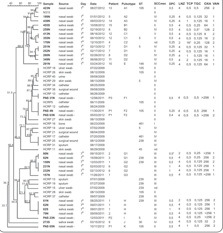
FIGURE 1 -Similarity dendrogram. Similarity was calculated with Bionumerics v.6.5 software using the Dice coeffi cient with 0.5% optimization
and 1.25% tolerance. ST: sequence typing; SCCmec: Staphylococcus cassette chromosome; DPC: daptomycin; LNZ: linezolide; TCP: teicoplanin;
TGC: tigecycline; OXA: oxacillin; VAN: vancomycin; N: nares; S: saliva; HCPs: healthcare providers. Isolates collected in this study are indicated in bold to differentiate them from those obtained in 2009, which were used only for clonality analysis.*resistance after 48h.
One isolate (176N) was identified as hVISA, which was confirmed by PAP (Figure 2B). According to CLSI breakpoints(3), this isolate exhibited an intermediate level of
resistance to teicoplanin after 24h of incubation (MIC = 16μg/ ml) and full resistance after 48h. However, it would be classifi ed as resistant after 24h based on EUCAST breakpoints(4).
MIC50 and MIC90 (μg/ml) of all antimicrobials were determined for each strain, and were as follows: 128/>256 for oxacilin, 4/4 for linezolid, 0.5/1 for teicoplanin, 0.125/0.5 for tigecycline, 0.5/0.5 for quinupristin/dalfopristin and daptomycin, and 1/2 for vancomycin. MICs for each isolate are shown in Figure 1.
LNZ TCP TGC OXA VAN
4 0,5 0,5 256 2
4 0,5 0,125 32 1 4 1 0,125 16 1 4 0,5 0,25 64 1 4 0,5 0,5* 128 2 4 0,5 0,125 8 2 4 0,5 0,125 16 2 2 16* 0,25 128 2 4 0,5 0,125 32 1 4 0,5 0,125 16 1 4 1 0,125 16 1 4 2 0,125 16 1 4 0,5 0,125 64 1
4 0,5 0,5 >256 1
4 0,5 0,5 256 1
4 0,5 0,5 >256 2
2 0,5 0,25 >256 1 4 0,5 0,25 256 2 4 0,5 0,125 256 2 4 0,5 0,125 256 1 4 0,5 0,125 256 1 4 0,5 0,125 >256 1
2 0,5 0,125 256 2 4 0,5 0,125 256 1 4 0,5 0,125 256 1 4 0,5 0,125 >256 1 4 0,5 0,25 >256 2 4 0,5 0,125 32 2 4 1 0,5 256 2 40 60 80 100
94.7 91.5 84.0 88.9 74.5 85.3 94.7 79.7 68.3 94.1 93.9 95.7 89.9 88.0 87.2 85.9 78.6 73.2 72.1 87.0 83.0 95.2 82.5 80.0 77.5 71.1 92.3 82.3 51.5 61.5 46.4 36.3 59.3 54.4 33.1
Sample Source Day Data Patient Pulsotype ST SCCmec DPC 423N nasal swab 1 08/27/2012 13 A1 105 II 0,5
N315 II
199N nasal swab 1 01/01/2012 5 A2 IV 0,25
439N nasal swab 1 09/03/2012 14 A3 IV 0,25
455S saliva swab 1 10/08/2012 15 B1 II 0,5
259N nasal swab 1 02/19/2012 8 B2 IV 0,5
413N nasal swab 1 08/16/2012 12 C1 V 0,5
355N nasal swab 1 06/10/2012 12 C1 5 V 0,5
176N nasal swab 7 12/10/2011 4 C2 nd 0,25
251N nasal swab 1 02/13/2012 7 D1 5 IV 0,25
252N nasal swab 7 02/17/2012 7 D1 II 0,25
260N nasal swab 7 02/26/2012 8 D2 IV 0,25
349N nasal swab 1 06/06/2012 11 D2 IV 0,5
291N nasal swab 1 03/24/2012 10 E 148 IV 0,25 HCRP 18 ulcer swab 07/22/2009 105 II
HCRP 26 skin swab 08/12/2009 105 II HCRP 40 urine 09/08/2009 II HCRP 29 skin swab 08/13/2009 5 IV HCRP 34 catheter 08/25/2009 II HCRP 38 surgical wound 09/08/2009 II HCRP 10 catheter 06/26/2009 II
PAS 31N nasal swab - 12/06/2011 P3 F1 II 0,5
HCRP5 06/11/2009 105 II HCRP 12 catheter
catheter
06/24/2009 II
PAS 4N nasal swab - 11/30/2011 P1 F2 105 II 0,25
PAS 63N nasal swab - 05/03/2012 P1 F2 II 0,4
HCRP 27 skin swab 08/13/2009 II HCRP 16 bone 06/23/2009 461 I HCRP 14 ulcer swab 07/02/2009 5 IV HCRP 21 surgical wound 08/04/2009 IV HCRP 17 catheter 07/20/2009 461 IV HCRP 25 surgical wound 08/12/2009 239 III HCRP 31 sputum 08/17/2009 IV HCRP 11 skin swab 06/29/2009 45 nd
80N nasal swab 09/15/2011 2 G1 III 0,5*
82N nasal swab 15/09/2011 3 G1 239 III 0,5
158N nasal swab 12/03/2011 1 G2 239 III 0,5
231N nasal swab 02/03/2012 6 G2 III 1
232N nasal swab 02/13/2012 6 G2 III 1
157N nasal swab t 11/26/2011 1 G3 III 0,5 HCRP 13 sputum 07/01/2009 239 III
HCRP 19 sputum 07/27/2009 239 III HCRP 15 ulcer swab 07/02/2009 239 nd HCRP 28 skin swab 08/13/2009 105 II HCRP 37 catheter 09/07/2009 239 III
61N nasal swab 08/25/2011 1 H 239 III 0,5
62N nasal swab 09/01/2011 1 H III 0,5
62S saliva swab 09/01/2011 1 H III 1
79N nasal swab 09/09/2011 2 H III 0,5
PAS 23N nasal swab - 12/03/2011 P2 I 5 IV 0,5
273S saliva swab 03/15/2012 9 J 30 IV 0,5
PAS 65N nasal swab - 10/11/2012 P1 K 105 II 0,5
10
9
6
4
2
0
L
o
g
1
0
(C
F
U
/m
L)
Daptomycin concentration (mg/L)
N315
mu3
mu50
80N
log10(CFU/
m
L
)
Vancomycin concent ration (mg/L)
176N
Mu3
N315 0 1 2 3 4 5 6 7
0 1 2 3 4 5 6 7 8 10
9
6
4
2
0
A
B
FIGURE 2 - Population analysis profi le to: A) daptomycin and B) vancomycin. CFU: colony forming units.
Only 2/29 isolates (6.9%) were PVL-positive (strains 199N and 273N), while 25 (86.2%) were fully hemolytic. Isolates from HIV patients showed considerable variation in terms of PFGE band patterns, which included eight pulsotypes (A-E, G, H, and J) in contrast to three pulsotypes (F, I, and K) among isolates from HCPs.
ST5, ST105, ST148, ST239, and ST30 were detected among isolates. Two of three HCPs were colonized by
ST105-SCCmecII, a single locus variant (SLV) of the New York/
Japan Clone, whereas one was colonized by ST5-SCCmecIV,
the Pediatric Clone. The following lineages were observed
in HIV patients: ST5-SCCmecIV, ST239-SCCmecIII,
ST30-SCCmecIV (Southern Pacifi c clone), ST5-SCCmecV,
ST105-SCCmecII, and ST148-SCCmecIV, all of which have been
previously described(16) (17). Different SCCmec types were
Only Patients #1 and #12 were readmitted to this hospital during the period of study. Isolates from these patients from days 1and 7 of hospitalization were indistinguishable. Although both isolates from Patient #1 collected at readmission were of the same lineage as that obtained at the fi rst hospitalization (ST239-SCCmecIII), they were of a different pulsotype (G instead of H), and the isolates from days 1 and 7 of readmission were of different subtypes (G3 and G2). Isolates obtained from Patient #12 over a two-month period were indistinguishable and MRSA was detected only on day 1 of both hospitalizations.
The fi rst P1 isolate (PAS 4N) was characterized by PFGE as pulsotype F. After decolonization(15), this HCP remained
colonized by the same MRSA clone (PAS 63N). A second decolonization procedure was carried out, and P1 was again found to be colonized, but this time by pulsotype K strain PAS 65N. Although the pulsotype had changed, all three isolates from P1 were of ST105-SCCmecII. There were no HIV patients
colonized by the same type of MRSA as determined by PFGE of HCP samples examined during the course of this study.
When MRSA isolated in 2009 from different infection sites from patients in different wards of the same hospital was compared to those of HIV patients and HCPs in the current study, we observed that with the exception of pulsotype H associated with Brazilian Epidemic Clone (BEC), all other HIV patient isolates were unrelated to those obtained from patients in 2009
(< 80% similarity) (Figure 1). However, HCPs P1 (isolates PAS
4N and PAS 63N) and P3 (isolate PAS 31N) were colonized in 2011 by clones that were indistinguishable from those isolated from infections at this hospital in 2009.
DISCUSSION
In this study, a variety of MRSA pulsotypes with few subtypes were isolated from HIV patients and HCPs. We observed no correlation between pulsotype and reduced susceptibility to antimicrobials. We found one hVISA isolate that also exhibited intermediate resistance to teicoplanin. The vanA gene responsible for complete glycopeptide resistance in S. aureus was not detected in any samples (data not shown). Teicoplanin resistance after vancomycin use has been reported(18); however, none of the
patients in our study were treated with vancomycin or teicoplanin. The mechanisms underlying daptomycin resistance are not well understood(19) (20), but it is known that decreased susceptibility to
daptomycin and vancomycin can co-occur both in vivo(20) and in
vitro(21)after selection with either antimicrobial(21). Although there
is evidence for cross-resistance(21),in this study, the hDRSA isolate
remained susceptible to vancomycin. Since none of the patients had been treated with daptomycin, the basis for resistance is unclear, but is currently under study. Heterogeneous type resistance to daptomycin has been previously observed, and is in part attributed to dltA overexpression. The detection of teicoplanin-intermediate
S. aureus, hVISA, and hDRSA is of great concern since these
represent last-line antimicrobials for treating MRSA infection.
ST105 and ST148 are SLVs of ST5 and account for the majority of isolates typed in this study. ST30 is a prototype of CC30 and is associated with community-acquired infection. The isolate ST30-SCCmecIV was positive for PVL, which is
consistent with MRSA strains found in the community(22).
Although many subtypes isolated from patients can be considered as closely related according to Tenover’s criteria(12),
only four pairs of patient strains yielded indistinguishable band patterns by PFGE. Interestingly, the isolate from P3 and the fi rst two isolates from P1 clustered with MRSA from different patients isolated 2 years prior-i.e., a clone related to ST5-SCCmecII that was circulating among patients in 2009. There was no evidence that these clones spread to the HIV patients studied in 2011. This clone is common in hospitals around the world, and has replaced BEC as the predominant MRSA clone in Brazil(23).
Although it would be prudent, there is currently no rule in Brazil requiring removal from the work environment of a
MRSA-positive HCP. British guidelines(24) (25) for the control
of MRSA in healthcare facilities include screening of staff for this bacterium. HCPs with colonized or infected hand lesions are required to be off work while receiving clearance therapy since they are a source of dissemination to non-colonized patients(24) (25). Moreover, HCPs work in different wards of
a hospital, which increases opportunities for dissemination. Security and hygienic practices should also be verifi ed more frequently, since HCPs can be colonized on different dates even after undergoing decolonization, as we observed here.
In conclusion, HCPs were found to be colonized by ST105-SCCmecII in 2011 and 2012 and this strain had infected other patients in 2009. Although the same pulsotype was detected, we cannot conclude that these HCPs were in direct contact with patients and that dissemination occurred. Finally, there was no broad dissemination of a specifi c MRSA clone among HIV patients up to the 7th day of hospitalization.
ACKNOWLEDGMENTS
The authors declare that there is no confl ict of interest. CONFLICT OF INTEREST
FINANCIAL SUPPORT
We thank Michael S. Gilmore and Juliana Sposto Avaca Crusca for critical review of the manuscript.
REFERENCES
1. Cole J, Popovich K. Impact of Community-associated methicillin-resistant Staphylococcus aureus on HIV-infected patients.
Curr HIV/AIDS Rep 2013; 10:244-253.
2. Ben-David MD, Mermel LA, Parenteau S. Methicillin-resistant
Staphylococcus aureus transmission: The possible importance of unrecognized health care worker carriage. Am J Infect Control 2008; 36:93-97.
3. Clinical and Laboratory Standards Institute. Performance standards for antimicrobial susceptibility testing; 23th Informational
Supplement. CLSI document M100-S23. Clinical and Laboratory Standards Institute, Wayne, Pennsylvania. 2013.
4. The European Committee on Antimicrobial Susceptibility Testing (EUCAST). Breakpoint tables for interpretation of MICs and zone
diameters. 2013. Version 3.1. [Internet] Available from: http://
www.eucast.orghttp://www.eucast.org.
5. Hiramatsu K, Aritaka N, Hanaki H, Kawasaki S, Hosoda Y,
Hori S, et al. Dissemination in Japanese hospitals of strains of
Staphylococcus aureus heterogeneously resistant to vancomycin. Lancet 1997; 350:1670-1673.
6. Cui L, Isii T, Fukuda M, Ochiai T, Neoh H, Camargo ILBC, et al. An rpoB mutation confers dual heteroresistance to daptomycin and vancomycin in Staphylococcus aureus. Antimicrob Agents Chemother 2010;54:5222-5233.
7. Howden BP, Davies JK, Johnson PDR, Stinear TP, Grayson ML. Reduced vancomycin susceptibility in Staphylococcus aureus, including intermediate and heterogeneous vancomycin-intermediate strains: resistance mechanisms, laboratory detection, and clinical implications. Clin Microbiol Rev 2010;23:99-139.
8. Cafi so V, Bertuccio T, Purrello S, Campanile F, Mammina C, Sartor A, et al. dltA overexpression: A strain-independent keystone of daptomycin resistance and vancomycin in Staphylococcus aureus. Int J Antimicrob Agents 2014; 43: 26-31.
9. Lina G, Piémont Y, Godail-Garnot F, Bes M, Peter MO, Gauduchon
V, et al. Involvement of Panton-Valentine leukocidin-producing
Staphylococcus aureus in primary skin infections and pneumonia. Clin Infect Dis1999; 29:1128-1132.
10. Kondo Y, Ito T, Ma XX, Watanabe S, Kreiswirth BN, Etienne J, et al. Combination of multiplex PCRs for staphylococcal cassette chromosome mec type assignment: rapid identifi cation system for mec, ccr, and major differences in junkyard regions.Antimicrob Agents Chemother2007; 51:264-274.
11. Tenover FC, Arbeit RD, Goering RV, Mickelsen PA, Murray BE,
Persing DH, et al. Interpreting chromosomal DNA restriction patterns produced by pulsed-fi eld gel electrophoresis: criteria for
bacterial strain typing. J Clin Microbiol 1995; 33:2233-2239.
12. McDougal LK, Steward CD, Killgore GE, Chaitram JM,
McAllister SK, Tenover FC. Pulsed-fi eld gel electrophoresis typing
of oxacillin-resistant Staphylococcus aureus isolates from the
United States: establishing a national database. J Clin Microbiol 2003; 41:5113-5120.
13. Enright MC, Day NPJ, Davies CE, Peacock SJ, Spratt BG. Multilocus sequence typing for characterization of methicillin-resistant and methicillin-susceptible clones of Staphylococcus aureus. J Clin Microbiol 2000; 38:1008-1015.
14. Multi locus sequence typing. [Internet] [cited 2015 May 20]. Available from: http://saureus.mlst.net/.
15. Rossini FP, Rodrigues B, Ferreira MVF, Silva MFI, Resuto TJO,
Silva MHA, et al. Protocolo para descolonização de pacientes
colonizados ou infectados por Staphylococcus spp multidroga por Staphylococcus spp multidroga resistentes (MDR). Revista
qualidade HC 2009; 41-44.
16. Shuenck RP, Nouér SA, Winter CO, Cavalcante FS, Scotti TD, Ferreira ALP, et al. Polyclonal presence of non-multiresistant methicillin-resistant Staphylococcus aureus isolates carrying SCCmec IV in health care-associated infections in a hospital
in Rio de Janeiro, Brazil. Diagn Microbiol Infect Dis 2009; 64:434-441.
17. Mimica MJ, Berezin EN, Damaceno N, Carvalho RB. SCCmec
type IV, PVL-negative, methicillin-resistant Staphylococcus aureus in cystic fi brosis patients from Brazil.Curr Microbiol 2011; 62:388-390.
18. Mainardi JL, Shlaes DM, Acar JF, Goldstein FW. Decreased teicoplanin susceptibility of methicillin-resistant strains of
Staphylococcus aureus. J Infect Dis1995; 171:1646-1650.
19. Pogliano J, Pogliano N, Silverman JA. Daptomycin-mediated reorganization of membrane architecture causes mislocalization of essential cell division proteins. J Bacteriol2012;194:4494-4504.
20. Hayden MK, Rezai K, Hayes RA, Lolans K, Quinn JP, Weinstein RA. Development of daptomycin resistance in vivo in methicillin-resistant Staphylococcus aureus. J Clin Microbiol2005; 43:5285-5287.
21. Camargo ILBC, Neoh H, Cui L, Hiramatsu K. Serial daptomycin selection generates daptomycin-nonsusceptible Staphylococcus aureus strains with a heterogeneous vancomycin-intermediate phenotype. Antimicrob Agents Chemother 2008;52:4289-4299.
22. Otter JA, French GL. Community-associated meticillin-resistant
Staphylococcus aureus strains as a cause of healthcare-associated infection.J Hosp Infect 2011;79:189-193.
23. Dabul ANG, Camargo ILBC. Clonal complexes of Staphylococcus aureus: all mixed and together. FEMS Microbiol Lett 2014;
351:7-8.
24. Ayliffe GAJ, Casewell MW, Cookson BD, Cox RA. Revised Guidelines for the control of methicillin-resistant Staphylococcus aureus infection in hospitals. J Hosp Infect 1998; 39:253-290.
25. Coia JE, Duckworth GJ, Edwards DI, Farrington M, Fry C,
Humphreys H, et al. Guidelines for the control and prevention of
meticillin-resistant Staphylococcus aureus (MRSA) in healthcare