Abstract—This paper investigates the performance of the
Source Affine Image Reconstruction (SAFFIRE) algorithm, on Magnetoencephalography (MEG) and Electroencephalography (EEG) simulated data. SAFFIRE is based on the direction of arrival estimation method known as the Reiterative Supperresolution (RISR) algorithm, which belongs to the family of iterative minimum norm estimate methods. The results are compared with non-iterative weighted minimum norm estimate (wMNE) technique to localize brain neural activity for MEG and EEG, and to reconstruct the original waveform. It is shown that advantages of the SAFFIRE are even more pronounced for EEG application than for MEG.
Index Terms—Inverse problem, minimum norm estimate,
RISR, SAFFIRE
I. INTRODUCTION
LECTROENCEPHALOGRAPHY (EEG) and
Magnetoencephalography (MEG) are two popular non-invasive electrophysiological techniques for recording brain activity. EEG measures the electric potential differences on the scalp, while MEG records weak magnetic fields outside the head. Brain neurons that produce the abovementioned electric and magnetic fields are usually modeled as current dipoles.
Using proper volume conductor models for the head, one can obtain the electric/magnetic fields that appear on/outside the scalp as a result of such neural activity. This derivation is known as the forward problem. The inverse problem, on the other hand, is locating neural activity of the brain from a specific set of measured bioelectric/magnetic signals. EEG and MEG studies are mainly concerned with the uppermost layer of the brain, which is the cerebral cortex.
The inverse problem of bioelectromagnetics is widely known as an ill-posed one, with no unique solution. Hence, it’s necessary to include a-priori knowledge about the current sources to constrain the solution. A famous approach which is called equivalent current dipole (ECD) model, assumes that neural activity can be represented by a few current dipoles [1]. A second approach is the distributed source modeling, which assumes the locations of a large number of dipoles to be fixed, while their amplitudes and orientations are estimated from the measured data [2]. A well known technique to constrain this highly underdetermined inverse problem is known as minimum
Manuscript received March 6, 2012.
T. Zarghami is with the Department of Electrical Engineering, American University of Sharjah, UAE (e-mail [email protected]).
H. Mir is with the Department of Electrical Engineering, American University of Sharjah, UAE (e-mail: [email protected]).
H. Al-Nashash is with the Department of Electrical Engineering, American University of Sharjah, UAE (phone: 6-5152935; fax: +971-6-5152979; e-mail: [email protected]).
norm estimate (MNE). This method searches for a current distribution solution with minimum norm. The main problem of MNE approach is the low resolution, and the bias it introduces towards superficial sources. Modifications of MNE have been proposed, known as weighted MNE (wMNE), which rectify the bias issue to some extent [3]. But low resolution of MNE and wMNE approaches remains a problem.
To improve spatial resolution, reweighted (iterative) minimum-norm algorithms have been developed, which do not require a prioriknowledge about the number of sources and yield sparse (focal) solutions. The recently developed Source Affine Image Reconstruction (SAFFIRE) algorithm belongs to this family of solutions [4]. The authors of [4] utilize the direction of arrival (DOA) estimation method of Reiterative Superresolution (RISR) algorithm [5] and apply it to MEG signals to retrieve brain electrical source location(s).
In this paper, the performance of SAFFIRE algorithm is inspected in comparison with non-iterative weighted minimum norm estimate (wMNE) method, for both MEG and EEG simulated data.
Section II of this paper explains the basis of the algorithm, the software used for solving the forward problem, and the simulation setup. In section III, simulation results are presented for MEG and EEG separately. Section IV concludes the findings of the paper.
II. METHODOLOGY
A. Source Affine Image Reconstruction (SAFFIRE) Algorithm [4]
The algorithm starts by a superposition assumption about the independent contributions of sources to the measured signals
(1)
where is an vector of measurements from sensors at time ; is the normalized lead-field matrix obtained from the forward problem solution; is
an vector representing amplitudes of sources at
time ; and is an vector of zero-mean additive Gaussian noise values at time . Usually
SAFFIRE searches for the adaptive filter bank that minimizes the mean square error (MSE) cost function
(2)
Setting the derivative of with respect to to zero, and assuming no correlation between signal and noise, MMSE filter bank is derived to be [4]
Recursive MEG and EEG Source Localization
Tahereh Zarghami, Hasan S. Mir, Hasan Al-Nashash
E
Proceedings of the World Congress on Engineering 2012 Vol I WCE 2012, July 4 - 6, 2012, London, U.K.
ISBN: 978-988-19251-3-8
ISSN: 2078-0958 (Print); ISSN: 2078-0966 (Online)
(3)
The spatial power distribution of the source is denoted as
. For L snapshots of the data, the algorithm
starts by computing the initial amplitude distribution using a matched filter using (4), and its corresponding initial average spatial power distribution using (5)
(4)
(5)
where is the Hadamard product. Next, the MMSE filter bank is determined using (3). This filter bank is in turn applied to the measured data to obtain the new amplitude distribution of sources
(6)
Steps (3), (5), (6) are repeated until the algorithm reaches a stable solution or a certain number of iterations. Once recursion stops, final amplitude distribution is obtained from the diagonal elements of average spatial power distribution.
(7)
The number of sources, their locations and amplitudes are estimated via the peaks in . This algorithm is robust to correlation between sources, requires low sample support, determines the model order intrinsically and can be extended to account for array modeling errors.
B. Software
There are several free academic softwares available for electromagnetic brain mapping. In a recent paper [6], Sylvain Baillet et al. presented a summarized comparison of the available features of existing free academic softwares for electromagnetic brain mapping using EEG and MEG.
In the present paper, forward problem calculations are performed using Brainstorm [7] for single sphere and multiple spheres head models. Source projection on the cortex is also demonstrated using the same software.
C. Simulation Setup
For both MEG and EEG forward problem calculations, the default head anatomy available in Brainstorm has been used. This anatomy is based on the Colin27 MRI volume provided by the Montreal Neurological Institute (MNI) with resolution of 1 mm [8]. The surfaces of the model include head, outerskull, innerskull and cortex. Cortex surface is divided to 15,010 tessellations, each of which can hold a current dipole as the source of neural activity. Each current dipole can have 3 strength components associated with it, in x, y, and z directions. However, since the pyramidal neurons (which constitute approximately 80% of the cortex) are organized normal to the local cortex surface, it is usually assumed that the dipole direction is perpendicular to its local cortex surface [9]. This assumption is also adopted in the simulations in this paper.
Due to the high number of possible source locations (15,010) and in order to make the localization process less
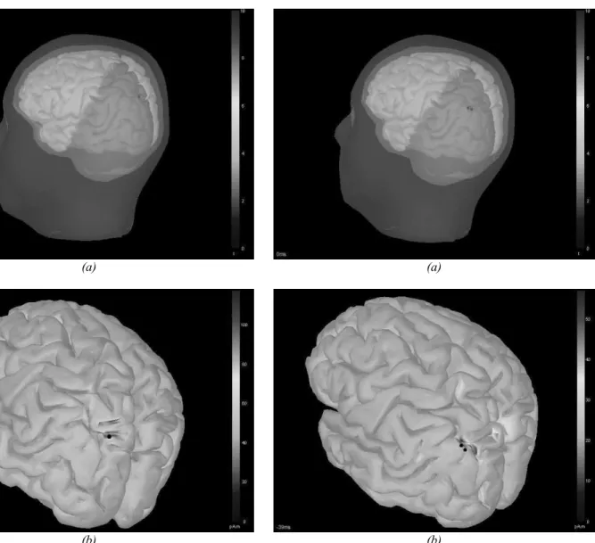
computationally exhaustive, it was assumed that we know which quarter of the cortex holds the active source(s) to be detected. This assumption can be justified because in most practical experiments, depending on the type of the stimulation and our knowledge of the cortical mapping (e.g. sensory area, motor area, and association area of the cortex), it is possible to estimate the primary region of activity in the cortex in a given evoked response experiment. The region assumed to hold the source is shown with color blue on the cortex surface in Fig. 1(a) and Fig. 2(a).
III. SIMULATION RESULTS
A. MEG
CTF Helmet with 151 axial gradiometers is used as the sensor array. Neural activity is simulated as a Guassian waveform with maximum amplitude of 30 nA.m. White Guassian noise with variance of 10fT is added to the simulated data. For localization purposes, only 3 snapshots (time samples) of simulated recordings are used in the SAFFIRE algorithm. Also the grid sampling factor of the algorithm is set to 20; i.e. the number of grid points included in the iterative search is 20 times the number of the elements in the sensor array.
Single Sphere Head Model
The forward problem is solved using a single sphere head model fitted to the cortex. MEG recordings are simulated using this head model, and applied to the algorithm to localize the underlying source activity. The search was conducted on a quarter of the cortex surface (blue). Fig. 1(a) shows the localization result for one source (located at -0.0494 mm, 0.0257 mm, 0.1004 mm on MRI coordinates) using SAFFIRE on the single sphere head model. The red spot is the location of the source correctly recovered.
With noise variance of 10fT for each channel and peak signal of 30nAm, the array SNR (over all electrodes) in this case is 2.7dB. Fig. 1(b) shows the result from localization of the same point using weighted minimum norm estimate method (wMNE). Even though wMNE is applied with privilege of lower noise value, more time samples of the recordings, and threshold value of 40%, still the result suffers from low resolution.
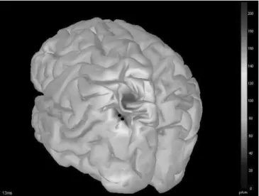
Next, in order to investigate robustness of the SAFFIRE algorithm to correlation of sources, and also to demonstrate high spatial resolution offered in this method, MEG recordings are simulated for two fully correlated Gaussian sources with a mere distance of 4mm. White Gaussian noise with variance of 10fT is added to each sensor, and the resulting noisy recordings are passed through SAFFIRE for localization. Correctly resolved proximate sources are depicted in Fig. 2 (a). In this case, overall (array) SNR is as low as -2 dB. At this SNR, no degradation due to correlation of sources is observed in the function of SAFFIRE. For lower SNRs, however, correlation of sources mandates a few more decibels of SNR for the same localization performance.
Proceedings of the World Congress on Engineering 2012 Vol I WCE 2012, July 4 - 6, 2012, London, U.K.
ISBN: 978-988-19251-3-8
ISSN: 2078-0958 (Print); ISSN: 2078-0966 (Online)
(a)
(b)
Fig. 1. Localization Results for a single source using (a) SAFFIRE, MEG/EEG. (b) wMNE, MEG.
Weighted minimum norm results are even further inferior for the case of correlated proximate sources (Fig. 2(b)). wMNE can not resolve the two sources; instead it finds a patch of the active cortex with an average distance of 9.5 mm from the first source and 9.1 mm from the second source.
Overlapping Spheres Head Model
This head model fits a sphere to the portion of the head that is closest to each MEG electrode when calculating the field value at that particular detector [10]. The same settings as in the previous section are applied. Source recovery results are also very similar to those of the previous section; hence not repeated here.
B. EEG
GSN 128 EEG cap is used as the sensor array. Similar to the MEG case, neural activity is simulated as a Guassian waveform with maximum amplitude of 30 nA.m. White Guassian noise with variance of 100 uV is added to the simulated data at each electrode. For localization purposes, only 3 snapshots of simulated readings are used in SAFFIRE algorithm, and the grid sampling factor of the algorithm was set to 30.
(a)
(b)
Fig. 2. Localization Results for 2 fully correlated proximate sources (4mm distance) using (a) SAFFIRE, MEG/EEG. (b) wMNE, MEG.
Three Spheres Head Model
The forward problem is solved using Brainstorm’s three spheres head model. In this model, three concentric spheres represent the cortex, skull and scalp, assuming homogeneous and isotropic conductivities for each layer. The relative conductivities of the three layers are set to the software’s default ratio of 1:1/80:1 (corresponding to 0.33, 0.004125, and 0.33 S/m). SNR value is set to 0 dB.
SAFFIRE is used to localize a single source using the simulated EEG recordings. The localization result is correct and similar to Fig. 1(a), which also formerly demonstrated the performance of SAFFIRE on MEG data. Next, wMNE is applied to the same set of EEG recordings. Fig. 3 shows the reconstructed low resolution patch of cortex using wMNE; black spot corresponds to the true source location, which SAFFIRE had successfully recovered. It’s also worth noting that performance of wMNE has deteriorated on EEG, compared to MEG data.
Subsequently, SAFFIRE is used on EEG readings to find two fully correlated sources, 4 mm apart, with overall SNR of -2 dB. The result in this case is similar to Figure 2(a), which shows that SAFFIRE resolves the two sources easily. Proceedings of the World Congress on Engineering 2012 Vol I
WCE 2012, July 4 - 6, 2012, London, U.K.
ISBN: 978-988-19251-3-8
ISSN: 2078-0958 (Print); ISSN: 2078-0966 (Online)
Fig. 3. Localization Results for a single source, using wMNE, for EEG.
Weighted MNE is then used to recover the same two sources. Even with higher SNR, more sample support and 40% threshold value, still the sources can not be resolved using wMNE. Also, both resolution and accuracy in this case are inferior to performance of wMNE on a similar scenario using MEG data.
Waveform Reconstruction
In each case (MEG/EEG/single source, two proximate correlated sources) SAFFIRE is used to reconstruct the original signals of the localized sources (using 26 samples of data). The original and reconstructed Gaussian waveforms for one of the recovered sources out of two, using EEG recordings, are depicted in Figure 5. Other scenarios produce similar results.
Fig. 5. Original and Reconstructed Waveforms using SAFFIRE
IV. CONCLUSION
SAFFIRE is an iterative MNE based method that can be applied to MEG or EEG recordings to yield sparse solutions for inverse problem of bioelectromagnetics. This algorithm is robust to correlation of sources, and requires low sample support. Comparing localization results with those of non-iterative wMNE shows that advantages of SAFFIRE are even more pronounced for EEG than MEG. The algorithm has also been applied to EEG readings for a realistic head model, including some calibration errors. The results will be presented in a future paper.
Fig. 4. Localization Results for 2 fully correlated proximate sources (4mm distance), using wMNE,for EEG.
REFERENCES
[1] A. C. Evans, D. L. Collins, and B. Milner, “An MRI-based stereotactic atlas from 250 young normal subjects”, J. Soc. For Neurosci. Abstr., vol. 18, p. 408, 1992.
[2] M. S. Hämäläinen and R. J. Ilmoniemi, "Interpreting magnetic fields of the brain: minimum norm estimates," Med. Biol. Eng. Comput., vol. 32, pp. 35-42., 1994.
[3] F.H. Lin, T. Witzel, S.P. Ahlfors, S.M. Stufflebeam, J.W. Belliveau,
and M.S. Hamalainen., “Assessing and improving the spatial accuracy
in MEG source localization by depth-weighted minimum-norm
estimates,” NeuroImage, vol. 31, no. 1, pp. 160-171, May 2006.
[4] M. Popescu, S.D. Blunt, and T. Chan, "Magnetoencephalography source localization using the source affine image reconstruction (SAFFIRE) algorithm," IEEE Trans. Biomed. Eng., vol. 57, no. 7, pp. 1652-1662, July 2010.
[5] S.D. Blunt, T. Chan, and K. Gerlach, "Robust DOA estimation: The re-iterative super resolution (RISR) algorithm," IEEE Trans. Aerosp. Electron. Syst. , vol. 47, no. 1, pp. 332-346, Jan. 2011.
[6] S. Baillet, K.J. Friston, and R. Oostenveld, “Academic software applications for electromagnetic brain mapping using MEG and
EEG,” Comp. Intell. Neurosc. 2011.
[7] F. Tadel, S. Baillet, J.C. Mosher, D. Pantazis, and R.M. Leahy,
“Brainstorm: A user-friendly application for MEG/EEG analysis,”
Comp. Intell. Neurosci. ,2011.
[8] P.L. Nunez, Electric Fields of the Brain: the Neurophysics of EEG, Oxford University Press, New York, 1981.
[9] M. Hämäläinen, R. Hari, R. Ilmoniemi, J. Knuutila, and O. V. Lounasmaa, "Magnetoencephalography - theory, instrumentation, and applications to noninvasive studies of the working human brain,"
Reviews of Modern Physics, vol. 65, no.2, pp. 413-497, Apr. 1993.
[10] M.X. Huang, J.C. Mosher, and R.M. Leahy, “A sensor-weighted overlapping-sphere head model and exhaustive head model
comparison for MEG,” Phys. Med. Bio., vol. 44, pp. 423-440. 1999.
Proceedings of the World Congress on Engineering 2012 Vol I WCE 2012, July 4 - 6, 2012, London, U.K.
ISBN: 978-988-19251-3-8
ISSN: 2078-0958 (Print); ISSN: 2078-0966 (Online)