Abstract
Submitted: September 1, 2016 Modiied: January 5, 2017 Accepted: March 10, 2017
Can whitening toothpastes maintain
the optical stability of enamel over
time?
Besides the effects on the health of individuals, cigarette smoking can also interfere with the appearance of their teeth. Objective: To evaluate the effect of cigarette smoking-toothbrushing-cycling (CSTC) with whitening toothpastes on the roughness and optical behavior of bovine enamel for eight weeks. Material and Methods: Thirty bovine dentin/enamel discs, 8.0 mm in diameter and 2.0 mm thick, were randomly divided into three groups according to the toothpastes: whitening (Colgate Luminous White – CW and Oral B 3D White – OW), and a non-whitening (Colgate – C). The roughness, color (CIE L*a*b* system), translucency and gloss were measured before and after the specimens were submitted to CSTC. The topography of the
specimens was analyzed by scanning electron microscopy. During the irst
week, the specimens were daily subjected to the consumption of 20 cigarettes and brushed (40 strokes/100 g) with the toothpastes’ slurries. Thereafter, the CSTC was weekly applied in an accumulated model (140 cigarettes/280 strokes) for seven weeks. The data were submitted to two-way ANOVA, Tukey’s HSD test, and paired-t test (α=0.05). Results: The three toothpastes
produced signiicant changes in roughness, color, translucency and gloss
(p<0.05). After eight weeks, the roughness and the gloss produced by the three toothpastes were similar (p>0.05), while OW produced the lowest color change and the translucency of C was lower than that of CW (p<0.05).
The three toothpastes produced a signiicant decrease in L* values and a signiicant increase in a* values after eight weeks (p<0.05). No signiicant
difference in the b* coordinate was found for OW (p=0.13) There were topographic changes in the enamel surfaces. Conclusions: The whitening toothpastes increased the roughness, changed the topography and were not able to maintain the optical stability of enamel exposed over eight weeks.
Keywords: Dental enamel. Toothbrushing. Toothpastes. Surface properties. Color.
Eduardo Moreira da SILVA1 Juliana Nunes da Silva Meirelles Dória MAIA1 Carine Gnatiuk MITRAUD1 Juliana do Espírito Santo RUSSO1 Laiza Tatiana POSKUS1 José Guilherme Antunes
GUIMARÃES1
Original Article
http://dx.doi.org/10.1590/1678-7757-2016-0460
1Universidade Federal Fluminense, Faculdade de Odontologia, Laboratório Analítico de Biomateriais Restauradores, Niterói, RJ, Brasil.
Corresponding address: Eduardo Moreira da Silva Faculdade de Odontologia -Universidade Federal Fluminense. Rua Mário Santos Braga, nº 30 Centro -Niterói - RJ - Brazil - 24040-110 Phone: 55 21 2629-9832 - Fax: 55 21 2622-5739
Introduction
Among others, cigarette smoking is one of the most deleterious habits that cause devastating effects, such as cancer, emphysema, bronchitis, and coronary disease, on individuals31. Unfortunately, according to
the World Health Organization (WHO), more than one billion people use tobacco around the world and six million die per year due to this habit32. This picture is
a matter of concern.
In addition to a great variety of toxic chemicals, e.g., naphthalene, hexane, formaldehyde, carbon monoxide, arsenic, ammonia, and toluene, tobacco also contains staining substances, such as tar and coffee, that may cause extrinsic discoloration of teeth3,
and restorative biomaterials1. From the point of view
of Dentistry, this aspect also represents an aesthetic
concern. Speciically in terms of tooth discoloration,
it has been shown that bleaching techniques that use
hydrogen peroxide and other substances are eficient
to remove intrinsic and extrinsic staining produced by different sources7,23. Clinically, the patient should avoid
the use of staining substances during the whitening protocols that use these chemical stain removers10.
However, this is not an easy task for smokers. Thus, the previously named whitening toothpastes seem to be an alternative path to these patients.
Besides the basic ingredients used in traditional
products, e.g., surfactants, thickening agents, lavor, and luorides, whitening toothpastes also contain higher
amounts of abrasives that are capable of removing or preventing the deposition of extrinsic stains on the tooth’s surface11. The most common abrasives used in
today’s whitening toothpastes include hydrated silica, calcium carbonate, dicalcium phosphate dihydrate, sodium bicarbonate, perlite, and alumina8. During
the toothbrushing, a three-phase system formed by the tooth surface, the toothbrush bristles, and the abrasives between these are responsible for stain removing14. However, depending on the hardness,
shape and the size of abrasives, toothbrushing may also wear the tooth surface and cause changes in color and roughness9.
Although the results presented in the scientiic
literature have added important aspects to the comprehension of the action of whitening toothpastes on the enamel surfaces, there is still a lack of sound information on their action over enamel submitted to cigarette smoking3. Therefore, the purpose of our
study was to conduct an in vitro investigation about
the inluence of a cigarette
smoking-toothbrushing-cycling (CSTC) by using whitening toothpastes on the roughness and the optical stability (color, translucency and gloss) of bovine enamel over a period of eight weeks. The null hypothesis tested was that no
toothpaste would inluence the roughness and the
optical stability of bovine enamel after eight weeks of exposure to cigarette smoking-toothbrushing-cycling.
Material and methods
Thirty bovine incisors selected according to similar color and maintained in a 0.2% thymol solution at 4°C for one week were used in this study. Before the specimens’ preparation, the teeth were examined
under a stereomicroscope at 10x magniication (SZ40,
Olympus, Tokyo, Japan) to identify the presence of any defects that could interfere with the obtained results. The roots were separated from the crowns and the teeth were sectioned through the pulp chamber using a low speed water-cooled diamond saw (Isomet 1000, Buehler, Lake Bluff, IL, USA) to obtain enamel/dentin slices from their labial surfaces. The enamel and dentin
surfaces of each slice were ground lat with 1200-,
2500-, and 4000-grit SiC papers (DPU-10, Struers, Copenhagen, Denmark), which was controlled with a digital caliper (MPI/E-101, Mitutoyo, Tokyo, Japan), until reaching a thickness of 2.0±0.1 mm (1.0 mm of dentin and 1.0 mm of enamel). Afterwards, disc-shaped enamel/dentin specimens with 8.0 mm in diameter were prepared from each slice by using a diamond bur (#3097, KG Sorensen, Cotia, SP, Brazil) in a
high-speed hand piece ixed in a special sample-aligning
device. The specimens were randomly divided into three groups of ten specimens according to the three
toothpastes analyzed (Figure 1) and kept in artiicial
saliva at 37°C before taking all measurements.
Baseline measurements
Roughness
formula:
where L is the length of the section and ƒ(x) is the displacement function.
Color and translucency
The color was measured according to the CIE L*a*b* system by using a spectrophotometer (model CM2600d, Konica Minolta Sensing Inc., Osaka, Japan). A D65 illuminant, under 100% UV energy and specular reflection included (SCI), was used with a 45° entrance angle and 0° observation angle geometry. We carried out the measurements using a small area view (SAV). Before each measurement session, the spectrophotometer was calibrated by using the white calibration standard supplied by the manufacturer. In order to guarantee the consistency of consecutive and repeated measurements of CIE L*a*b* parameters, they were carried out over white and black spectrophotometry ceramic standards (Konica Minolta Sensing Inc., Osaka, Japan) that were precisely attached to the base unit of the spectrophotometer by using a customized jig with a central hole where the specimens were positioned. This procedure allowed the color to be consistently measured in the central area and at the same position for all the specimens. The L*, a* and b* values of each specimen were separately measured in triplicate against the white and the black backgrounds.
Gloss
Gloss was measured by using a small-area
glossmeter (ZGM 1110, Zehntner testing instruments,
Sissach, Switzerland), with a square measurement area of 2 mm x 2 mm and 60° geometry. A custom-made jig was used to place the specimen over the aperture of the glossmeter at the same position at each time
of measurement. The gloss, expressed in gloss units (GU), was measured in triplicate for each specimen.
Cigarette Smoking-Toothbrushing-Cycling –
CSTC
After the baseline measurements, the specimens were submitted to CSTC (Figure 2). During the
irst week, the specimens were daily exposed to 20
cigarettes (Hollywood, Souza Cruz, Cachoeirinha, RS, Brazil) by using a cigarette smoking machine. This consisted of a hermetically closed acrylic box
with ive holes on each side to it the cigarettes and
internal supports that allowed the specimens to be positioned with the enamel surfaces facing up. The smoking machine was connected to a vacuum pump by a silicone tube that caused a negative pressure enough to consume and aspirate the smoke released by the cigarettes. The specimens were exposed to smoke produced simultaneously by 10 cigarettes for 10 min. Then, each specimen was brushed [20 strokes/100 g + toothpaste slurry in a proportion of 1:2 by weight
(18 g of each toothpaste and 36 mL of artiicial saliva)]
in a brushing machine (MEV2, Odeme Biotechnology, Joaçaba, SC, Brazil). After that, the specimens were again exposed to 10 cigarettes and brushed using the same parameters. Between the daily cycles, the
specimens were stored in artiicial saliva (KCl, NaCl,
MgCl, CaCl, Nipagin, CNC, Sorbitol, and deionized water – pH=7) at 37°C. The CSTC was repeated every day for
seven days. After the irst week, the specimens were maintained in artiicial saliva at 37°C and resubmitted
to CSTC once a week for a period of eight weeks in a cumulative model (7x20 cigarettes + 7x 40 strokes).
Reevaluation of properties
All the properties (roughness, color, translucency, and gloss) were reevaluated after each CSTC (daily
during the irst week and weekly from the second
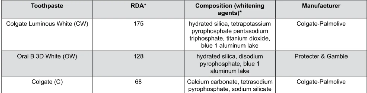
Toothpaste RDA* Composition (whitening
agents)*
Manufacturer
Colgate Luminous White (CW) 175 hydrated silica, tetrapotassium pyrophosphate pentasodium triphosphate, titanium dioxide,
blue 1 aluminum lake
Colgate-Palmolive
Oral B 3D White (OW) 128 hydrated silica, disodium pyrophosphate, blue 1
aluminum lake
Protecter & Gamble
Colgate (C) 68 Calcium carbonate, tetrasodium
pyrophosphate, sodium silicate Colgate-Palmolive
*According to manufacturers' information
to the eighth week). The color change (∆E) for each
specimen was calculated from the mean ∆L*, ∆a*, and ∆b* values, which were obtained against the white
background, by using the following formula:
where ∆L*, ∆a*, ∆b* are the differences in L*, a*
and b* coordinates obtained before and after each subsequent CSTC.
The L*, a* and b* coordinates obtained on the irst
and second day of evaluation were used to calculate the ∆E at baseline.
The translucency parameter (TP) for each specimen after each day (when measured daily) and each week (when measured weekly) was calculated using the following formula:
where the subscript B and W letters represent the measurements against the black and white backgrounds, respectively, in each subsequent CSTC.
Topographic analysis
Two specimens from each group, randomly selected, were analyzed by scanning electron microscope (SEM) at baseline and after the eighth week. The specimens were mounted in a charge reduction sample holder and observed under SEM (Phenom ProX, Phenom World, Eindhoven, Netherlands) operating in backscattered mode in a low vacuum environment. The SEM images
were taken by employing 15 Kv, at a magniication of
x2500.
Statistical analysis
We analyzed the obtained data using Statgraphics Centurion XVI software (STATPOINT Technologies, Inc., Warrenton, VA, USA). The normal distribution of errors and the homogeneity of variances were preliminarily checked by Shapiro-Wilk’s and Levene’s tests. Based on these analyses, roughness, color, translucency and gloss were separately analyzed by two-way ANOVA repeated measures and Tukey’s HSD post hoc test. We used paired-t test to analyze the differences in L*, a*,
and b* coordinates at baseline and after eight weeks
of CSTC. All analyses were performed at a signiicance level of α=0.05.
Results
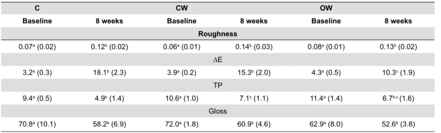
The mean values of roughness, ∆E, TP, and gloss at baseline and after eight weeks of CSTC are presented
in Table 1. The three toothpastes produced signiicant
changes in roughness, ∆E, TP, and gloss after eight weeks of CSTC (p<0.05). For roughness, the values after eight weeks were statistically similar (p>0.05). The ∆E produced by OW after eight weeks was lower than those produced by C and CW (p<0.05), which were not different from each other (p>0.05). After eight weeks, C produced the lower alteration in enamel translucency (p<0.05), but with no difference from that produced by OW. After eight weeks of CSTC, the gloss produced by the three toothpastes was similar (p>0.05).
The L*, a*, and b* color coordinates of enamel at baseline and after eight weeks are shown in Table 2.
The three toothpaste groups presented a signiicant decrease in L* values and a signiicant increase in a*
values after eight weeks (p<0.05). Conversely, only C
and CW showed a signiicant increase in b* coordinate
after eight weeks (p=0.1323).
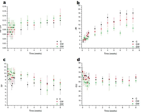
Figure 3 shows the evolution of all properties from the baseline to the eighth week of the CSTC. For the three toothpastes, roughness (a) increased uniformly from the second to the eighth week. Color
(∆E) (b) presented a remarkable change until the ifth
week, modifying in a subtle way from the ifth to the
eighth week. The translucency (TP) (c) decreased in an irregular way, with values ranging up and down.
Gloss (d) presented the greatest changes in the irst
week, and was relatively stable from the second to the eighth week.
Representative SEM micrographs of enamel before (a) and after (b, c, and d) eight weeks of CSTC are depicted in Figure 4. Regardless of the toothpaste used, CSTC produced topographic changes in the enamel surfaces, with the enamel prisms being more evident in the specimens brushed with the whitening toothpastes: CW (c) and OW (d).
C CW OW
Baseline 8 weeks Baseline 8 weeks Baseline 8 weeks
Roughness
0.07a (0.02) 0.12b (0.02) 0.06a (0.01) 0.14b (0.03) 0.08a (0.01) 0.13b (0.02) ∆E
3.2a (0.3) 18.1b (2.3) 3.9a (0.2) 15.3b (2.0) 4.3a (0.5) 10.3c (1.9) TP
9.4a (0.5) 4.9b (1.4) 10.6a (1.0) 7.1c (1.1) 11.4a (1.4) 6.7b,c (1.6) Gloss
70.8a (10.1) 58.2b (6.9) 72.0a (1.8) 60.9b (4.6) 62.9a (8.0) 52.6b (3.8)
In rows, means followed by the same lowercase letter are similar (Tukey HSD test, p>0.05)
Table 1- Mean values (±SD) of roughness (μm), ∆E, TP and Gloss at baseline and after 8 weeks of cigarette-smoking-toothbrushing-cycle (CSTC)
C CW OW
Baseline 8 weeks Baseline 8 weeks Baseline 8 weeks
L*
85.73a (1.87) 70.91b (7.11) 84.75a (1.44) 72.85b (4.31) 84.62a (1.24) 77.09b (4.15) a*
0.99a (0.21) 6.68b (1.39) 0.58a (0.13) 6.73b (1.50) 0.69a (0.18) 5.22b (1.10)
b*
11.98a (1.34) 20.68b (4.55) 12.82a (2.02) 20.04b (3.87) 13.84a (2.05) 17.68a (2.95)
In rows, for each toothpaste, means followed by the same lowercase letter are similar (Paired-t test, p>0.05)
Discussion
In a recent study on the prevalence of smoking and cigarette consumption in 187 countries, Ng, et al.21
(2014) showed that, in 2012, 34 countries presented
an average of cigarettes consumption per smoker
per day of less than 10. In 78 countries, this number ranged from 10 to 20 cigarettes, and was greater than 20 in the last 75 countries. This was the basis to use a protocol of the consumption of 20 cigarettes per day in this study, analyzing the responses accessed here
Figure 3- Curves of evolution of properties vs. time for the three toothpastes: roughness (a), color (b), translucency (c) and gloss (d)
by using a worst-case scenario. A previous study has shown that moderate (10-20 cigarettes/day) and heavy (>20 cigarettes/day) smokers presented a habit of toothbrushing twice a day25. This was the rationale to
the strategy of consumption of 10 cigarettes followed
by brushing two times in the irst week. The idea was
to simulate, as closely as possible, a common day in a smoker’s life. The number of 40 strokes per day to brush the specimens was based on an estimation that a tooth is brushed for 20 s in each daily toothbrushing of 2 min. Thus, considering that a heavy smoker brushes their teeth twice a day25, this means that each tooth
will be submitted, on average, to 40 strokes daily. We can note in Table 1 that after eight weeks of
CSTC, the three tested toothpastes produced signiicant
alterations on roughness and optical stability [color
(∆E), translucency and gloss] of bovine enamel. Thus,
the null hypothesis of our study was rejected. From the clinical point of view, surface roughness presents crucial importance due to two aspects. First, it exerts
a great inluence on bacterial adhesion forces18, an
aspect that may increase the bioilm accumulation
and, therefore, produce demineralization of enamel6,
and periodontal diseases24. Second, roughness implies
in sulcus formation in the enamel surfaces that can favor the accumulation of oral pigments, e.g., coffee, tea and tobacco, which may interfere with the optical appearance of enamel. This is an aesthetic matter. In
our study, we found a signiicant increase in roughness
from the baseline to the eighth week (Table 1), with the baseline and eight week values of roughness nicely agreeing with others previously published30.
Moreover, Figure 3a shows that this increase in roughness was gradual, characterizing a cumulative effect of toothbrushing on this response. On the other
hand, it is noteworthy that the inal absolute values
of roughness, ranging from 0.12 to 0.14 mm, were below 0.2 mm, which is the crucial number in terms of bacteria accumulation4. Thus, these roughness values
would not represent a clinical issue from the periodontal and enamel demineralization point of view.
The abrasiveness of toothpastes is strongly
inluenced by characteristics of the abrasive particles
included in their formulations, i.e., hardness, size, shape and size distribution5,11,14. This was considered
to analyze the three toothpastes used in this study. Colgate (RDA=68) is a regular toothpaste with less abrasive calcium carbonate particles19. Conversely, the
whitening toothpastes (CW – RDA = 175 and OW – RDA
= 128) have hydrated silica, an intermediate abrasive agent19. CW also presents titanium dioxide, which has
moderate abrasiveness. Based on these differences, the similarity among the values of roughness of eight weeks produced by the three toothpastes was unexpected. However, some previous studies
can support these indings. Hilgenberg and others9
(2011) showed similar bovine enamel roughness after 1,600 toothbrushing strokes with a regular calcium carbonate-based (low abrasiveness) and two whitening silica-based toothpastes. Moreover, an analysis of the roughness of human enamel brushed with different toothpastes in situ (42 days in the oral environment) by Melo, Manfroi and Spohr19 (2014) also showed
no differences on enamel roughness produced by a calcium carbonate-based and two hydrated silica-based whitening toothpastes. This last study is noteworthy because, even in the oral environment, a dynamic system and toothpastes with different RDAs produced no differences on enamel roughness.
According to Pascaretti-Grizon, Mabilleau and Chappard22 (2013), even having different abrasives,
toothpastes can produce similar abrasiveness due to the differences in size and other characteristics of these particles. Thus, we assume that while the three toothpastes analyzed in our study present different abrasives (Table 1), the synergism produced by their size, hardness and or distribution could have
inluenced the inal values of roughness observed here.
The features presented in Figure 4 could reinforce this thought, that is, although CW and OW present hydrated silica as abrasive, it seems that the sulcus produced on the enamel surfaces by CW (Figure 4c) were somewhat wider than those produced by OW (Figure 4d), suggesting that the former presents greater abrasive particles.
Different from previous studies3,15,28, the whitening
toothpastes in this study were not capable of removing the staining and maintaining the color stability of enamel surfaces after eight weeks (Table 1). Most probably, this result can be explained by the differences between the experimental protocols used in previous and present studies, that is, in those cited
studies, the enamel specimens were irst exposed
only to staining solutions (black tea15 or coffee28) or
to a coffee solution plus cigarette smoking3 and then
removed by the subsequent toothbrushing2. In this
study, the enamel surfaces were alternately submitted to cigarette smoking and toothbrushing during the entire experimental protocol. Thus, it is possible that in each CSTC, the products derived from the cigarette smoke could have impregnated the sulcus produced by toothpaste abrasives (Figure 4) and the dissimilarities
between the diameter of the toothbrush ilaments and
the width of those sulcus could have prevented the abrasive particles from reaching these deeply stained areas14.
Another important aspect observed here is that all the values of ∆E after eight weeks were greater than 3.3, which is the clinically acceptable value for color changing26. Moreover, from the data shown in
Figure 3b, the progressive DE was clearly a cumulative phenomenon, reinforcing that none of the toothpastes was capable of maintaining the color stability over time. Regardless, the curves depicted in Figure 3 show that during the eighty weeks the ∆E of CW and OW were lower than those of C. Most probably, the optical whitening agents present in CW and OW (Table 1)
inluenced this behavior. Blue 1 aluminum lake is an
optical whitening agent proposed to be deposited onto the tooth surfaces and to create a blue color perception that increases the whitening effect.
The CIE L*a*b* system represents a three-dimensional color space that provides a representation for the perception of color stimuli, where the L* axis measures the lightness of the object, ranging from 0 (black) to 100 (white) and the a* and b* axes represent the degree of green-red and blue-yellow color, respectively12. In our study, all toothpastes
produced a signiicant decrease in L* values after eight weeks (Table 2). From this inding, we can interpret
that the enamel underwent a reduction in lightness,
regardless of the toothpaste used. Also, the signiicant
increase in a* and b* values indicates a tendency to discoloration to dark brown and dark yellow and reinforces the fact that no toothpaste was capable of removing the staining produced by the cigarette smoking. Furthermore, the fact that OW did not present
a signiicant increase in b* value and presented a
higher numerical L* coordinate after eight weeks can be possibly explained by a greater amount of Blue 1 aluminum lake in its composition.
Translucency can be deined as the relative quantity of light transmission or diffuse relection from a material
surface through a turbid medium. In enamel, this
phenomenon is inluenced by its complex microstructure
(crystals and prisms)27, and, among other things, by
micromorphological surface modiications13. In this
ield, Ma, et al.16 (2009) showed that the translucency
of enamel was reduced after 14 days of bleaching with
carbamide peroxide and linked this inding to enamel
surface alterations produced by the bleaching protocol. This behavior is corroborated by the study of Vieira, Arakaki and Caneppele29 (2008). In our study, the
three toothpastes produced a progressive decrease
in enamel translucency from the irst to the eighth
week (Figure 3c), reaching signiicantly lower TPs at
the end of the experimental protocol (Table 1). Most
likely, these results were inluenced by the increase
in roughness after each CSTC (Figure 3a), which
could have increased the diffuse relectance onto the
enamel surfaces, thereby lowering its translucency13.
This decrease in translucency observed here could
have also inluenced the results of color evaluation.
Tooth color is the result of diffuse relectance from the
inner dentine through the outer translucent enamel17.
Thus, if the translucency of the enamel was reduced, clearly, less light from the dentine was captured by the spectrophotometer. This thought is supported by the study by Ma, et al.17 (2011) who showed that the
tooth color change was inluenced by the lowering of
the translucency of enamel after bleaching.
Although the three toothpastes produced a
statistically signiicant difference on gloss between the irst and the eighth week (Table 1), this was the
optical property least affected by the CSTC in our study. In fact, the overall changes in gloss happened in the
irst week, being relatively stable from the second to
the eighth week of the CSTC (Figure 3d). Muñoz, et
al.20 (2004) compared the eficacy of a low-abrasive
calcium, phosphate, and sodium bicarbonate-based dentifrice with a high-abrasive silica-containing dentifrice in situ and showed that, after three months, the former improved the roughness and the gloss of enamel surfaces. Considering that gloss is result of the interaction between the light and the morphology of a surface, it seems safe to claim that the decrease in gloss in our study was due to the increase in the light scattering on the rougher enamel surfaces produced by the CSTC.
mind that it has several limitations. The use of bovine teeth, only two whitening toothpastes and one type of cigarette, and the short time of evaluation (eight weeks) are among the limitations. These and other aspects should be addressed in future investigations.
Conclusions
Within the limitations of our study, we can conclude that the three toothpastes increased the surface roughness and were not capable of maintaining the optical stability (color, translucency and gloss) of enamel over a period of eight weeks submission to a cigarette smoking-toothbrushing-cycling. These results suggest that the therapy of using whitening
toothpastes could be not totally eficient when used
by heavy smokers.
References
1- Alandia-Roman CC, Cruvinel DR, Sousa AB, Pires-de-Souza FC,
Panzeri H. Effect of cigarette smoke on color stability and surface roughness of dental composites. J Dent. 2013;41(Suppl3):e73-9.
2- Alshara S, Lippert F, Eckert GJ, Hara AT. Effectiveness and mode of action of whitening dentifrices on enamel extrinsic stains. Clin Oral
Investig. 2014;18(2):563-9.
3- Bazzi JZ, Bindo MJ, Rached RN, Mazur RF, Vieira S, Souza EM. The
effect of at-home bleaching and toothbrushing on removal of coffee and cigarette smoke stains and color stability of enamel. J Am Dent
Assoc. 2012;143(5):e1-7.
4- Bollen CM, Lambrechts P, Quirynen M. Comparison of surface
roughness of oral hard materials to the threshold surface roughness for bacterial plaque retention: a review of the literature. Dent Mater.
1997;13(4):258-69.
5- Borges AB, Santos LF, Augusto MG, Boniette D, Hara AT, Torres
CR. Toothbrushing abrasion susceptibility of enamel and dentin bleached with calcium-supplemented hydrogen peroxide gel. J Dent.
2016;49:54-9.
6- Cross SE, Kreth J, Wali RP, Sullivan R, Shi W, Gimzewski JK. Evaluation
of bacteria-induced enamel demineralization using optical proilometry.
Dent Mater. 2009;25(12):1517-26.
7- Dantas AA, Bortolatto JF, Roncolato A, Merchan H, Floros MC, Kuga MC, et al. Can a bleaching toothpaste containing Blue Covarine
demonstrate the same bleaching as conventional techniques? An in
vitro, randomized and blinded study. J Appl Oral Sci. 2015;23:609-13. 8- Hattab FN, Qudeimat MA, al-Rimawi HS. Dental discoloration: an overview. J Esthet Dent. 1999;11(6):291-310.
9- Hilgenberg SP, Pinto SC, Farago PV, Santos FA, Wambier DS. Physical-chemical characteristics of whitening toothpaste and evaluation of its
effects on enamel roughness. Braz Oral Res. 2011;25(4):288-94. 10- Joiner A. The bleaching of teeth: a review of the literature. J Dent.
2006;34:412-9.
11- Joiner A. Whitening toothpastes: a review of the literature. J Dent.
2010;38:Suppl 2:e17-24.
12- Joiner A, Philpotts CJ, Alonso C, Ashcroft AT, Sygrove NJ. A novel optical approach to achieving tooth whitening. J Dent. 2008;36
Suppl1:S8-14.
13- Kwon SR, Wertz PW. Review of the mechanism of tooth whitening.
J Esthet Restor Dent. 2015;27(5):240-57.
14- Lewis R, Dwyer-Joyce RS, Pickles MJ. Interaction between
toothbrushes and toothpaste abrasive particles in simulated tooth cleaning. Wear. 2004;257(3-4):368-76.
15- Lima DA, Silva AL, Aguiar FH, Liporoni PC, Munin E, Ambrosano GM, et al. In vitro assessment of the effectiveness of whitening dentifrices for
the removal of extrinsic tooth stains. Braz Oral Res. 2008;22(2):106-11.
16- Ma X, Jiang T, Sun L, Wang Z, Zhou Y, Wang Y. Effects of tooth
bleaching on the color and translucency properties of enamel. Am J Dent. 2009;22:324-8.
17- Ma X, Li R, Sa Y, Liang S, Sun L, Jiang T, et al. Separate contribution
of enamel and dentine to overall tooth colour change in tooth bleaching.
J Dent. 2011;39:739-45.
18- Mei L, Busscher HJ, van der Mei HC, Ren Y. Inluence of surface
roughness on streptococcal adhesion forces to composite resins. Dent Mater. 2011;27:770-8.
19- Melo CF, Manfroi FB, Spohr AM. Microhardness and roughness of enamel bleached with 10% carbamide peroxide and brushed with
different toothpastes: an in situ study. J Int Oral Health. 2014;6(4):18-24.
20- Muñoz CA, Stephens JA, Proskin HM, Ghassemi A. Clinical eficacy
evaluation of calcium, phosphate, and sodium bicarbonate on
surface-enamel smoothness and gloss. Compend Contin Educ Dent. 2004;25(9 Suppl 1):32-9.
21- Ng M, Freeman MK, Fleming TD, Robinson M, Dwyer-Lindgren L, Thomson B, et al. Smoking prevalence and cigarette consumption in
187 countries, 1980-2012. JAMA. 2014;311(2):183-92.
22- Pascaretti-Grizon F, Mabilleau G, Chappard D. Abrasion of 6
dentifrices measured by vertical scanning interference microscopy. J Appl Oral Sci. 2013;21(5):475-81.
23- Públio JC, D’Arce MB, Brunharo NM, Ambrosano GM, Aguiar
FH, Lovadino JR, et al. Inluence of surface treatments on enamel
susceptibility to staining by cigarette smoke. J Clin Exp Dent. 2013;5(4):e163-8.
24- Quirynen M, Bollen CM. The inluence of surface roughness and
surface-free energy on supra- and subgingival plaque formation in
man. A review of the literature. J Clin Periodontol. 1995;22(1):1-14. 25- Santos A, Pascual A, Llopis J, Giner L, Kim DM, Levi P Jr. et al.
Self-reported oral hygiene habits in smokers and nonsmokers diagnosed with periodontal disease. Oral Health Prev Dent. 2015;13(3):245-51.
26- Stober T, Gilde H, Lenz P. Color stability of highly illed composite
resin materials for facings. Dent Mater. 2001;17(1):87-94.
27- Ten Bosch JJ, Coops JC. Tooth color and relectance as related to
light scattering and enamel hardness. J Dent Res. 1995;74:374-80.
28- Torres CR, Perote LC, Gutierrez NC, Pucci CR, Borges AB. Eficacy
of mouth rinses and toothpaste on tooth whitening. Oper Dent.
2013;38:57-62.
29- Vieira GF, Arakaki Y, Caneppele TM. Spectrophotometric assessment
of the effects of 10% carbamide peroxide on enamel translucency. Braz Oral Res. 2008;22(1):90-5.
30- Vieira-Junior WF, Lima D, Tabchoury C, Ambrosano G, Aguiar F, Lovadino JR. Effect of toothpaste application prior to dental bleaching
on whitening effectiveness and enamel properties. Oper Dent. 2016;41(1):E29-38.
31- Vu T, Jin L, Datta PK. Effect of cigarette smoking on epithelial to mesenchymal transition (EMT) in lung cancer. J Clin Med. 2016;5(4).
pii: E44.
32- World Health Organization. Tobacco [online]. 2017 May. [cited 2017 Jan 5]. Available from: http://www.who.int/mediacentre/factsheets/