Anatomical organization of aortic arch variations
in the India: embryological basis and review
Organização anatômica das variações do arco aórtico na população indiana:
base e revisão embriológica
Soubhagya R. Nayak, Mangala M. Pai, Latha V. Prabhu, Sujatha D’Costa, Prakash Shetty*
* Department of Anatomy, Centre for Basic Sciences, Kasturba Medical College, Bejai, Mangalore, Karnataka, India.
Article submitted March 17, 2006, accepted June 06, 2006.
J Vasc Bras 2006;5(2):95-100.
Copyright © 2006 by Sociedade Brasileira de Angiologia e de Cirurgia Vascular.
95
Abstract
Objectives: To determine the percentage and type of aortic arch
variations in Indian subjects and their clinical and surgical importance and embryological basis.
Patients and methods: In our investigation, branching patterns
of the aortic arch were studied in 62 formalin-fixed cadavers of both sexes of Indian origin, aged 45-79. T he dissections were carried out in formalin-preserved cadavers and the aortic arch variations were observed after exposing the thoracic and cervical region during routine dissection of undergraduate students of Indian origin in Kasturba Medical College, Mangalore.
Results: T he usual three-branched aortic arch was found in 56 cadavers (91.4%); variations were found in six cadavers (9.6%); 4.8% presented common origin of the carotid arteries; 1.6% had bi-innominate sequence, and the same specimen had left coronary artery arising from arch of aorta directly; 1.6% presented right subclavian artery arising directly from the aorta; 1.6% had left vertebral artery a branch of aortic arch. Five out of six cadavers with anomalous aortic arch branching pattern were females. One male cadaver presented anomalous origin of left vertebral artery directly from the arch.
Conclusion: T he wide spectrum of variations in the anatomical
arrangements of the aortic arch branches in the Indian population was at par with other populations of the world. Although anomalous origins of the aortic arch branches are merely anatomic variants, accurate information about them is vital for vascular surgery in the thorax, head and neck region.
Key words: Aortic arch, vertebral artery, brachiocephalic trunk,
variations.
Resumo
Objetivos: Determinar a porcentagem e o tipo de variações do
arco aórtico em indivíduos indianos, bem como sua importância clí-nica e cirúrgica e base embriológica.
Pacientes e métodos: Em nossa investigação, os padrões de
rami-ficação do arco aórtico foram estudados em 62 cadáveres fixados em formalina de ambos os sexos, de origem indiana e com idade entre 45 e 79 anos. As dissecações foram realizadas em cadáveres preservados em formalina, e as variações do arco aórtico foram observadas após a exposição das regiões torácica e cervical durante a dissecação de rotina de alunos da graduação do Kasturba Medical College, Mangalore, Índia.
Resultados: O arco aórtico normal de três ramificações foi en-contrado em 56 cadáveres (91,4%); as variações foram encontradas em seis cadáveres (9,6%); 4,8% apresentavam origem comum das ar-térias carótidas; 1,6% tinham seqüência binominada, e o mesmo es-pécime tinha a origem da artéria coronária esquerda diretamente no arco aórtico; 1,6% apresentavam a origem da artéria subclávia direita diretamente na aorta; 1,6% tinham como ramificação do arco aórtico uma artéria vertebral esquerda. Cinco de seis cadáveres com padrão de ramificação anômalo do arco aórtico eram do sexo feminino. Um cadáver do sexo masculino apresentou origem anômala da artéria ver-tebral esquerda diretamente no arco.
Conclusão: O amplo espectro de variações nos padrões
anatômi-cos das ramificações do arco aórtico na população indiana estava em concordância com outras populações mundiais. Embora as origens anômalas das ramificações do arco aórtico sejam meramente varia-ções anatômicas, informavaria-ções precisas sobre elas é essencial para a cirurgia vascular na região do tórax, cabeça e pescoço.
Palavras-chave: Arco aórtico, artéria vertebral, tronco
braquio-cefálico, variações.
Introduction
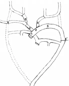
The great arteries of the thoracic region are well known for their variations. The aortic arch is one of them, with well known variations. The arrangement regarded as normal for humans is actually found more frequently than other types combined. In specimens of normal variety, the branches leave the arch in the following succession from left to right: left subclavian, left common carotid (LCC) and brachiocephalic trunk (with right common carotid and right subclavian as its derivatives). The variations of branches arising from aortic arches (Figure 1) are well known and documented by several authors in different races, such as Barry,1
Barewell,2 Birmingham,3 Brodie,4 Brown et al.,5 Degaris et al.,6 Harley,7 Lize,8 Grande et al.,9 Bhatia et al.10
Some authors, such as Bernardi et al.,11 hypothesize that anomalous origins and the distribution of the large aortic arch vessels can cause changes in cerebral hemodynamics that may lead to cerebral abnormalities. The true value of detecting anomalous origins is the diagnostic gain prior to the surgery of supraaortic arteries. For cases in which the vertebral artery originates from the carotid artery or its branches, the ligation of
Figure 1 - Derivation of major arteries from aortic arches in normal case
1, 2, 3) Development into aortic arch. 4) Brachiocephalic trunk. 5) Common carotid arteries. 6) Subclavian artery.
the common carotid artery may cause a compromise of the posterior fossa blood supply, as reported by Flynn et al.12 Detailed knowledge of an anomalous origin of supraaortic arteries is also important for patients who have to undergo four-vessel angiography in an emergency, in order to rule out, for example, the possibility of intracranial aneurysm after subarachnoid hemorrhage. If the detection of a vertebral artery in the normal position is not possible, the presence of such a
variant must be taken into consideration.13 T he
anatomic and morphologic variations of the aortic arch and its branches are significant for diagnostic and surgical procedures in the thorax and neck. If a right retroesophageal subclavian artery is diagnosed during aortic arch repair, corrective surgery should be considered. Intensive care patients should be screened before long term placement of nasogastric tube, in order to avoid fistulization and fatal hemorrhage.14 Momma et al. described that aortic arch anomalies are also associated with chromosome 22q11 deletion.15
The present study describes the aortic arch branching pattern in Indian subjects and discusses the findings according to their embryological, clinical and surgical implications.
Materials and methods
During routine dissection conducted over 62 cadavers of both sexes (aged 45-79) of Indian origin at Kasturba Medical College, Mangalore, the mediastinum was opened and observations were made for abnormalities of the branching pattern of the aortic arch.
Results
In six cases (9.6%), the abnormalities were found in the branching pattern of the aortic arch. Of these three cases, 4.8% presented a common abnormality (the LCC artery as a branch of brachiocephalic trunk with different degrees of branching).
Cases I, II, and III
In case III, the brachiocephalic trunk also gave rise to the LCC artery, but the origin was higher than the previous two cases, the division of the right subclavian and right common carotid artery was even higher.
Case IV
In this case (Figure 5) there were only two trunks arising from the arch, left and right innominate arteries. Each one was giving rise to the subclavian and common carotid arteries of the respective sides. The left coronary artery was arising from the posterior aspect of the aortic arch.
Case V
In this case (Figure 6) the vertebral artery was a branch of the aortic arch, between the origins of LCC and left subclavian artery.
Figure 3 - Aortic arch variations, case II
BCT = brachiocephalic trunk; LCCA = left common carotid artery; LSA = left subclavian artery.
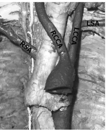
Figure 2 - Aortic arch variations, case I
BCT = brachiocephalic trunk; LCCA = left common carotid artery; LSA = left subclavian artery; RCCA = right common carotid artery; RSA = right subclavian artery.
Figure 4 - Aortic arch variations, case III
BCT = brachiocephalic trunk; LCCA = left common carotid artery; LSA = left subclavian artery; RCCA = right common carotid artery; RSA = right subclavian artery.
Figure 5 - Aortic arch variations, case IV. A) Anterior view.
B) Posterior view
Figure 6 - Aortic arch variations, case V
BCT = brachiocephalic trunk; LCCA = left common carotid artery; LSA = left subclavian artery; LVA = left vertebral artery.
Figure 7 - Aortic arch variations, case VI
LCCA = left common carotid artery; LSA = left subclavian artery; RCCA = right common carotid artery; RSA = right subclavian artery.
Case VI
Anomalous origin of right subclavian artery from the descending aorta, which courses behind the trachea and esophagus to reach the right axilla (Figure 7).
Discussion
The true value of detecting anomalous origins is in the diagnostic gain before vascular surgeries of supraaortic arteries, as variations of the branches of the aortic arch are likely to occur as a result of the altered development of certain branchial arch arteries during the embryonic period of gestation.10 The approximation of the LCC artery to the brachiocephalic trunk is an important observation while invading the aortic arch and its branches with instruments, since all cases are susceptible to surgical attack.10 Non-recognition of a critical aortic arch branch variation at surgery may cause fatal consequences.16 The angiographic detection
of common brachiocephalic trunk may be a marker for the presence of accompanying congenital cardiac defects and coronary arterial abnormalities. Understanding the pathophysiological effects of the common brachiocephalic trunk is important when planning the palliative or corrective procedures, and when assessing the potential benefit of the surgical repair over the long term.17
According to Anson et al.,18 the normal three-branched arrangement of the aortic arch is found in 64.9%. An arrangement distinguished by reduction in the number of stems to two, both common carotid arteries arising from the innominate occurs in 27.1%. The right subclavian artery passes dorsal to the esophagus and the last branch of the aortic arch reaching the right upper extremity in 0.5%. A bi-innominate sequence in which paired vessels are the only derivatives of the aortic arch is found in 1.2%. The left vertebral artery is the additional vessel arising from the arch in 2.5%. In our study we found the following variations with different percentages: 4.8, 1.6, 1.6, 1.6% respectively.
Embryological basis of the variations in our study
Case I, II, III
Case I. The left limb of the aortic sac normally forms the part of the arch that intervenes between the origins of the brachiocephalic trunk and the LCC artery. If the aortic sac fails to bifurcate into right and left limbs, then the LCC artery will connect to the aortic sac directly. That results in a common origin of the carotid arteries (COCA).
the brachiocephalic trunk will be abnormal as in the above three cases.
Case IV
The abnormal left brachiocephalic trunk was formed by the fusion of the proximal part of the left third arch artery and left seventh intersegmental artery into the left fourth arch artery, which was responsible for the left subclavian artery and LCC artery arising from a common trunk. The left coronary artery may bud from the aortic arch instead of the ascending aorta. T his was an additional anomaly.
Case V
The left subclavian artery normally develops from left seventh intersegmental artery and the first part of the vertebral artery develops from the dorsal ramus of the seventh intersegmental artery (proximal to post-costal anastomosis). The left subclavian artery is only developed from the left seventh intersegmental artery. In the present case the vertebral artery developed from the persistent sixth cervical intersegmental artery.
Case VI
This anomaly was caused by the degeneration of the right fourth aortic arch, so that the right seventh cervical intersegmental artery and the right dorsal aorta caudal to it are continued as the right subclavian artery. Anatomical and morphological variations of the vertebral artery are of great importance in surgery,
angiography and all non-invasive procedures.19
According to Bernardi & Delton, the abnormal origin of vertebral artery may favor cerebral disorders due to alterations in cerebral hemodynamics.11
Sometimes aortic arch anomalies are clinically useful. Catheterization of the LCC artery arising from an innominate artery can be achieved without catheter exchange.20 When three of the four primary sources of
cerebral blood flow arise from a single aortic branch, such as in the case of common origin of common carotid arteries, stenosis or occlusion of a common
trunk can cause severe ischemic consequences.21
Komiyana et al. reported the incidence of arterial dissection of the vertebral artery of aortic origin and vertebral artery of subclavian origin. According to their studies, the left vertebral artery of aortic origin was associated with a significantly higher incidence of
vertebral artery dissection than left vertebral artery of left subclavian artery origin and right vertebral artery of right subclavian origin.22
In our study, the aortic arch branch variations were observed in six cases (9.6%). The percentage of aortic arch branch variations was not as high as the data provided by Grande et al.9 in Portuguese population and Bhatia et al.10 in South Australian population of
European descent.
Conclusion
The wide spectrum of variations in the anatomical arrangements of the aortic arch branches in Indian population was at par with other populations of the world, but the percentage of anatomical variants was less comparative to other populations. Although anomalous origins of the aortic arch branches are merely anatomic variants, accurate information about them is vital for vascular surgery in the thorax, head and neck region.
References
1. Barry A. T he aortic arch derivatives in the human adult. Anat Rec. 1951;111(2):221-38.
2. Barwell. Abnormal origin of arteries from the aortic arch. T rans Pathol Soc Lond. 1867;18:68.
3. Birmingham A. Extreme anomaly of the heart and great vessels. J Anat Physiol. 1893;27:139-50.
4. Brodie G. Rare abnormality of the aortic arch. Lancet. 1888;2:971.
5. Brown JD, Brown FJ. Abnormal origin of the vessels from the arch of the aorta. Brit Med J. 1868;1:632.
6. De Garis CF, Black IH, Riemenschneider EA. Patterns of the aortic arch in American white and Negro stocks, with comparative notes on certain other mammals. J Anat. 1933;67:599-619.
7. Harley HR. T he development and anomalies of the aortic arch and its branches. Br J Surg. 1959;46:561-73.
8. Lize I. Abnormal origin of the great blood vessels from the aortic arch. Folia Morphol (Warsaw). 1970;29:355-7. 9. Grande NR, Costa e Silva A, Pereira AS, Aguas AP. Variations
in the anatomical organization of the human aortic arch. A study in a Portuguese population. Bull Assoc Anat (Nancy). 1995;79(244):19-22.
10. Bhatia K, Ghabriel MN, Henneberg M. Anatomical variations in the branches the human aortic arch: a recent study of a South Australian population. Folia Morphol (Warsz). 2005;64(3):217-23.
Correspondence: Soubhagya R. Nayak
Department of Anatomy – Kasturba Medical College 575004 – Bejai, Mangalore, Karnataka, India E-mail: [email protected]
12. Flynn RE. External carotid origin of the dominant vertebral artery. Case report. J Neurosurg.1968;29:300-1.
13. Lemke AJ, Benndorf G, Liebig T , Felix R. Anomalous origin of the right vertebral artery: review of the literature and case report of right vertebral artery origin distal to the left subclavian artery. AJNR Am J Neuroradiol. 1999;20:1318-21. 14. Fazan VPS, Ribeiro RA, Ribeiro JAS, Rodrigues Filho OA.
Right retroesophageal subclavian artery. Acta Cir Bras. 2003;18:54-6.
15. Momma K, Matsuoka R, T akao A. Aortic arch anomalies associated with chromosome 22q11 deletion (CAT CH 22). Pediatr Cardiol. 1999;20:97-102.
16. Satyapal KS, Singaram S, Partab P, Kalideen JM, Robbs JV. Aortic arch branch variations – case report and arteriographic analysis. S Afr J Surg. 2003;41:48-50.
17. Moskowitz WB, T opaz O. T he implications of common brachiocephalic trunk on associated congenital cardiovascular defects and their management. Cardiol Young. 2003;13: 537-43.
18. Anson BV, Mcvay CB. Surgical anatomy. 5th ed. Philadelphia: WB Saunders; 1971.
19. Matula C, T rattnig S, T schabitscher M, Day JD, Koos WT . T he course of the prevertebral segment of the vertebral artery, anatomy and clinical significance. Surg Neurol. 1997;48(2): 125-31.
20. Carlson DH, McDonald DG. Simplified catheterization of a left common carotid artery arising from the innominate trunk. Radiology. 1982;144:419.
21. Azakie A, McElhinney DB, Messina LM, Stoney RJ. Common brachiocephalic trunk: strategies for revascularization. Ann T horac Surg. 1999;67(3):657-60.