ELECTRONIC PAPERS
Coexistence of coronary cameral fistulae and cor
triatriatum sinister in an elderly patient
Se
´rgio Nabais
1,2*, Nuno Salome
´
1, Aida Branda
˜o
1, Alda Simo
˜es
1, Jorge Marques
1, Joa
˜o Costa
1,
Luı´s Basto
1, Anto
´nio Costeira
1, and Adelino Correia
11Cardiology Department, Hospital de S. Marcos, Apartado 2242, 4701-965 Braga, Portugal; and2Life and Health Sciences
Research Institute, School of Health Sciences, University of Minho, Braga, Portugal
Received 28 September 2007; accepted after revision 21 March 2008; online publish-ahead-of-print 27 April 2008
Coronary cameral fistulae are unusual congenital or acquired anomalous communications between an epicardial coronary artery and a cardiac chamber. There are no reported cases of the association of cor-onary cameral fistulae and cor triatriatum, a rare congenital cardiac anomaly in which a fibromuscular membrane divides the left atrium into two chambers. We report the case of an 82-year-old man present-ing with recurrent anterior chest pain. Echocardiographic examination identified non-obstructive cor triatriatum, mitral valve prolapse resulting in significant mitral regurgitation, dilated coronary arteries, and established the entry site of coronary artery fistulae at the apex of the left ventricle (Figure 1). Coronary angiography confirmed the existence of a plexiform fistula between the left anterior descend-ing coronary artery and the left ventricle. Tetrofosmine scintigraphy revealed the presence of stress-induced ischaemia in the apex. To our knowledge, we report the oldest person with coronary cameral fistulae presenting with angina only at this stage, and the interesting case of the coexistence of two, although unconnected, congenital conditions in an elderly patient. In addition, this report highlights the important role of transthoracic and transoesophageal echocardiography to the characterization of these unusual anomalies, and the complementary information offered by three-dimensional trans-thoracic echocardiography.
KEYWORDS Coronary artery fistulae; Cor triatriatum; Congenital heart disease; Three-dimensional
echocardiography; Angina;
Coronary artery disease
Introduction
Coronary cameral fistulae are unusual congenital or acquired anomalous communications between an epicardial coronary artery and a cardiac chamber. There are no reported cases of the association of coronary cameral fistulae and cor tria-triatum, a rare congenital cardiac anomaly in which a fibro-muscular membrane divides the left atrium (LA) into two chambers. We report the unique case of an 82-year-old man presenting with chest pain, in whom coexistent coron-ary cameral fistulae, cor triatriatum, and mitral valve pro-lapse with severe mitral regurgitation were evaluated by echocardiography and other imaging modalities.
Case report
An 82-year-old man was referred to our department for evaluation of recurrent episodes of anterior chest discom-fort at rest as well as during exercise in the last 4 months,
with more frequent episodes within the previous week. Mild exertional dyspnoea was also noted. He had no classic cardiovascular risk factors except a history of cigarette smoking from the age of 20 to 45 years. Atrial fibrillation had been detected 6 months before presentation to our department in a routine electrocardiogram (ECG); a pre-vious ECG performed 3 years earlier showed sinus rhythm and LA anomaly.
Cardiovascular system examination revealed a holosysto-lic murmur (Levine grade III/VI) at the cardiac apex with radiation to the axilla. The remainder of the cardiovascular examination was unremarkable. The 12-lead ECG showed atrial flutter with variable atrioventricular conduction (3:1 or 4:1, mean ventricular rate 80 bpm), left anterior hemi-block, and mild ST-segment depression in leads I, aVL, V4– V6. On serial testing, there was no elevation of cardiac injury markers. Transthoracic echocardiographic examin-ation in the apical view showed a thin echo-dense mem-brane that divided a moderately dilated LA into two distinct chambers. Interestingly, multiple channels with turbulent flow draining to the left ventricle near the apex were seen using colour Doppler imaging (Figure 1, see
*Corresponding author. Tel:þ351 933 406 200/þ351 253 209 155; fax:
þ351 253 209 091.
E-mail address: sergionnabais@gmail.com
Published on behalf of the European Society of Cardiology. All rights reserved.&The Author 2008. For permissions please email: journals.permissions@oxfordjournals.org.
European Journal of Echocardiography (2008)9, 712–715
doi:10.1093/ejechocard/jen140
at 50600 Hosptial CUF on February 17, 2012
http://ehjcimaging.oxfordjournals.org/
Supplementary material online, Video 1). Pulsed and continuous wave Doppler interrogation, and colour Doppler M-mode imaging of this area revealed predominant diastolic flow draining to the left ventricle, suggestive of coronary flow (Figure 2). In addition, there was left ventricle enlarge-ment (left ventricular end diastolic diameter: 65 mm) with preserved left ventricle systolic function. Mild systolic prolapse of the anterior leaflet of the mitral valve and sig-nificant mitral regurgitation were also present. Transoeso-phageal echocardiography (TEE) revealed an incomplete membrane across the LA, separating a postero-superior chamber receiving the pulmonary veins from an antero-inferior chamber in connection with the LA appen-dage and mitral valve (Figure 3A). There was a large postero-inferior opening between the two chambers without a significant pressure gradient or a mosaic pattern of turbulent flow across the membrane by continuous wave or colour Doppler study. Dilated left main coronary artery (Figure 3B) and left circumflex coronary artery were also seen. Further anatomic characterization of the mitral valve revealed diffusely thickened leaflets with maximum thickness of 6 mm at the tip of the anterior leaflet, suggesting myxomatous degeneration. Systolic prolapse of the anterior leaflet resulted in a highly eccentric and
disorganized mitral regurgitation jet. On colour Doppler, the main component of the mitral regurgitation jet was swirling along the membrane in the antero-inferior chamber and penetrated through the large opening into the postero-superior chamber (Figure 3C, see Supplemen-tary material online, Video 2). Mitral regurgitation was graded as severe based on the combination of overall find-ings, including significant colour Doppler regurgitant jet dimensions (considering the effects of flow constraint by the atrial walls), largevena contracta (7 mm), dense con-tinuous wave Doppler spectral signal, and reversal of systolic flow in the pulmonary veins.
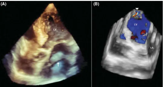
Three-dimensional (3D) echocardiography was performed using a commercial scanner (iE33, Philips, Andover, MA, USA). Using a cut plane, the LA membrane was visualized from multiple views, with a communication between the two LA chambers (Figure 4). In addition, 3D colour Doppler imaging showed multiple small shunts crossing the apex of the left ventricle.
Selective coronary angiography demonstrated dilated left main coronary artery, and dilated and tortuous left anterior descending coronary artery, left circumflex coronary artery, and right coronary artery, with variable degree of non-significant stenotic lesions. The first diagonal branch of the left anterior descending coronary artery was draining into a plexiform fistula that was opening into the left ventricle (Figure 5). The patient underwent an exercise treadmill-stress test with tetrofosmine scintigraphy, which revealed the presence of stress-induced ischaemia in the apex.
The patient was discharged on carvedilol, transdermal nitrates, lisinopril, simvastatin, warfarin, and clopidogrel. He was referred to cardiac surgery consultation for evalu-ation of possible mitral valve surgery to correct severe mitral valve regurgitation, but the patient eventually refused the possibility of any surgical intervention. On follow-up he denied recurrence of chest pain, and is in class II of the New York Heart Association classification of functional capacity.
Discussion
Congenital fistulous connections between the coronary arteries and a cardiac chamber appear to represent persist-ence of embryonic intertrabecular spaces and sinusoids.
Figure 1 Two-dimensional transthoracic echocardiography of apical ‘five-chamber view’ shows left atrium divided into antero-inferior and postero-superior chambers by membranous septum (arrow). Colour Doppler interrogation shows drainage of fis-tulae to the left ventricle near the apex (arrow head).
Figure 2 (A) Pulsed wave Doppler, and (B) colour Doppler M-mode examination showing diastolic flow through the myocardium draining to the left ventricle.
Coexistence of coronary cameral fistulae and cor triatriatum sinister 713
at 50600 Hosptial CUF on February 17, 2012
http://ehjcimaging.oxfordjournals.org/
Acquired causes of coronary fistulae include coronary atherosclerosis, Takayasu arteritis, trauma, and invasive cardiac procedures.1 The majority of coronary fistulae arise from the right coronary artery or the left anterior
descending coronary artery. Right-sided chambers are the most common sites of drainage, while fistulous communi-cations to the left-sided chambers are less frequent, especially those emptying into the left ventricle.2 Most adult patients are usually asymptomatic and the fistulae are discovered during cardiac catheterization for congenital heart anomalies or coronary artery disease. A continuous murmur is often present, but was not detected in our patient. Other possible clinical presentations include con-gestive heart failure, endocarditis, arrhythmias, pulmonary hypertension, premature arteriosclerotic changes within the fistulae, thromboembolic events, and rupture.3Angina is uncommon and myocardial infarction rare; it is postulated that these ischaemic symptoms are caused by coronary ‘steal’ phenomena.4 Our patient presented with anterior chest discomfort that could be related to decreased coron-ary blood flow distal to the fistula causing apex ischaemia, as demonstrated by cardiac scintigraphy.
In cor triatriatum sinister a fibromuscular membrane divides the LA into a postero-superior chamber receiving the pulmonary veins and an antero-inferior chamber containing the mitral valve and the LA appendage.5 Cor triatriatum may result from incomplete incorporation of the embryonic common pulmonary vein into the LA.6
Figure 3 (A) Transoesophageal echocardiography showing a membrane (arrow head) dividing the left atrium (LA) into two chambers. (B) Dilated left main coronary artery (arrow), and LA membrane (arrow head) are shown. (C) Colour Doppler imaging reveals mitral regur-gitation jet swirling around LA membrane (arrow head) and penetrating through large communication from antero-inferior into postero-superior chamber.
Figure 4 (A) Live three-dimensional (3D) transthoracic echocardiography of four-chamber apical view shows an incomplete membrane (arrow) in the mid-portion of the left atrium (LA); (B) 3D colour Doppler imaging shows a small shunt crossing the apex of the left ventricle (LV) (arrow head).
Figure 5 Angiogram with catheter tip in left coronary artery. Fistula from first diagonal branch of left coronary artery to the left ventricle is evident.
S. Nabaiset al.
714
at 50600 Hosptial CUF on February 17, 2012
http://ehjcimaging.oxfordjournals.org/
Depending on the size of the opening in the membrane, the obstruction to left ventricular inflow can manifest clinically from infancy to late adulthood, with symptoms of pulmonary venous congestion that imitate mitral stenosis. In rare cases, patients with cor triatriatum may remain asymptomatic and be diagnosed incidentally,7 whereas others may have late onset of symptoms, possibly related to fibrosis and calcifica-tion of the orifice in the anomalous membrane, or the devel-opment of mitral regurgitation or atrial fibrillation.8 Importantly, in our patient with non-obstructive cor triatria-tum, exertional dyspnoea was probably related to the devel-opment of significant mitral regurgitation and atrial fibrillation/flutter. Apparent myxomatous thickening of the mitral valve leaflets with associated systolic prolapse of the anterior leaflet resulted in a highly eccentric and disor-ganized mitral regurgitation jet. An impinging wall jet in an abnormal LA chamber limited the use of the common Doppler parameters and challenged the quantification of mitral regurgitation. Colour Doppler area of an eccentric jet such as this will underestimate the regurgitant volume by 40% when compared with an identical regurgitant
volume that is centrally located.9 In addition, accurate evaluation of regurgitant flow by the proximal isovelocity area method was precluded by the geometry of the proximal regurgitant jet. Systolic retrograde flow in the pulmonary veins remained an appropriate marker in the grading of severity of mitral regurgitation, considering there was no pressure gradient between the two LA chambers. Surgical intervention was indicated, but was eventually refused by the patient after thoughtful explanation.
Although other cardiac abnormalities may appear associ-ated with coronary fistulae (e.g. critical pulmonary stenosis, aortic atresia) and cor triatriatum (e.g. secundum atrial septal defect), the association of coronary fistulae and cor triatriatum in the same patient may be considered just a ‘finding’ without any pathophysiological implications. It is, however, interesting that the patient developed angina only at 82 years old, which is not the usual case of a conge-nital coronary abnormality.
Generally, TEE is superior to transthoracic imaging to diagnose cor triatriatum, providing better imaging of the LA, LA appendage, morphology of the dividing membrane and the degree of obstruction caused by this structure.5 Three-dimensional echocardiography, however, is a modality that offers additional information not offered by 2D echo-cardiography. It provides better spatial orientation, and visualization of the size and number of openings in the divid-ing membrane.5Regarding coronary fistulae, various cardiac imaging modalities are used for diagnosis and for planning surgical or percutaneous interventions if closure is indi-cated. A significantly enlarged coronary artery and turbulent flow pattern at the shunt entry site can usually be detected by 2D echocardiography. Multiplane TEE can define more precisely the origin, course, and drainage site of coronary fistulae, and its relation to the neighbouring structures.10 New imaging modalities, such as cardiac CT angiography or magnetic resonance imaging can be used as adjunct to coronary angiography, allowing 3D imaging of the anatomic relations of coronary fistulae to other structures, and their course.
Management of cor triatriatum depends on the degree of obstruction between the LA chambers. Surgical resection of the accessory membrane is indicated in symptomatic patients with obstructive cor triatriatum. The management of coronary fistulae is controversial.1 Patients can be treated by transcatheter or surgical closure of the communi-cation. Our patient was not considered an optimal candidate for percutaneous approach considering the morphology of the fistulous tracts. Furthermore, anti-anginal drug therapy relieved his chest pain and consequently no inter-vention was recommended.11
In conclusion, this case highlights the possible late presentation of coronary fistulae and cor triatriatum, and the important role of echocardiography to their characterization.
Supplementary data
Supplementary data are available atEJECHOonline. Video 1Two-dimensional transthoracic echocardiography of apical ‘five-chamber view’ showing left atrium divided into antero-inferior and postero-superior chambers by membranous septum. Colour Doppler interrogation in the same view shows drainage of fistulae to the left ventricle near the apex.
Video 2 Transoesophageal echocardiography with colour Doppler imaging reveals mitral regurgitation jet swirling around left atrium membrane and penetrating through large communication from antero-inferior into postero-superior chamber.
Conflict of interest:none declared.
References
1. Gowda RM, Vasavada BC, Khan IA. Coronary artery fistulas: clinical and
therapeutic considerations.Int J Cardiol2006;107:7–10.
2. Levin DC, Fellows KE, Abrams HL. Hemodynamically significant primary
anomalies of the coronary arteries: angiographic aspects. Circulation
1978;58:25–34.
3. Hauser M. Congenital anomalies of the coronary arteries.Heart2005;91:
1240–5.
4. Cheng OC, Washington MD. Left coronary artery-to-left ventricular
fistula: demonstration of coronary steal phenomenon. Am Heart J
1982;104:870–1.
5. Jacobs A, Weinert LC, Goonewardena S, Gomberg-Maitland M, Lang RM. Three-dimensional transthoracic echocardiography to evaluate cor
tria-triatum in the adult.J Am Soc Echocardiogr2006;19:468.e1–4.
6. Parsons CG. Cor triatriatum: concerning the nature of an anomalous
septum in the left auricle.Br Heart J1950;12:327–38.
7. Tanaka F, Itoh M, Esaki H, Isobe J, Inoue R. Asymptomatic cor triatriatum
incidentally revealed by computed tomography.Chest1991;100:272–4.
8. Chen Q, Guhathakurta S, Vadalapali G, Nalladaru Z, Easthope RN, Sharma AK. Cor triatriatum in adults: three new cases and a brief
review.Tex Heart Inst J1999;26:206–10.
9. Chen C, Thomas JD, Anconina J, Harrigan P, Mueller L, Picard MHet al.
Impact of impinging wall jet on color Doppler quantification of mitral
regurgitation.Circulation1991;84:712–20.
10. Krishnamoorthy KM, Rao S. Transesophageal echocardiography for the
diagnosis of coronary arteriovenous fistula.Int J Cardiol2004;96:281–3.
11. Papazoglou PD, Mitsibounas D, Nanas JN. Left anterior descending coron-ary artery – left ventricular fistula presenting as unstable angina and
syncope.Int J Cardiol2004;96:121–2.
Coexistence of coronary cameral fistulae and cor triatriatum sinister 715
at 50600 Hosptial CUF on February 17, 2012
http://ehjcimaging.oxfordjournals.org/