341
Radiol Bras. 2017 Set/Out;50(5):338–348Letters to the Editor
http://dx.doi.org/10.1590/0100-3984.2016.0059
Bruno Niemeyer de Freitas Ribeiro1, Edson Marchiori2
1. Instituto Estadual do Cérebro Paulo Niemeyer, Rio de Janeiro RJ, Brazil. 2. Universidade Federal do Rio de Janeiro (UFRJ), Rio de Janeiro, RJ, Brazil. Mailing address: Dr. Bruno Niemeyer de Freitas Ribeiro. Instituto Estadual do Cérebro Paulo Niemeyer – Departamento de Radiologia. Rua do Rezende, 156, Centro. Rio de Janeiro, RJ, Brazil, 20231-092. E-mail: bruno.niemeyer@ hotmail.com.
cades of life(6,7), with a slight predilection for males and prefer-ential involvement of the sacrococcygeal region (50%), followed by the spheno-occipital region (35%), cervical spine, and lumbar spine, occurring only rarely in the dorsal spine and posterior mediastinum(6–8). Symptoms often appear only after the lesion has reached large proportions, with local invasion affecting neu-rovascular structures. Local recurrence is common when com-plete resection was not possible.
The differential diagnoses of chordoma include metastases, chondrosarcoma, multiple myeloma, neurogenic tumors, among others. Although imaging methods help delineate the lesion, the diagnosis is made on the basis of the histopathological analysis(7).
On MRI, most chordomas show isointense or hypointense signals in T1-weighted sequences, whereas they show hyperin-tense signals in T2-weighted and short-tau inversion-recovery sequences, relecting their high water content, some lesions con -taining ibrous septa and therefore showing low signal intensity in T2-weighted sequences(6–8). Gadolinium contrast enhance-ment tends to be moderate and heterogeneous(6,8). Lesions are often accompanied by bone erosion, which was not observed in the case reported here. Recent studies have highlighted the use of diffusion-weighted imaging in the differentiation between chordomas and chondrosarcomas, reporting that the latter show higher apparent diffusion coeficients(9,10).
In addition to an unusual site of involvement, our patient presented the peculiarity of a synchronous lesion. Although some authors have reported similar cases(7,8,11,12), there is no speciic criterion for differentiating between a multicentric chordoma and metastatic dissemination. We believe that our case could represent dissemination to the cerebrospinal luid, because there was involvement of the vertebral canal.
The treatment of choice for chordoma is surgical resection with adjuvant radiotherapy, resulting in a disease-free period approximately 2.5 years longer than that achieved after surgi-cal treatment alone(7). Because chordoma is resistant to con-ventional radiotherapy, other modalities, such as stereotactic radiosurgery, are used. Chordoma does not respond well to che-motherapy, antitumor activity having been observed, in small studies, only with the use of imatinib mesylate(13).
Albeit rare, a diagnosis of chordoma should be considered in patients with lesions affecting the posterior mediastinum. In
addition, the possibility of synchronous lesions should be inves-tigated in such patients.
REFERENCES
1. Guimaraes MD, Hochhegger B, Koenigkam-Santos M, et al. Magnetic resonance imaging of the chest in the evaluation of cancer patients: state of the art. Radiol Bras. 2015;48:33–42.
2. Pessanha LB, Melo AMF, Braga FS, et al. Acute post-tonsillectomy nega -tive pressure pulmonary edema. Radiol Bras. 2015;48:197–8.
3. Barbosa BC, Marchiori E, Zanetti G, et al. Catamenial pneumothorax.
Radiol Bras. 2015;48:128–9.
4. Nishiyama KH, Falcão EAA, Kay FU, et al. Acute tracheobronchitis
caused by Aspergillus: case report and imaging indings. Radiol Bras.
2014;47:317–9.
5. Fernandes GL, Teixeira AA, Antón AGS, et al. Churg-Strauss syndrome: a case report. Radiol Bras. 2014;47:259–61.
6. Rodallec MH, Feydy A, Larousserie F, et al. Diagnostic imaging of solitary tumors of the spine: what to do and say. Radiographics. 2008;28:1019–41. 7. Aydin AL, Sasani M, Oktenoglu T, et al. A case of chordoma invading
multiple neuroaxial bones: report of ten years follow up. Turk Neuro-surg. 2013;23:551–6.
8. Lim JJ, Kim SH, Cho KH, et al. Chordomas involving multiple neuraxial bones. J Korean Neurosurg Soc. 2009;45:35–8.
9. Yeom KW, Lober RM, Mobley BC, et al. Diffusion-weighted MRI:
dis-tinction of skull base chordoma from chondrosarcoma. AJNR Am J Neu -roradiol. 2013;34:1056–61.
10. Freeze BS, Glastonbury CM. Differentiation of skull base chordomas
from chondrosarcomas by diffusion-weighted MRI. AJNR Am J Neuro
-radiol. 2013;34:E113.
11. Badwal S, Pal L, Basu A, et al. Multiple synchronous spinal extra-osseous intradural chordomas: is it a distinct entity? Br J Neurosurg.
2006;20:99–103.
12. Simon SL, Inneh IA, Mok CL, et al. Multiple epidural lumbar chordo-mas without bone involvement in a 17-year-old female: a case report.
Spine J. 2011;11:e7–10.
13. Casali PG, Stacchiotti S, Sangalli C, et al. Chordoma. Curr Opin Oncol.
2007;19:367–70.
Esthesioneuroblastoma
Dear Editor,
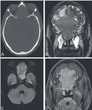
A 64-year-old male presented with nasal obstruction, anos-mia, and a reduction in visual acuity over the last few months, together with weight loss and a two-year history of headache. Computed tomography (CT) of the brain (Figure 1A) showed an expansile lesion with poorly deined borders, occupying the ethmoid cells, sphenoid sinuses, and the anterior cranial fossa, accompanied by edema of the frontal lobes. On magnetic reso-nance imaging (MRI) scans (Figures 1B, 1C, and 1D), the le-sion showed restricted diffule-sion and intense enhancement after contrast administration. A biopsy was performed, and analysis of the biopsy sample revealed hyperchromatic cells organized around a ibrillar stroma, forming rosettes, consistent with a di-agnosis of olfactory neuroblastoma. The lesion was staged his-tologically as grade I in the Hyams grading system. There was no evidence of cervical involvement or distant metastases. The patient died 15 days after undergoing the examinations.
Olfactory neuroblastoma, also known as esthesioneurob-latoma, is a rare malignant neoplasm of neuroectodermal origin and accounts for 3–6% of all malignant tumors of the paranasal sinuses. It has a bimodal age distribution, being most common among adults in the second or ifth decades of life(1). It is believed
that the neoplasm arises from the olfactory epithelium, origi-nating in the superior portion of the nasal cavities, ascending across the cribriform plate, and extending into the anterior cra-nial fossa(2).
Clinically, olfactory neuroblastoma manifests as nasal ob-struction or epistaxis. It can show indolent behavior, promote local invasion, and generate distant metastases. It tends to in-vade the paranasal sinuses, orbits, and anterior cranial fossa. The most common metastases are to the lymph nodes of the neck, lungs, liver, and bone, such dissemination at the time of diagnosis being the main predictor of survival(2). Although there
342
Radiol Bras. 2017 Set/Out;50(5):338–348 Letters to the EditorAline de Araújo Naves1, Luiz Gonzaga da Silveira Filho1, Renata Etchebehere1, Hélio Antônio Ribeiro Júnior1, Francisco Valtenor A. Lima Junior2
1. Universidade Federal do Triângulo Mineiro (UFTM), Uberaba, MG, Brazil. 2. Hospital das Clínicas da Faculdade de Medicina de Ribeirão Preto da Uni-versidade de São Paulo (HCFMRP-USP), Ribeirão Preto, SP, Brazil. Mailing address: Dra. Aline de Araújo Naves. Universidade Federal do Triângulo Mi-neiro. Avenida Getúlio Guaritá, 130, Nossa Senhora da Abadia. Uberaba, MG, Brazil, 38025-440. E-mail: [email protected].
A indicates that the tumor is limited to the nasal cavity; stage B indicates that it involves only the nasal cavity and paranasal sinuses; and stage C indicates that it extends beyond the stage B limits. The staging system proposed by Dulguerov employs the tumor-node-metastasis classii cation(3,4).
Bone destruction and calcii cation within the lesion can be characterized by CT(5). An MRI scan provides more accurate
information on the extent of the tumor, especially in terms of intracranial and orbital involvement. On MRI, the majority of olfactory neuroblastomas present a signal that is (in relation to that of muscle tissue) hypointense in T1-weighted sequences
and hyperintense in T2-weighted sequences, as well as showing intense enhancement in contrast-enhanced sequences(6,7). MRI
is also superior to CT in the evaluation of recurrence after cra-niofacial resection, because of its greater ability to differentiate i brous scar tissue from residual or recurring neoplasia(6). Cysts
in the intracranial margin of the tumor have been reported in cases of olfactory neuroblastoma. Another relevant aspect is a dumbbell-like morphology, the tumor mass being divided be-tween the anterior cranial fossa and the nasal cavity, the cribri-form plate cribri-forming the “waist”(5).
The main differential diagnoses of olfactory neuroblastoma include: squamous cell carcinoma, typically in the maxillary an-trum, with bone erosion; sinonasal adenocarcinoma, with het-erogeneous enhancement, which has been associated with occu-pational exposure to wood dust; undifferentiated sinonasal carci-noma, which affects older patients; and dural-based invasive me-ningioma, with poorly dei ned borders and areas of necrosis(8).
REFERENCES
1. Ferreira MCF, Tonoli C, Varoni ACC, et al. Estesioneuroblastoma. Rev
Ciênc Méd. 2007;16:193–8.
2. Howell MC, Branstetter BF 4th, Snyderman CH. Patterns of regional spread for esthesioneuroblastoma. AJNR Am J Neuroradiol. 2011;32:929–
33.
3. Van Gompel JJ, Giannini C, Olsen KD, et al. Long-term outcome of esthe-sioneuroblastoma: Hyams grade predicts patient survival. J Neurol Surg B
Skull Base. 2012;73:331–6.
4. Tajudeen BA, Arshi A, Suh JD, et al. Importance of tumor grade in
esthe-sioneuroblastoma survival: a population-based analysis. JAMA
Otolaryn-gol Head Neck Surg. 2014;140:1124–9.
5. Mendonça VF, Carvalho ACP, Freitas E, et al. Tumores malignos da cavi-dade nasal: avaliação por tomograi a computadorizada. Radiol Bras. 2005;
38:175–80.
6. Li C, Yousem DM, Hayden RE, et al. Olfactory neuroblastoma: MR evalu-ation. AJNR Am J Neuroradiol. 1993;14:1167–71.
7. Schuster JJ, Phillips CD, Levine PA. MR of esthesioneuroblastoma (olfac-tory neuroblastoma) and appearance after craniofacial resection. AJNR Am J Neuroradiol. 1994;15:1169–77.
8. Baptista AC, Marchiori E, Boasquevisque E, et al. Comprometimento
órbito-craniano por tumores malignos sinonasais: estudo por tomograi a
computadorizada. Radiol Bras. 2002;35:277–85. Figure 1. CT of the brain (A), with a bone window, showing an expansile lesion
occupying ethmoid cells and containing calcii cations, with bone destruction.
MRI demonstrated that the lesion was extra-axial, with lobulated contours, lo-cated in the upper portion of the nasal cavity, and extended to the anterior cranial fossa, facial sinuses, and orbits. A coronal T2-weighted sequence (B) shows that the expansile lesion presented an isointense signal, although a hyperintense signal (edema) can be seen in the brain parenchyma in the fron-tal lobe, mainly on the left. An axial diffusion-weighted imaging sequence (C) shows a hyperintense signal (restricted diffusion). A contrast-enhanced coronal T1-weighted sequence (D) shows intense enhancement.
http://dx.doi.org/10.1590/0100-3984.2015.0206
Giant ovarian teratoma: an important differential diagnosis of pelvic mas-ses in children
Dear Editor,
An 8-year-old female patient presented with diffuse ab-dominal pain accompanied by progressive distension. Physical examination revealed a large abdominal mass, predominantly in the mesogastrium, that was depressible and painless on palpa-tion. Ultrasound showed a solid-cystic formation extending from the epigastrium to the hypogastrium, with a calcium component and an air-l uid level (Figure 1). Computed tomography (CT) showed a massive solid-cystic formation, with a fat component and soft tissue, as well as calcii cations, measuring 12.6 × 19.2
× 20.8 cm, exerting a signii cant mass effect, displacing the small intestine, aorta, and inferior vena cava, as well as causing slight compression of the pancreas, kidneys, and ureters, with no apparent signs of ini ltration (Figure 2). Intraoperatively, the mass was seen to be adhered to the left fallopian tube and to the greater omentum (Figure 1). The tumor was excised without complications, and the patient was discharged i ve days later. A follow-up abdominal ultrasound revealed no changes.