Letters to the Editor
Radiol Bras. 2015 Set/Out;48(5):333–340
338
http://dx.doi.org/10.1590/0100-3984.2014.0078
Chordoid glioma of the third ventricle
Glioma cordoide do terceiro ventrículo
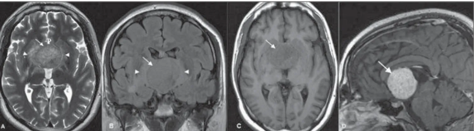
Figure 1. Axial MRI T2-weighted (A) and coronal FLAIR (B) sequences reveal a slightly hyperintense, well-defined hypothalamic/third ventricular tumor (arrows), with perilesional vasogenic edema (arrowheads). C: Axial MRI T1-weighted sequence reveals a predominantly isointense tumor (arrow). D: Gadolinium-enhanced sagittal MRI T1-weighted sequence reveals the tumor with uniform contrast enhancement (arrow).
REFERENCES
1. Reis F, Schwingel R, Nascimento FBP. Central nervous system lym-phoma: iconographic essay. Radiol Bras. 2013;46:110–6.
2. Barreira Junior AK, Moura FC, Monteiro MLR. Linfoma não-Hodgkin bilateral do seio cavernoso como manifestação inicial da síndrome de imunodeficiência adquirida: relato de caso. Arq Bras Oftalmol. 2011;74: 130–1.
3. Osborn AG. Encéfalo de Osborn. Imagem, patologia e anatomia. Porto Alegre, RS: Artmed; 2014.
4. Rocha AJ, Vedolin L, Mendonça RA. Encéfalo. Série CBR. São Paulo, SP: Elsevier; 2012.
Arthur Henrique de Aquino Dultra1, Fabio Noro2, Alessandro Severo Alves de Melo3, José Alberto Landeiro4, Edson Marchiori4, Marilene Filgueira do Nascimento5
1. Hospital Copa D’Or, Rio de Janeiro, RJ, Brazil. 2. Universidade Federal do Rio de Janeiro (UFRJ), Rede D’Or, Rio de Janeiro, RJ, Brazil. 3. Universidade Federal Fluminense (UFF), Niterói, RJ, Hospital Barra D’Or, Rio de Janeiro, RJ, Brazil. 4. Universidade Federal Fluminense (UFF), Niterói, RJ, Brazil. 5. Instituto Nacional de Câncer (INCA), Rio de Janeiro, RJ, Brazil. Mailing Address: Dr. Arthur Henrique de Aquino Dultra. Rua Real Grandeza, 281, Botafogo. Rio de Janeiro, RJ, Brazil, 22281-035. E-mail: [email protected].
Dear Editor,
A previously healthy 27-year-old man was referred with an 8-month history of headaches, memory loss, progressive weight gain (obesity), hyperphagia and behavior changes.
Computed tomography (CT) scans revealed the presence of a midline, solid, and homogeneously enhancing mass involving the anterior aspect of the third ventricle.
Brain magnetic resonance imaging (MRI) (Figure 1) showed a well-defined, rounded mass in the third ventricle, measuring about 4.0 cm in the craniocaudal axis. The tumor was slightly heterogeneous, predominantly isointense at T1- and T2-weighted MRI sequences, presenting with diffuse enhancement after ga-dolinium injection. Perilesional vasogenic edema, compression and subsequent displacement of midbrain and hypothalamic structures were observed.
A subtotal resection of the tumor was microsurgically per-formed by interhemispheric transcallosal approach to the third ventricle.
The tumor was histologically classified as a chordoid glioma. The mass showed nests of regular epithelioid cells with large nuclei, prominent nucleoli, and abundant eosinophilic cytoplasm, within a myxoid stroma. Sparse lymphocytic infiltrate was present. Im-munohistochemical studies demonstrated diffuse cytoplasmic expression for glial fibrillary acidic protein, vimentin, and CD34. The patient died three months after surgery as a conse-quence of massive hypothalamic invasion combined with pneu-monia.
Chordoid glioma is an unusual, noninvasive and slow-grow-ing tumor that arises from the anterior third ventricle, frequently adherent to the hypothalamus(1). There are reports in the
litera-ture about chordoid gliomas in other locations, such as the tem-poroparietal region, left thalamus and the corona
radiata/thala-mus(2,3), most of them affecting children(2).
It is typically a well-circumscribed, round or oval-shaped tu-mor, with greatest diameter in the craniocaudal direction. The tumor is hyperdense to the gray matter at CT, isointense at MRI T1-weighted sequences, and isointense to slightly hyperintense at MRI long-TR, with strong, uniform enhancement after con-trast agent administration(1,2,4–6). Cystic changes and necrosis
may be present(2,5,7). Calcifications are usually rare(2,5,7). Usually,
bilateral and symmetric perilesional vasogenic edema may also be observed(3–5).
Given the tumor location, patients usually present with signs and symptoms related to obstructive hydrocephalus, such as nau-sea and headache, although endocrine imbalance, visual distur-bances, behavior disorders and autonomic dysfunction are also reported in the literature(1,4–6).
The histological and immunohistochemical features of these tumors are very typical and uniform, characterized by cords of oval to polygonal epithelioid cells with abundant eosinophilic cytoplasm and avid staining for glial fibrillary acidic protein and vimentin(1,2,4).
The differential diagnosis includes masses of suprasellar re-gion, such as pituitary macroadenoma, craniopharyngioma, optic and hypothalamic pilocytic astrocytoma, meningioma, ependy-moma and lymphoma(2,4).
Currently, the treatment of choice is complete surgical re-section of the tumor(1,4,6). Adjuvant radiotherapy has been used
following subtotal resection(2).
Despite being a low-grade tumor, the prognosis is usually poor because of its location and the difficulty in obtaining complete surgical resection without causing severe hypothalamic symp-toms(4). On the other hand, partial resection of the tumor is
Letters to the Editor
Radiol Bras. 2015 Set/Out;48(5):333–340
339
Marília Henrique Destefani1, Alessandro Spanó Mello2, Ricardo Santos de Oliveira3, Gustavo Novelino Simão2
1. Cedirp – Radiologia e Diagnóstico por Imagem, Ribeirão Preto, SP, Brazil. 2. Hospital das Clínicas – Faculdade de Medicina de Ribeirão Preto da Universidade de São Paulo (HCFMRP-USP), and Cedirp – Radiologia e Diagnóstico por Imagem, Ribeirão Preto, SP, Brazil. 3. Hospital das Clínicas – Faculdade de Medicina de Ribei-rão Preto da Universidade de São Paulo (HCFMRP-USP), RibeiRibei-rão Preto, SP, Brazil. Mailing Address: Dra. Marília Henrique Destefani. Avenida Professor João Fiusa, 2055, Jardim Irajá. Ribeirão Preto, SP, Brazil, 14024-260. E-mail: [email protected].
http://dx.doi.org/10.1590/0100-3984.2014.0125 REFERENCES
1. Ortega-Martínez M, Cabezudo JM, Bernal-García LM, et al. Glioma cordoide del III ventrículo. Nuevo caso y revisión de la literatura. Neuro-cirugía. 2007;18:115–22.
2. Desouza RM, Bodi I, Thomas N, et al. Chordoid glioma: ten years of a low-grade tumor with high morbidity. Skull Base. 2010;20:125–38. 3. Ni HC, Piao YS, Lu DH, et al. Chordoid glioma of the third ventricle:
four cases including one case with papillary features. Neuropathology. 2013;33:134–9.
4. Pomper MG, Passe TJ, Burger PC, et al. Chordoid glioma: a neoplasm unique to the hypothalamus and anterior third ventricle. AJNR Am J Neuroradiol. 2001;22:464–9.
5. Smith AB, Smirniotopoulos JG, Horkanyne-Szakaly I. From the radio-logic pathology archives: intraventricular neoplasms: radioradio-logic-patho- radiologic-patho-logic correlation. Radiographics. 2013;33:21–43.
6. Zarghouni M, Vandergriff C, Layton KF, et al. Chordoid glioma of the third ventricle. Proc (Bayl Univ Med Cent). 2012;25:285–6.
7. Glastonbury CM, Osborn AG, Salzman KL. Masses and malformations of the third ventricle: normal anatomic relationships and differential diagnoses. Radiographics. 2011;31:1889–905.
Enteroenteric intussusception in an adult caused by an ileal angiomyolipoma
Intussuscepção entero-entérica em um adulto causada por um angiomiolipoma ileal
Dear Editor,
A white, 32-year-old man was admitted to the emergency de-partment with severe pain principally in the right inferior quad-rant of the abdomen, abdominal distension and vomiting for one day.
Abdominal radiography, ultrasonography and computed to-mography demonstrated small bowel loops distension (Figure 1A) and signs of ileo-ileal invagination associated with intraluminal
nodule with fat content compatible with “intussusception head” (Figures 1B, 1C and 1D). Option was made for surgical treatment. Anatomopathological study in association with immunohis-tochemical analysis diagnosed angiomyolipoma (AML) as follows:
Macroscopy: Bowel loop containing a non-encapsulated de-limited, submucosal, polypoid yellowish lesion measuring 3.0 × 2.5 × 2.3 cm, with no sign of malignancy.
Microscopy: Masson’s trichrome staining diagnosed AML
compromising the entire intestinal wall, from the serosa to the mucosa.
Immunohistochemical analysis: Desmin, HHF 35, CD31,
CD34, protein S100, smooth muscle actin 1 to 4 = positive. Intussusception is the invagination of a proximal intestinal segment with its mesenteric fold with the corresponding