cop
yr
ight
© ABE&M todos os direitos reser
v
ados
clinical case report
LUCIANA COSENTINODE MACEDO
FERNANDA CAROLINE SOARDI
NAYLA ANANIAS
VERA MARIA SANTORO BELANGERO
SUMARA ZUAZANI PINTO RIGATTO
MARICILDA PALANDI DE-MELLO
LÍLIA D’SOUZA-LI
Pediatric Endocrinology Laboratory, Center for Investigation in Pediatrics (LCM, NA, LSL); Department of Pediatrics. Faculty of Medical Science (VMSB, SZPR, LSL); Center of Molecular Biology and Genetic Engineering (CBMEG) (MPM); State University of Campinas (Unicamp), Campinas, SP, Brazil.
Received in 25/8/2008 Accepted in 14/10/2008
ABSTRACT
Mutations in the vitamin D receptor (VDR) are associated to the hereditary 1,25-dihydroxivitamin D-resistant rickets. The objectives of this work are: search for mutations in the VDR and analyze their functional consequences in four Brazilian children presented with rickets and alopecia. The coding region of the VDR was amplifi ed by PCR e direct sequenced. We identifi ed three mu-tations: two patients had the same mutation in exon 7 at aminoacid position 259 (p.Q259E); one patient had a mutation in exon 8 at codon 319 (p.G319V) and another one had a mutation in exon 3 leading to a truncated protein at position 73 (p.R73X). Functional studies of the mutant receptors of fi broblast primary culture, from patients’ skin biopsy treated with increasing doses of 1,25(OH)2 vitamin D showed that VDR mutants were unable to be properly activated and presented a reduction in 24-hydroxylase expression level. (Arq Bras Endocrinol Metab 2008; 52/8:1244-1251)
Key words: Rickets; Bone deformities; Hypocalcemia; Vitamin D.
RESUMO
Mutações no Gene do Receptor de Vitamin D em Quatro Pacientes com Raquitismo Hereditário Resistente a 1,25-Dihydroxyvitamin D.
Mutações no receptor de vitamina D (VDR) são associadas a raquitismo hereditário resistente a 1,25-dihidroxivitamina D. Os objetivos deste trabalho foram procurar mutações no VDR e analisar suas conseqüências funcionais em quatro pacientes com raquitismo e alopécia. A região codifi cadora do VDR foi amplifi cada por PCR e seqüenciada diretamente. Identifi camos três mutações: dois pacientes apresentavam a mesma mutação no éxon 7 na posição protéica 259 (p.Q259E); um paciente apresentava uma mutação no éxon 8 no códon 319 (p.G319V) e o outro apresentava uma mutação no exon 3 resultando em uma proteína truncada na posição 73 (p.R73X). O estudo funcional dos receptores mutados nos extratos de culturas de fi broblasto primárias obtidas de biópsia de pele dos pacientes, tratados com doses cres-centes de 1,25(OH)2 vitamina D demonstraram que os receptores mutantes não apresentam ativação adequada apresentando expressão reduzida de 24-hidroxilase. (Arq Bras Endocrinol Metab 2008; 52/8:1244-1251)
Descritores: Raquitismo; Deformidades ósseas; Hipocalcemia; Vitamina D.
INTRODUCTION
cop
yr
ight
© ABE&M todos os direitos reser
v
ados
427 amino acid protein. At the amino terminal portion of the molecule there is a Cysteine rich region that in-teracts with the VDRE in the DNA, through two zinc fi ngers motifs (2). The ligand binding domain has high affi nity to the 1,25 dihydroxyvitamin D (1,25(OH)2vit D) and is located at the carboxi-terminal portion of the VDR. The 1,25(OH)2vit D activates the VDR, which by heterodimerazing with the Retinoic acid receptor X (RXR) interacts with specifi c sequences in the DNA of target genes denominated VDR response elements (VDRE) (2). The VDRE is composed of two sequen-ces of direct repeats (RGKTCA) separated by three base pairs. More than 40 mutations in the VDR have been described in approximately seventy families pre-senting HVDRR (3). Clinically, patients presented bone deformities in the lower limbs, due to rickets du-ring the fi rst years of life. In addition, the majority of affected individuals have alopecia that appears soon af-ter birth denoting the importance of the VDR in the natural development of hair follicle. Mutations that affects the DNA ligand domain usually is associated with a more severe phenotype. The objective of this study is to perform a molecular analysis of the VDR in four patients with rickets and alopecia.
SUBJECTS AND METHODS
Patients
We studied four patients with clinical diagnosis of HVDRR presenting alopecia and rickets, followed at the Pediatrics Nephrology Outpatient Clinics from the Department of Pediatrics, at the Faculty of Medical Science of the State University of Campinas. Patient 1 is a 9.5 years old boy followed since he is two years-old. He presented alopecia since 2 months old and de-velop deformities of the lower limbs at the age of 1 year. He responded remarkably well to the treatment with Calcitriol (0.25μg/day), improving bone defor-mities. Patient 2 is a 19.2 years old man that at age of 2 years presented severe malnourishment, rickets, alope-cia and several upper and lower respiratory infections. He responded well to the treatment with vitamin D3 (vit D) (3500 IU/day), latter changed to Calcitriol (0.25 µg/day), associated with calcium and phosphate solution, normalizing his weight and growth. Patient 3 is a 20.6 years old woman that presented alopecia since birth and coarse bone deformities of lower limbs. She
started the treatment at the age of seven with vitamin D3 (7000 IU/day), latter changed to Calcitriol (0.25µg/day) and phosphate solution. Patient 4 is a 6.7 years old boy, who presented alopecia since 2 mon-ths of age and by the age of 2 he had severe rickets with coarse bone deformities, of upper and lower limbs not deambulating. His parents are fi rst degree cousins. He underwent treatment with increasing doses of Calci-triol up to 1.5 µg/day and phosphate solution. After 6 months of treatment,his condition improved, become able to walk but persisted hypocalcemic and with high serum alkaline phosphatase levels. Patients 1 and 3 were apparently not related, although both are from the same region (Bahia State). Both patients refer cou-sins presenting similar features, with alopecia and ri-ckets. This study was approved by the local Ethical Committee of the Faculty of Health Science (CEP n° 330/02) and to the National Ethics in Research Coun-cil (CONEP n° 4623).
Sequence analysis of the VDR gene
Leukocyte DNA was isolated from blood from the four patients after written informed consent, and used as a template for the PCR reactions. All coding and fl anking regions from exons 2 to 9 from the VDR gene were PCR amplifi ed using 7 pairs of primers. Due to its pro-ximity and small sizes, a single pair of primers were used to amplify exon 7 and 8, with the forward primer loca-ted upstream exon 7 and the reverse primer downstre-am exon 8. The PCR products were gel purifi ed and directly sequenced using a semi-automated laser detec-tion system (ABI Prism 377sequencer, Applied Biosys-tems). The same primers from the PCR amplifi cation were used in the Big Dye reaction (Applied Biosystem, version 2). All sequences were compared to the human VDR sequence (GenBank gi|3617739|gb|AC004466.1). The mutations were confi rmed by digesting the PCR fragments with specifi c restriction enzymes.
Fibroblast primary culture
cop
yr
ight
© ABE&M todos os direitos reser
v
ados
(DMEM, Invitrogen, CA, USA) supplemented with 10% fetal bovine serum (Invitrogen) and penicillin/ streptomycin (complete medium).
1,25(OH)2 vit D induction of 24-hydroxylase
mRNA
For VDR functional studies, we analyzed the 24hydroxylase [24(OH)ase] expression from the patient’s fi -broblast primary culture. When the primary cultures were confl uent they were treated with different doses of 1,25(OH)2vit D (10-7, 10-8 and 10-9 M) in a serum free media supplemented with 1% BSA for 6 hours. Controls, without treatment were also included. At the end of 6 hours, cells were harvested for total RNA and nuclear extraction.
RT-PCR
Total RNA was extracted from the fi broblast primary cultures by TRIzol (Invitrogen) as manufacturer’s pro-tocol. Five-microgram RNA samples were reverse-transcribed with recombinant SuperScript III™ , reverse transcriptase (Invitrogen) using random pri-mers in a total volume of 20 μl. Five microliters of the RT mixture were used in the PCR with primers specifi c to amplify the 24(OH)ase gene. A water control from the RT and RNA from fi broblast primary culture from normal individual were used as negative and positive controls, respectively. The PCR products were analyzed by agarose gel electrophoresis.
Real time PCR
For a quantitative measurement of mutant receptor response, 100 ng of cDNA from control and patient’s fi -broblast with and without 10-8M of 1,25(OH)
2vit D treatment, were used to perform a real time PCR expe-riment. The amount of mRNA from the 24(OH)ase gene were measured using specifi c primers labeled with FAM, and controlling its expression with the 18S house keeping gene. The degree of expression was quantifi ed by real time PCR using the detection system ABI Prism sequencer 5700 (PE).
Nuclear protein extraction
For the nuclear protein extraction, cells were harvested in solution A (10 mM HEPES, pH 7.9, 1.5 mM MgCl2, 10 mM KCl, 0.5 mM DTT, 0.5 mM PMSF, 2.5 µg/ml of aprotinin), centrifuged for 5 minutes at 1,000 rpm, at 4oC and the pellet was re-suspended in 200µl of
so-lution A plus 0.1% Nonidet P-40, passed through and syringe gauge with fi ne needle for 15 times and centri-fuged 5 min, at 12,000 x g at 4 oC. The nuclear mem-brane in the pellet was lysed with osmotic shock with solution B (20 mM HEPES, pH 7.9, 25% glycerol, 1.5 mM MgCl2, 0.42 M NaCl, 0.2 mM EDTA, 0.5 mM DTT, 0.5 mM PMSF, 2.5 µg/ml of aprotinin and leu-peptin, and 1 µg/ml of pepstatin). After 30 min incu-bation at 4 °C under agitation, cells were centrifuged for 10 min, 4°C, 12,000 x g. The supernatants, contai-ning the nuclear fraction, were collected and stored at -70 °C. The protein concentration were determined by Bradford assay.
Western Blotting
The expression of the mutant receptor compared to wild type was analyzed by Western Blot. Nuclear pro-tein extract was subjected to 12% SDS-PAGE and the separated proteins were electrotransferred and immo-bilized on PVDF membranes. After blocking unspecifi c binding with PBS plus Tween 20 (PBS-T) and 5% skim milk, the membranes were incubated with primary an-tibody VDR specifi c diluted 1:100 (Santa Cruz) for twelve hours followed by a secondary antibody (anti-mouse) conjugated to horseradish peroxidase (Biorad; diluted 1:5,000) for two hours. For detection of the proteins, ECL chemiluminescence system (GE Health-care) was used.
polyacrylami-cop
yr
ight
© ABE&M todos os direitos reser
v
ados
de gels using 0.5x Tris borate/EDTA buffer and the complexes were visualized in Hyperfi lm MP autoradio-graphies (Amersham Biosciences/GE, USA).
RESULTS
Patients
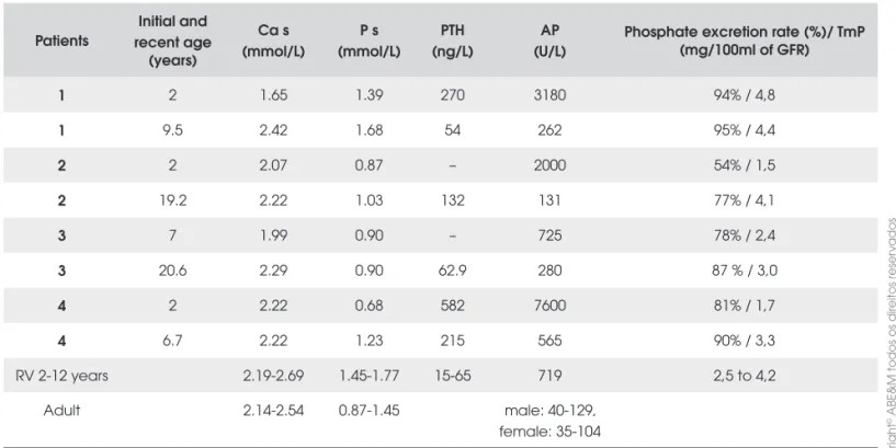
The biochemical levels of all patients at the beginning of the treatment and the most recent evaluations are in table 1. Except for patient 4, all patients responded well to the treatment with supra physiological doses of vita-min D and or Calcitriol, calcium and phosphate. In pa-tient 1 and 2 there was a better control of the disease between the age of six to eight years of age, requiring discontinuation of the vitamin D due to hypercalciuria. Even in patient 4 that had a more severe form of rickets compared to the other patients, became at the age of six clinically and biochemically stable. All patients have normal renal functions and after correction of the phos-phate depletion, presented a normal phosphos-phate excre-tion rate.
VDR Mutations
We identifi ed VDR-gene mutations in all the patients studied. All the mutations were in homozygous state. In patients 1 and 3 we identifi ed a novel missense mutation in exon 7 changing a C to a G at nucleotide 775 (c.775 C>G) (Figure 1A). Resulting in a change in amino acid from the Glutamine to Glutamic acid at protein position 259 (p.Q259E), on the ligand binding domain of the receptor. This mutation was confi rmed by restriction en-zyme analysis as it disrupts an Ava II site (data not sho-wn). Patient 2 presented a novel mutation in exon 8 at nucleotide 955, resulting in a G to T transversion (c.955 G>T), changing of the amino acid from Glycine (hydro-philic) to Valine (hydrophobic) at codon 319 (p.G319V) (Figure 1B). This mutation was confi rmed by restriction enzyme analysis as it creates a Rsa I site (data not sho-wn). In patient 4, a nonsense mutation in exon 3 at nu-cleotide 217, changing C to T(c.217C>T) was identifi ed, resulting in change of Arginine to a termination codon at position 73 (p.R73X) (Figure 1C). This mutation was confi rmed by restriction enzyme analysis using a mutant primer that creates an Pvu II site (data not shown).
Table 1. Age and biochemical parameters before the beginning of the treatment and at the most recent follow up in all four
patients.
Patients
Initial and recent age
(years)
Ca s (mmol/L)
P s (mmol/L)
PTH (ng/L)
AP (U/L)
Phosphate excretion rate (%)/ TmP (mg/100ml of GFR)
1 2 1.65 1.39 270 3180 94% / 4,8
1 9.5 2.42 1.68 54 262 95% / 4,4
2 2 2.07 0.87 – 2000 54% / 1,5
2 19.2 2.22 1.03 132 131 77% / 4,1
3 7 1.99 0.90 – 725 78% / 2,4
3 20.6 2.29 0.90 62.9 280 87 % / 3,0
4 2 2.22 0.68 582 7600 81% / 1,7
4 6.7 2.22 1.23 215 565 90% / 3,3
RV 2-12 years 2.19-2.69 1.45-1.77 15-65 719 2,5 to 4,2
Adult 2.14-2.54 0.87-1.45 male: 40-129,
female: 35-104
cop
yr
ight
© ABE&M todos os direitos reser
v
ados
Figure 1. Direct sequence results of control samples (left
panel) and affected patients (right panels). Arrow indicates the nucleotide change. The underlined triplet indicates the altered codon. A) Mutation found in patient 1 and 3 showing a homozygous mutation at nucleotide c. 775. C>G. B) Muta-tion found in patient 2 with a homozygous mutaMuta-tion at nucle-otide c.955 G>T. C) Mutation found in patient 4. The homozygous mutation at c.217C>T creates a premature ter-mination codon at protein position 73 (p.R73X). The consan-guineous parents were heterozygous for the mutation.
VDR functional analysis
The expression of 24(OH)ase gene increases through the activation of the VDR by vit D (5). For functional analyses, fi broblast primary culture from the patients and a normal individual were treated with increasing doses of 1,25(OH)2 vit D for 6 hours. The 24(OH)ase expression was strongly induced in control cells, even with lower doses (Figure 2). Intermediate doses showed variable responses in the fi broblast cultures from the patients while all patients responded to 10-7M 1,25(OH)
2 vit D treatment, despite the different types of mutation.
Real time PCR
In order to quantify the differences in response of the mutant receptors, the amount of mRNA from the 24(OH)ase gene was measured, by real time PCR, from control and patients’ fi broblast with and without 10-8M of 1,25(OH)2vit D treatment. 18S house keeping gene expression was used to normalized the 24(OH) gene expression level. Unlike the patients, the control sho-wed 24(OH)ase expression even in the basal state, and it responded well to the vit D treatment with a fi ve fold increase in the gene expression level (Figure 3A). Only patient 2 and 1 presented a signifi cant increase in 24(OH)ase gene expression after 1,25(OH)2 vit D tre-atment (Figure 3A) showing an increase of 38 and 53 fold in 24(OH)ase gene expression after 1,25(OH)2 vit D treatment respectively (Figure 3B). Patient 3 and 4 were not able to increase 24(OH)ase gene expression to a good level after 1,25(OH)2 vit D treatment (Figu-re 3A). However, when compa(Figu-red the basal level, they presented an increase of 35 and 5 fold in 24(OH)ase gene expression respectively (Figure 3B), with the truncated receptor presenting the worse response.
Figure 2. Increase of 24(OH)ase gene expression (upper fi gure) and β-actin (lower fi gure) after treatment with 10-7, 10-8 e 10-9 nM
cop
yr
ight
© ABE&M todos os direitos reser
v
ados
3 A
2
1
0
1 2 3 4 5 6 7 8 9 10
Relative quantification
24 OH( )ase gene expression in fibroblasts
100%
1 2 3 4 5 B
80%
60%
40%
20%
0%
24(OH)ase gene expression fibroblasts
Expression ratio of V
it. D
treatment over basal
Figure 3. Real time PCR from control and patients. A) Amount of mRNA from the 24(OH)ase gene was measured, by real time PCR, from control and patient’s fi broblast with and with-out 10-8M of 1,25(OH)
2vit D treatment. The 18S house keeping
gene was used to normalized the expression level. Columns 1 and 2- control sample; columns 3 and 4- patient 2; columns 5 and 6- patient 1; columns 7 and 8- patient 3; columns 9 and 10- patient 4. Odd columns show the expression of 24(OH) ase gene in fi broblast not treated and even columns show the expression of 24(OH)ase gene in fi broblast treated with 10-8M of 1,25(OH)
2vit D. B) Folds increase in 24(OH)ase gene
expression compared to the basal expression level. Column 1- control sample; column 2- patient 2; column 3- patient 1; columns 4- patient 3; columns 5- patient 5.
Western Blotting
We analyzed the receptor expression in the nuclear ex-tract from the patients culture comparing to the con-trol. Three bands were visualized: a 50 kDa band corresponding to the mature VDR (427 amino acid), and a 38 and 20 kDa band. In a nuclear extract from fi broblast culture of all patients, the 50 kDa band was the most reduced when compared to the wild type (Fi-gure 4). The VDR antibody was raised against a pepti-de corresponding to the VDR amino acids 344-424 of the human protein. Since the truncated receptor, with only 73 amino acids, lost its epitope for the antibody, it was not detected.
Control Patient 1 Patient 2
Patient 3 Patient 4
50kDa
Figure 4. Western blot analysis of fi broblast nuclear protein extract from control e patients. Lane 1- Control; Lane 2- pa-tient 1; Lane 3- papa-tient 2; Lane 4- papa-tient 3; Lane 5- papa-tient 4. The 50 kDa band represents the mature VDR protein stained with the VDR monoclonal antibody raised against position 344-424 of the human VDR protein.
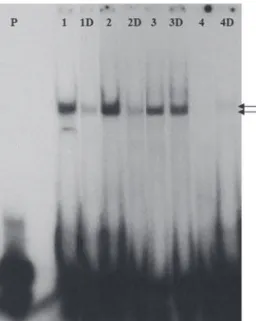
Electrophoretic Mobility Shift Assay (EMSA) Nuclear extract from control fi broblast showed two shifts: a lower shift and a more abundant upper shift (Figure 5, lane1). Interesting, the lower shift disappea-red with vit D treatment (Figure 5, lane 1D). Nuclear extract from patients 1, 2 and 3 showed a similar pat-tern, with only the upper shift similar to the control, and the shift patterns did not changed with vit D treat-ment (Figure 5). As expected, for patient 4 with p. R73X mutation it was not observed a shift (Figure 5, lane 4), however when treated with vitamin D a diffe-rent shift slightly higher than the control sample was detected (Figura 5, lane 4D).
DISCUSSION
cop
yr
ight
© ABE&M todos os direitos reser
v
ados
compared to the wild type. However, it did not showed reduction in the DNA interaction. This was expected since the mutation is located in the ligand binding do-main. The change in the amino acid charge from hydro-phobic to acid negatively charged, probably changed the ligand binding pocket impairing agonist-receptor interaction. This mutation is located in a very conser-ved region in the E1 domain involconser-ved in the VDR-RXR heterodimerization (6). The presence of the alopecia also suggests that the mutation impairs the receptor signaling and this was confi rmed by the poor response to vit D treatment. Despite having the same mutation, patient 1 presented a better treatment response than patient 3. One of the reasons is probably the difference in age of the treatment onset. Patient 1 started the treat-ment with 1 year old, while patient 3 at 7 years of age. Furthermore, on the in vitro functional analyses patient 1 responded better to vit D treatment than patient 3. This suggests that other factors are also involved in the VDR activation and function. In the same position another mutation is described (p.Q259P) in a consan-guineous Indian family where the affected members presented alopecia (5). Interesting, the affected
bro-thers of this family also responded well to iv calcium with complete healing of the rickets and correction of the biochemical abnormalities (5).
The novel mutation p.G319V found in patient 2 is also located on the ligand binding domain and in the three-dimensional model based on the structure of the RXRα apo-ligand binding domain, exon 8 represents the α-helix 7 and 8 (7). Upon ligand binding, a confor-mational change and a reorganization of the helices oc-cured, allowing heterodimer formation (7). The presence of mutation in one of these helices may alter its organization and compromise the interaction of the receptor with the ligand (8). This patient presented with severe clinical manifestations, however, responded to a modest dose of vitamin D (3500 UI/day) with normalization of the biochemical levels and of the gro-wth. This is in accordance to the functional studies, which showed a good VDR-DNA interaction as well as a good response to the vit D treatment.
These three patients with mutations located in the ligand binding domain showed less severe phenotypes, and responded to treatment with vit D, calcium and phosphate. Correction of the inorganic phosphorus with phosphate solution was crucial since its depletion may impair vit D treatment, specially under high meta-bolic state such as the intense growth during the fi rst years of life and during puberty. In addition, the vit D defi ciency induces an increase in PTH which in turn will worse the renal phosphate excretion. Although most of the patients with HVDRR do not respond even to supraphysiologic doses of calcitriol, there are several reports in the literature of patients responding well to treatment, with complete healing of the rickets and correction of biochemical abnormalities (5,8-13).
In patient 4, the mutation results in a truncated receptor at position 73 (p.R73X) in the DNA binding domain just after the second zinc fi nger. This portion of the receptor is highly conserved and mutations in this region result in reduction of the DNA affi nity bin-ding. Clinically, this patient presents a much more seve-re clinical manifestations than the other patients, confi rming that the VDR in this patient has little if any activity. However, he improved upon supraphysiologi-cal doses of supraphysiologi-calcitriol, suggesting that some of the dise-ase manifestations are in part VDR independent (14). Functional studies confi rmed the inability of this recep-tor to interact with DNA under basal conditions. In fact it is debatable if this truncated protein is produced, as cells effi ciently identify nonsense codons and induce
Figure 5. EMSA analysis. The assay was performed with
cop
yr
ight
© ABE&M todos os direitos reser
v
ados
mRNA decay. As expected, under basal conditions, the truncated receptor did not presented DNA interaction. Surprisingly, after vit D treatment, a shift was detected, indicating that in the presence of vit D, a interaction of a protein with the VDRE occurs. However, the com-plex size of the shift was slightly above the expected shift, suggesting that this truncated receptor may form a different complex and that the vit D allowed some kind of binding. Whether this binding is dependent of the mutant VDR remains unclear. This mutation was described in the literature in a Greek boy (5) with no family history of consanguinity and also in a Marocan patient (15), both with poor response to alfacalcidol and oral calcium with persistence of severe rickets. In another Brazilian family a nonsense mutation at position 30 was described, resulting in a truncated receptor in the DNA binding domain in the fi rst zinc fi nger (16).
Functional analysis of the mutations showed that all the mutant receptors function was impaired. The re-ceptor truncated in the DNA ligand domain was with worse response to vit D treatment. In vitro studies con-fi rmed the loss of function of the truncated receptor. When we evaluated the receptors with mutations in the ligand binding domain their expression and their func-tion were impaired, however, their ability to bind to DNA was preserved. In conclusion we report three mutations in the VDR. Two novel mutations in the li-gand binding domain and one mutation in the DNA binding domain that resulted in a truncated receptor. All mutations result in reduction of receptor expression and function.
Acknowledgments: Thank you Jussara Rehder, for technical as-sistance. Special acknowledgments to the patients who had collaborated with the research and to the grant agency Fapesp Disclosure: None. No potential confl ict of interest relevant to this article was reported.
REFERENCES
1. Hughes MR, Malloy PJ, Kieback DG, Kesterson RA, Pike JW, Feldman D, O’Malley BW. Point mutations in the human vita-min D receptor gene associated with hypocalcemic rickets. Science. 1988;242:1702-5.
2. Mangelsdorf DJ, Evans RM. The RXR heterodimers and or-phan receptors. Cell 1995;83:841-50.
3. Liberman UA. Vitamin D-resistant diseases. J Bone Miner Res. 2007;22 Suppl 2:V105-7.
4. Sambrook J, Fritsch EF, Maniatis TE. Molecular cloning, a la-boratory manual. New York: Cold Spring Harbor 1989. 5. Cockerill F, Hawa NS, Yousaf N, Hewison M, O´Riordan LH,
Farrow SM. Mutations in the vitamin D receptor gene in three kindreds associated with hereditary vitamin D resistant rickets. J Clin Endocrinol Metab. 1997;82:3156-60.
6. Jin CH, Kerner SA, Hong MH, Pike JW. Transcriptional activa-tion and dimerizaactiva-tion funcactiva-tions in the human vitamin D recep-tor. Mol Endocrinol. 1996;10:945-57.
7. Bourguet W, Ruff M, Chambon P, Gronemeyer H, Moras D. Crystal structure of the ligand binding domain of the human nuclear receptor RXR-α. Nature. 1995;375:377-82.
8. Malloy PJ, Pike JW, Feldman D. The vitamin D receptor and the syndrome of hereditary 1,25-dihydroxyvitamin D-resistant rickets. Endocr Rev. 1999;20:156-88.
9. Malloy PJ, Xu R, Peng L, Peleg S, Al-Ashwal A, Feldman D. Hereditary 1,25-dihydroxyvitamin D resistant rickets due to a mutation causing multiple defects in vitamin D receptor func-tion. Endocrinology. 2004;145(11):5106-14.
10. Rosen JF, Fleischman AR, Finberg L, Hamstra A, DeLuca HF. Rickets with alopecia: An inborn error of vitamin D metabo-lism. J Pediat. 1979;94(5): 729-35.
11. Liberman UA, Halabe A, Samuel R, Kauli R. End-organ resis-tance to 1,25-Dihydroxicholecalciferol. Lancet. 1980;8:504-7. 12. Hochberg Z, Gilhar A, Haim S, Friedman-Birnbaum R, Levy J,
Benderly A. Calcitriol-resistance rickets with alopecia. Arch Dermatol. 1985;121: 646-7.
13. Takeda E, Yokota I, Kawakami I, Hashimoto T, Kuroda Y, Arase S. Two siblings with vitamin-D-dependent rickets type II: no recurrence of rickets for 14 years after cessation of therapy. Eur J Pedatr. 1989;149: 54-7.
14. Panda DK, Miao D, Bolivar I, Li J, Huo R, Hendy GN, Goltzman D. Inactivation of the 25-hydroxyvitamin D 1α-hydroxylase
and vitamin D receptor demonstrates independent and inter-dependent effects of calcium and vitamin D on skeletal and mineral homeostasis. J Biol Chem. 2004;279:16754-66. 15. Wiese RJ, Goto H, Prahl JM, Marx SJ, Thomas M, al-Aqeel A,
DeLuca HF. Vitamin D-dependency rickets type II: truncated vitamin D receptor in three kindreds. Mol Cell Endocrinol. 1993;90(2):197-201.
16. Mechica JB, Leite MO, Mendonca BB, Frazzatto ES, Borelli A, Latronico AC. A novel nonsense mutation in the fi rst zinc fi n-ger of the vitamin D receptor causing hereditary 1,25-dihydro-xyvitamin D3-resistant rickets. J Clin Endocrinol Metab. 1997;82:3892-4.
Correspondence to:
Lília D’Souza-Li
Center for Investigation in Pediatrics Rua Tessália Vieira de Camargo, 126
Cidade Universitária “Zeferino Vaz” – Caixa postal 6111 13083-887 Campinas, SP, Brazil.


