Materials Research, Vol. 12, No. 2, 225-227, 2009 © 2009
to 200 °C for 20 minutes so that auto-combustion would take place. Finally, the product was annealed in a furnace for 2 hours at differ-ent temperatures.
2.2. Measurements
X ray diffraction patterns were obtained using an XPert Pro Panalitical diffractometer with Cu Kα radiation (λ = 1.5418 Å). The average particle size was calculated from line broadening using the TOPAS application3, academic edition.
Mössbauer spectra were recorded at several temperatures between 25 K and room temperature in a homemade instrument using a source of 57Co(Rh) with an activity of about 50 mCi.
3. Experimental Results and Analysis
Table 1 shows the average particle sizes, as calculated from the X ray spectra shown in Figure 1, for samples as prepared and after annealing at three different temperatures.
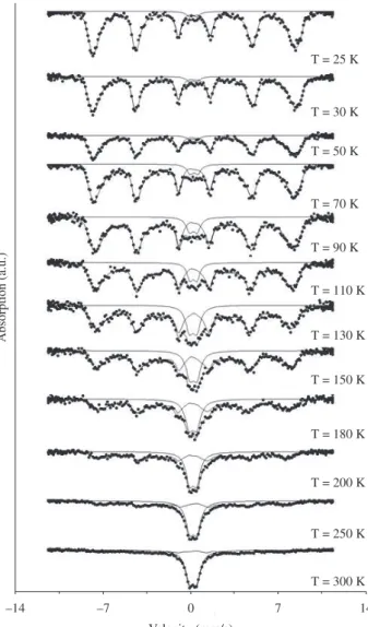
The Mössbauer spectra of sample 2 in Table 1 are shown in Figure 2 for several different measurement temperatures. While at room temperature there is a doublet due to superparamagnetic relaxa-tion,at lower temperatures one sees a sextet which is characteristic of bulk magnesioferrite4. By taking the ratio of the area under the doublet
to the area under the sextet, it is possible to estimate the volume frac-tion of unblocked particles for each measurement temperature. The
1. Introduction
Nanosized ferrites display physical and chemical properties which may be quite different from those of their bulk counterparts. Magnesioferrite (MgFe2O4) is one of the most interesting, because, due to its small magnetocrystalline anisotropy, superparamagnetic properties are still present at relatively low temperatures and/or high magnetic fields. The purpose of this work was to investigate the influence of annealing on the average particle size and particle size distribution of MgFe2O4 nanoparticles prepared using the sol-gel/ combustion method1, a fast and relatively inexpensive technique.
The average particle size was determined by X ray diffraction, while the particle size distribution was obtained by measuring at several temperatures the relative intensity of the Mössbauer spectra due to superparamagnetic particles and to ferrimagnetic particles, a method that has already been applied successfully2 to another nanosized
ferrite, CoFe2O4.
2. Experimental Procedure
2.1. Sample preparation
Analytical grade Fe(NO3)3.9H2O, Mg(NO3)2.6H2O, and C6H8O7.H2O were dissolved in deionized water to obtain the start-ing solution. The concentrations of ferric nitrate, magnesium nitrate and citric acid were 0.5, 0.25 and 0.75 M. The solution was stirred for 4 hours at 70 °C, heated to 90 °C and kept at this temperature until the sol turned into a transparent gel. The gel was then heated
Influence of Annealing on Magnesioferrite Nanoparticles
Synthesized by a Sol-Gel/Combustion Method
Ronaldo Sergio de Biasia*, Lúcia Helena Guimarães Cardosoa*, José Brant de Camposb*,
Dalber Ruben Sanchezc*, João Batista Marimon da Cunhad*
a
Seção de Engenharia Mecânica e de Materiais, Instituto Militar de Engenharia,
Pr. Gen. Tibúrcio, 80, SE/4, Urca, 22290-270 Rio de Janeiro - RJ, Brazil
b
Divisão de Processamento e Caracterização de Materiais, Instituto Nacional de Tecnologia,
Av. Venezuela, 82, Saúde, 20081-312 Rio de Janeiro - RJ, Brazil
c
Instituto de Física, Universidade do Estado do Rio de Janeiro,
R. São Francisco Xavier, 524, Maracanã, 20550-013 Rio de Janeiro - RJ, Brazil
d
Instituto de Física, Universidade Federal do Rio Grande do Sul,
Av. Bento Gonçalves 9500, 91501-970 Porto Alegre - RS, Brazil
Received: September 18, 2008; Revised: April 2, 2009
Nanocrystalline particles of magnesioferrite (MgFe2O4) were prepared by a sol-gel/combustion method using iron nitrate, Fe(NO3)3.9H2O, magnesium nitrate, Mg(NO3)2.6H2O, and citric acid, C6H8O7.H2O, and annealed for 2 hours at 400, 500 and 600 °C. The average particle size, determined by X ray diffraction, was found to depend on the annealing temperature and varied from 〈D〉 = 8.1 to 〈D〉 = 17.8 nm. By measuring at several temperatures the relative intensity of the Mössbauer spectra due to superparamagnetic particles and to ferrimagnetic particles, we determined the size distribution of the nanoparticles in the samples. It was found to be a log-normal distribution with a most probable diameter that varied from Dm = 6.4 to 17.2 nm and a full width at half-height ∆D in the 5-6 nm range.
Keywords: nanoparticles, mössbauer spectroscopy, magnesioferrite
*e-mail: [email protected], [email protected], [email protected], [email protected], [email protected]
226 Biasi et al. Materials Research
result is shown in Figure 3, where the dots are the experimental points and the line is a fit to a cumulative log-normal function
f T( ) =C +C erf ln -T 2
1 2 µ
δ
(1)
where T is the absolute temperature, erf(T) is the error function, C1 and C2 are constants and µ and δ are adjustable parameters. The best fit was obtained with C1 = 0.64, C2 = 0.61, µ = 5.70 and δ = 0.64.
The distribution of unblocking temperatures of the system is given by5,6
P T C
T
df T dT
( ) = 1 ( )
1/3
(2)
where C is a normalization constant.
The temperature dependence of Equation 2 may be converted to a dependence on particle diameter (thus yielding the particle size distribution) using the relation5,6
D T D T
Tc ( ) =
1/3
(3)
where D is the particle diameter, 〈D〉 is the average particle diameter, as estimated from the X ray results, and 〈Tc〉 is the average blocking temperature, given by
Table 1. Properties of MgFe2O4 nanoparticles as prepared and annealed at
three different temperatures.
Sample Ta (°C) 〈D〉 (nm) Dm (nm) ∆D (nm)
1 as prepared 8.1 ± 0.1 6.4 5.7
2 400 13.2 ± 0.1 11.6 5.5
3 500 13.4 ± 0.1 12.5 5.3
4 600 17.8 ± 0.1 17.2 6.5
Figure 1. X ray diffraction patterns of MgFe2O4 samples as prepared and
annealed at different temperatures.
Figure 2. Mössbauer spectra of sample 2 at several temperatures.
Figure 3. Temperature dependence of the volume fraction of unblocked
Vol. 12, No. 2, 2009 Influence of Annealing on Magnesioferrite Nanoparticles Synthesized by a Sol-Gel/Combustion Method 227
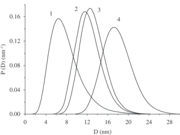
Figure 4. Particle size distributions of MgFe2O4 samples as prepared and
annealed at different temperatures. The numbers refer to the annealing condi-tions given in Table 1: 1, as prepared; 2, annealed at 400 °C; 3, annealed at 500 °C, 4, annealed at 600 °C.
4. Conclusions
The sol-gel/combustion technique has been used to prepare MgFe2O4 nanoparticles, which were annealed at different temperatures. X ray diffraction patterns show broad peaks in the positions corre-sponding to the crystal structure of magnesioferrite. Mössbauer spectra exhibit superparamagnetic behavior, confirming that the particles are in the nanometric range. Analysis of the broadening of the X ray lines shows that the average particle size increases with increasing annealing temperature. Analysis of the temperature dependence of the Mössbauer spectra of as prepared and annealed samples yields fairly narrow log-normal distributions of particle sizes. The results suggest that the sol-gel/ combustion technique may be used to synthesize nanosized MgFe2O4 powders whose average size can be controlled by subsequent annealing without appreciably changing the distribution of particle sizes.
References
1. Huang Y, Tang Y, Wang J, Chen Q. Synthesis of MgFe2O4 nanocrystallites under mild conditions. Materials Chemistry and Physics. 2006;
97(2-3):394-397.
2. de Biasi RS, Figueiredo ABS, Fernandes AAR, Larica C. Synthesis of cobalt ferrite nanoparticles using combustion waves. Solid State Communications. 2007; 144(1):15-17.
3. Bruker AXS. DIFFRACplus TOPAS. Available from: <http://www.bruker-axs.de/topas.html>. Access in: 02/04/2009.
4. Chen Q, Rondinone AJ, Chakoumakos BC, Zhang ZJ. Synthesis of superparamagnetic MgFe2O4 nanoparticles by coprecipitation. Journal of Magnetism and Magnetic Materials. 1999; 194(1-3):1-7.
5. de Biasi RS, Folly WSD. Use of ferromagnetic resonance to determine the size distribution of magnetic particles. Physica B. 2002; 321(1-4):117-119.
6. de Biasi RS, Gondim EC. Use of ferromagnetic resonance to determine the size distribution of gamma-Fe2O3 nanoparticles. Solid State Communications. 2006; 138(6):271-274.
Tc TP T dT
P T dT
= ( )
( ) 0
0
∞ ∞
∫
∫ (4)