No n-ne uro nal ce lls are no t the lim iting
facto r fo r the lo w axo nal re ge ne ratio n
in C5 7 BL/6 J m ice
Departamentos de 1Anatomia and 2Fisiologia e Biofísica,
Universidade Estadual de Campinas, Campinas, SP, Brasil A.L.R. O liveira1
and F. Langone2
Abstract
Peripheral axonal regeneration was investigated in adult male mice of the C57BL/6J (C), BALB/cJ (B) and A/J (A) strains and in their F1 descendants using a predegenerated nerve transplantation model. Four types of transplants were performed: 1) isotransplants between ani-mals of the C, B and A strains; 2) donors of the C strain and recipients of the C x B and C x A breeding; 3) donors of the B strain and recipients of the C x B breeding, and 4) donors of the A strain and recipients of the C x A breeding. Donors had the left sciatic nerve transected and two weeks later a segment of the distal stump was transplanted into the recipient. Four weeks after transplantation the regenerated nerves were used to determine the total number of regen-erated myelinated fibers (TMF), diameter of myelinated fibers (FD) and myelin thickness (MT). The highest TMF values were obtained in the groups where C57BL/6J mice were the donors (C to F1 (C x B) = 4658 ± 304; C to F1 (C x A) = 3899 ± 198). Also, A/J grafts led to a significantly higher TMF (A to F1 (C x A) = 3933 ± 565). Addition-ally, isotransplant experiments showed that when the nerve is previ-ously degenerated, C57BL/6J mice display the largest number of myelinated fibers (C to C = 3136 ± 287; B to B = 2759 ± 170, and A to A = 2835 ± 239). We also observed that when C57BL/6J was the graft donor, FD was the highest and MT did not differ significantly when compared with the other groups. These morphometric results reinforce the idea that Schwann cells and the nerve environment of C57BL/6J provide enough support to the regenerative process. In this respect, the present results support the hypothesis that the non-neu-ronal cells, mainly Schwann cells, present in the sciatic nerve of C57BL/6J mice are not the main limiting factor responsible for low axonal regeneration.
Co rre spo nde nce A.L.R. O liveira
Departamento de Anatomia Instituto de Biologia, UNICAMP Caixa Postal 6109
13083-970 Campinas, SP Brasil
Fax: + 55-19-289-3124 E-mail: alroliv@ unicamp.br
Research supported by CNPq. Publication supported by FAPESP.
Received April 3, 2000 Accepted August 14, 2000
Ke y wo rds
·Nerve regeneration
·Schwann cells
·Isogenic mice
·Nerve transplantation
·Tubulization
Intro ductio n
After axotomy, a sequence of events oc-curs distally to the lesion, resulting in nerve degeneration (1-4). This process, called Wallerian degeneration, is initiated by a lo-cal inflammation when several signal
participates in the myelin and axonal clear-ance. Finally, the Schwann cells will support and guide the regenerating axons to the tar-get organs by lining themselves up, forming the bands of Büngner (8,9,12,13). They also produce neurotrophic factors such as NGF, BDNF, NT-3, NT-4 and CNTF that support neuronal survival (14-18).
In the literature concerning nerve regen-eration, the importance of non-neural cells, such as Schwann cells and macrophages, has been emphasized (19-23). Less attention has been given to the intrinsic regenerative neu-ronal characteristics, which are also an impor-tant limiting factor for the nerve regenerative process (24,25). In this regard, neuronal sur-vival after axotomy is a prerequisite for regen-eration and is facilitated by various trophic factors (21). Also, axotomized neurons must switch from a transmitting mode to a growth mode, expressing growth-associated proteins, such as GAP-43, as well as different neu-ropeptides and cytokines (26). Axonal sprouts must reach the distal nerve stump at a time when its growth support is optimal (27,28). If such coordination does not occur, nerve re-generation may be diminished and neuronal death may occur. In this respect, an interesting example is the C57BL/6J mouse strain, which shows delayed axonal regeneration after a peripheral lesion (29-31).
Lu et al. (29,30) observed that C57BL/6J mice have a lower axonal regeneration po-tential after a crush lesion compared with BALB/cJ, A/J, C3H/HeJ and DBA/1J mice. These results were confirmed by Lainetti et al. (31) in a study of axonal regeneration in the same strains, using the nerve tubulization technique. They also observed that C57BL/ 6J displays extensive dorsal root ganglion death after lesion.
The first hypothesis to explain the low axonal regeneration of C57BL/6J mice was to relate it to its intrinsic deficiency in mac-rophage response to injury. Nevertheless, this was not experimentally confirmed (29). Also, Lu et al. (30) investigated the genetics
of this deficiency in recombinant mouse strains, and proposed that it could be a result of a temporary deficiency rather than a per-manent neuronal impairment, which seems to involve mainly sensitive neurons.
Based on the facts reported above, it is still not well understood if the regenerative deficiency of C57BL/6J mice is mainly a result of the intrinsic neuronal characteris-tics or if it is due to the nerve microenviron-ment and non-neural cell features. In the present study we propose an in vivo model based on predegenerated allo- and isograft transplants using the C57BL/6J (C), A/J (A), BALB/cJ (B) strains, and their F1 descen-dants. This method was used in order to determine the relative importance of neu-ronal and non-neuneu-ronal C57BL/6J cells in peripheral nerve regeneration after sciatic nerve transection.
Mate rial and Me tho ds
Anim als
In this study, fifty-one 8 to 10-week-old male mice were used. These mice were from C57BL/6J (N = 15), BALB/cJ (N = 12), A/J (N = 12) isogenic strains, and their F1 de-scendants: F1 (C57BL/6J x A/J) [F1 (C x A)] (N = 6) and (C57BL/6J x BALB/cJ) [F1 (C x B)] (N = 6). Three additional male mice from C57BL/6J, A/J and BALB/cJ isogenic strains were used for the predegenerated nerve graft morphological study.
Two main groups were studied: the first was based on sciatic nerve allotransplants and C57BL/6J mice were used as graft donor for both F1 (C x A) (N = 3) and F1 (C x B) (N = 3). Also, A/J and BALB/cJ mice were used as donors for F1 (C x A) (N = 3) and F1 (C x B) (N = 3), respectively.
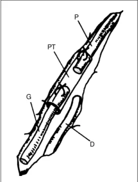
Surgical pro ce dure s (Figure 1)
Under deep anesthesia (pentobarbital, 50 mg/kg, ip), donors received a left sciatic nerve transection at the obturator tendon level and the proximal stump was ligated to avoid regen-eration. Two weeks later, the donor transected nerve was exposed and the distal stump was dissected out for transplantation. One milli-meter of the proximal end of the predegenerated donor nerve was introduced and attached to a 3-mm long polyethylene tube. At the same time, the recipients sciatic nerve was exposed and transected at the mid-thigh level and its proximal stump was inserted and sutured into the polyethylene tube containing the donor predegenerated nerve, with a 1-mm gap left between the stumps.
Spe cime n pre paratio n and mo rpho lo gic and
m o rpho m e tric analysis
Four weeks after transplantation, recipi-ents were perfused transcardially with Kar-novsky solution (2% glutaraldehyde and 1% paraformaldehyde) and the regenerated sci-atic nerves were dissected out. The speci-mens were post-fixed with 2% osmium tetrox-ide and processed for Araldite embedding. Transverse semithin sections (0.5 µm thick) were obtained at the midpoint of the polyeth-ylene tube and stained with toluidine blue and the total number of regenerated axons (TMF) was counted. Also, the TMF in the contralateral sciatic nerve (CL) was obtained and a regeneration rate (RR) was calculated (RR = TMF/CL). Transverse ultrathin sec-tions were obtained and 4 fields of each specimen were photographed with the elec-tron microscope (2000X) covering an area of at least 30% of the cross-sectional area of the nerve. Sampling bias was avoided by spreading the micrographs systematically over the entire cross-section according to the scheme proposed by Mayhew and Sharma (32). The negatives were copied (X3) and the diameter of myelinated fibers and myelin
thickness were measured on a digitizing table (SummaSketch® II Professional) using Sigma
Scan Measurement®
software. Axon diam-eter (D) was calculated from the perimdiam-eter (P) by applying the formula D = P/p. Data are reported as means ± SD. One-way ANOVA and the Newman-Keuls test (P<0.05) were used for statistical analysis.
Re sults
Mo rpho lo gical o bse rvatio ns
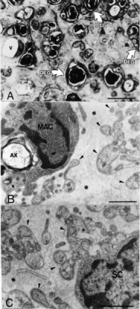
Predegenerated nerves (Figure 2). Two weeks after transection, predegenerated sci-atic nerves from the three isogenic mice strains showed slight morphologic differ-ences. The endoneural microenvironment was characterized by the presence of many groups of myelinated fibers undergoing Wallerian degeneration. These degenerating fibers showed accumulated mitochondria and membranous electron-dense bodies shrunk together with the disorganized myelin sheath in the Schwann cell cytoplasm. The Schwann cells were lined up into the so-called bands of Büngner, identified ultrastructurally by
P
PT
G
D
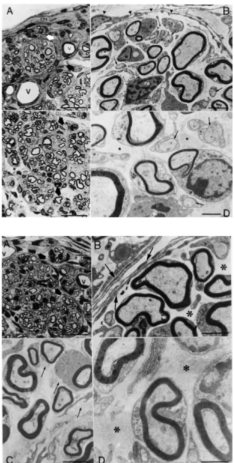
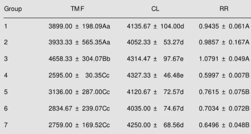
ously transplanted graft. Transverse semi-thin sections of the regenerated nerves at the tube midpoint showed regenerated myeli-nated fibers organized into small bundles surrounded by perineural-like cells and fi-broblasts. Both isografts and allografts showed the same morphologic aspect when examined with the electron and light micro-scopes. The epineurium was composed of multiple layers of flattened cells separated by collagen fibrils. These cells resembled those of the bundles but did not have a basal lamina. The myelinated fibers were of dif-ferent dimensions typically associated with just one Schwann cell, while the nonmyeli-nated axons were surrounded by several Schwann cell cytoplasmic projections. In the endoneural microenvironment we iden-tified fibroblasts, macrophages and eventu-ally mast cells. No immunological reaction resembling graft rejection was observed.
Mo rpho m e tric re sults
Number of regenerated axons (Table 1, Figure 5). The TMF 4 weeks after graft transplantation surgery is shown in Table 1. The allotransplant group in which C57BL/6J was the donor and F1 (C x B) the recipient showed the highest number of regenerated axons, followed by the groups in which C57BL/6J and A/J were donors (P<0.05, Newman-Keuls test). On the other hand, when contralateral myelinated fiber num-bers were computed for RR calculation, no statistical difference was observed between groups 1, 2 and 3 (P>0.05, Newman-Keuls test). The other groups did not differ con-cerning the number of regenerated axons.
Axonal diameter and myelin thickness (Table 2)
The diameters of myelinated fibers dif-fered significantly between groups 4 weeks after graft transplantation (P<0.05, Newman-Keuls test). When C57BL/6J was the al-lograft donor (groups 1 and 3), a significant-Figure 2 - A, Transverse section
of a predegenerated nerve 2 w eeks after sciatic nerve tran-sect ion. The endoneural m i-croenvironment is enlarged and there are m any degenerating axons (DEG). A blood vessel is also observed (V). Bar = 10 µm.
B, Electron microscopic trans-verse section of a sciatic nerve 2 w eeks after transection. A mac-rophage (M AC) is observed de-grading a myelinated fiber (AX) in the endoneural microenviron-ment. Also, many Schw ann cell projections (arrow heads) sur-rounded by the basal lamina are observed betw een the endoneu-ral collagen fibrils (* ). Bar = 1 µm. C, Detail of a Schw ann cell (SC) w ith its cytoplasmic projec-tions (arrow heads). The w hole cell is surrounded by the basal lamina and endoneural collagen fibrils (* ). Bar = 1 µm.
their characteristic basal lamina. Also mac-rophages were observed and distinguished from Schwann cells by their lack of basal lamina. These cells were usually associated with myelin debris and degenerating axons. The dimensions of the epineurium, perineu-rium and endoneuperineu-rium were increased mainly due to cell proliferation and overproduction of extracellular matrix components such as collagen fibrils.
previ-Figure 3 - A, Transverse semithin section of a regener-ated nerve 4 w eeks after graft transplantation. Donor: C57BL/6J, recipient: F1 (C x A). The epineurium (EPI) is composed of multiple layers of flattened cells and the regenerated nerve fibers are organized into small bundles (arrow s). Blood vessels (V) are also observed. Bar = 12.5 µm. B, Electron microscopic view of a regenerated nerve 4 w eeks after graft transplantation. Donor: BALB/cJ, recipient: F1 (C x B). Detail of a nerve bundle surrounded by perineural cells organized in con-centric layers (arrow heads). M yelinated fibers, nonmy-elinated fibers (arrow s) and a fibroblast (FB) are ob-served in the endoneural microenvironment. Bar = 2 µm. C, Transverse semithin section of a regenerated nerve 4 w eeks after graft transplantation. Donor: C57BL/6J, recipient: F1 (C x B). The axons are orga-nized into bundles containing many regenerated nerve fibers (arrow s). Bar = 12.5 µm. D, Electron microscopic view of a regenerated nerve 4 w eeks after graft trans-plantation. Donor: C57BL/6J, recipient: F1 (C x B). De-tail of the endoneural microenvironment show ing my-elinated and nonmymy-elinated regenerated axons (ar-row s) associated w ith Schw ann cells and surrounded by collagen fibrils (* ). Bar = 1 µm.
Table 1 - Total number of regenerated axons (TM F) obtained at the polyethylene tube midpoint level 4 w eeks after graft transplantation.
The number of myelinated fibers counted in the contralateral nerve (CL) and the regeneration rate (RR) are also presented. 1) Donor: C57BL/6J (C), recipient: F1 (C x A); 2) donor: A/J (A), recipient: F1 (C x A); 3) donor: C57BL/6J, recipient: F1 (C x B); 4) donor: BALB/cJ (B), recipient: F1 (C x B); 5) donor: C57BL/6J, recipient: C57BL/6J; 6) donor: A/J, recipient: A/J; 7) donor: BALB/cJ, recipient: BALB/cJ. The values are reported as means ± SD. The capital letters indicate the comparison betw een the means of the 7 experimental groups, and the small letters show the comparison betw een each experimental group and its normal group. Different paired letters indicate statistical difference (P<0.05, New mann-Keuls test). N = 3 for all groups.
Group TM F CL RR
1 3899.00 ± 198.09Aa 4135.67 ± 104.00d 0.9435 ± 0.061A
2 3933.33 ± 565.35Aa 4052.33 ± 53.27d 0.9857 ± 0.167A
3 4658.33 ± 304.07Bb 4314.47 ± 97.67e 1.0791 ± 0.049A
4 2595.00 ± 30.35Cc 4327.33 ± 46.48e 0.5997 ± 0.007B
5 3136.00 ± 287.00Cc 4120.67 ± 72.57d 0.7615 ± 0.075B
6 2834.67 ± 239.07Cc 4035.00 ± 74.67d 0.7034 ± 0.072B
7 2759.00 ± 169.52Cc 4250.00 ± 68.56d 0.6496 ± 0.048B
Table 2 - Fiber diameter (FD) and myelin thickness (M T) obtained at the polyethylene tube midpoint 4 w eeks after graft transplantation.
Both values obtained for the regenerated and contralateral nerves are presented. 1) Donor: C57BL/6J (C), recipient: F1 (C x A); 2) donor: A/J (A), recipient: F1 (C x A); 3) donor: C57BL/6J, recipient: F1 (C x B); 4) donor: BALB/cJ (B), recipient: F1 (C x B); 5) donor: C57BL/6J, recipient: C57BL/6J; 6) donor: A/J, recipient: A/J; 7) donor: BALB/cJ, recipient: BALB/cJ. The values are reported as means ± SD. The capital letters indicate the comparison betw een the means of the 7 experimental groups, and the small letters show the comparison betw een each experimental group and its normal group. Different paired letters indicate statistical difference (P<0.05, New mann-Keuls test). N = 3 for all groups.
Group FD M T
Regenerated Contralateral Regenerated Contralateral
1 2.30 ± 0.88Aa 3.21 ± 1.21c 0.29 ± 0.10Aa 0.66 ± 0.25b
2 2.09 ± 0.86Bb 3.21 ± 1.21c 0.30 ± 0.09Aa 0.66 ± 0.25b
3 2.24 ± 0.92Aa 3.65 ± 1.60d 0.29 ± 0.09Aa 0.72 ± 0.30c
4 2.05 ± 0.86Bb 3.65 ± 1.60d 0.31 ± 0.09Aa 0.72 ± 0.30c
5 1.98 ± 0.86Bb 3.55 ± 1.61d 0.31 ± 0.10Aa 0.70 ± 0.29c
6 2.00 ± 0.76Bb 3.27 ± 1.30c 0.30 ± 0.08Aa 0.68 ± 0.29b
7 1.87 ± 0.81Bb 3.27 ± 1.45c 0.30 ± 0.08Aa 0.67 ± 0.26b
R
e
g
e
n
e
ra
ti
o
n
r
a
te
1.2
1.0
0.8
0.6
0.4
0.2
0.0
* *
*
C/CA A/CA C/CB B/CB A/A C/C B/B Experimental groups
Figure 5 - Regeneration rate obtained by the division of total number of axons in the regenerated nerves by the num ber in the contralateral nerves. Experim ental groups: C/CA - donor: C57BL/6J, recipient: F1 (C x A); A/CA - donor: A/J, recipient: F1 (C x A); C/CB - donor: C57BL/6J, recipient: F1 (C x B); B/CB - donor: BALB/cJ, recipient: F1 (C x B); C/C - donor: C57BL/6J, recipient: C57BL/6J; A/A - donor: A/J, recipient: A/J; B/B - donor: BALB/cJ, recipient: BALB/cJ. * P<0.05 compared to the other groups (New man-Keuls test).
ly larger axonal diameter was observed (P<0.05). The other experimental groups did not differ significantly from one another. In all groups, axonal diameter was significantly smaller than control. Myelin thickness did not differ between the experimental groups but was always smaller than control (P<0.05).
D iscussio n
macro-phage migration and activity in C57BL/6J was demonstrated not to be statistically dif-ferent from the other strains, there is the possibility that Schwann cells may be in-volved. In this regard, several studies have emphasized the contribution of Schwann cells to the degenerative and regenerative pro-cesses after peripheral nerve injury (9,10,22). One way to investigate this hypothesis would be to transplant segments from a pe-ripheral nerve of C57BL/6J mice to recipi-ents from another strain with a higher regen-erative potential. Nevertheless, the direct al-lotransplant between strains would result in graft rejection since they have a different major histocompatibility complex (34). Con-sidering this fact, in the present study we have proposed the utilization of F1 mice obtained by breeding C57BL/6 with A/J and BALB/cJ as graft recipients. Also, in order to have a higher number of Schwann cells in the graft already organized into the bands of Büngner, we have performed predegenera-tion of the donor nerves by transecting them two weeks before transplantation (5,6,35-37). The morphologic results obtained two weeks after transplantation showed no rejec-tion of the nerve grafts, since immunocom-petent cells were almost absent.
Myelin thickness did not differ between experimental groups. Also, axonal diameter was significantly wider in the allotransplant groups where the C57BL/6J strain was the donor. These facts demonstrate that C57BL/ 6J Schwann cells do not differ in myelin synthesis compared to those from the A/J and BALB/cJ strains. Also, the C57BL/6J nerve microenvironment is able to support axonal regeneration.
The TMF was higher in the allotransplant groups where the C57BL/6J and A/J strains were donors. Additionally, the group in which C57BL/6J mice were donors and F1 (C x B) mice were the recipients showed the highest TMF. Since the TMF counted for the con-tralateral nerves was not even for all groups (Table 1), we calculated the ratio between
the ipsilateral and contralateral sides (RR). In this regard, no statistical RR differences were found between F1 recipients that re-ceived C57BL/6J or A/J grafts. This ratio was higher than observed in F1 recipients, which received BALB/cJ grafts.
These results reinforce the hypothesis that the nerve microenvironment of C57BL/ 6J mice does provide support to axonal generation. Nevertheless, the low axonal re-generation previously reported indicated a substantial sensory neuron death, a fact that would contribute to establishing a perma-nent deficit (31). It is possible that the de-scribed neuronal death after sciatic nerve transection is the result of a mismatch in the timing of the neurons needed for neurotrophic substances and its production by the cells in the distal stump. The predegeneration of the graft used in our model may compensate for such delay, explaining the higher number of regenerated axons in the groups where C57BL/6J mice were donors.
Interestingly, basically using a similar transplantation model in Trembler mice, Pol-lard and McLeod (38) have demonstrated that the Schwann cells were responsible for the demyelinating neuropathy of this strain (39,40). Based on the facts reported above, we believe that the experimental model used in our study would be able to show eventual Schwann cell abnormalities, which could be morphologically and morphometrically ob-served. In this respect, such cells and the nerve microenvironment are probably not the main factor contributing to the low ax-onal regeneration of C57BL/6J mice.
these data, it is possible that the non-neu-ronal cell response to injury is delayed in C57BL/6J mice. Such delay was reduced because of the isograft predegeneration, al-lowing early Wallerian degeneration steps to occur already before transplantation. On the other hand, soon after injury, the capacity of C57BL/6J neurons to recognize extracellu-lar stimuli may be impaired, resulting in an initial delay in axonal regeneration. In this respect, apoptotic mechanisms may be in-volved that might contribute to explaining
the previously described dorsal root gan-glion death after sciatic nerve transection (Oliveira ALR, unpublished results).
Ackno wle dgm e nts
We are grateful to Prof. Dr. Mary Anne H. Dolder for a careful review of the English text, and to Mr. Marco Aurélio and Mr. Norivaldo Celestino for excellent technical assistance.
Re fe re nce s
1. Lunn ER, Brow n M C & Perry VH (1990). The pattern of axonal degeneration in the peripheral nervous system varies w ith dif-ferent types of injury. Journal of Neuro-science, 35: 157-165.
2. Cabaud HE, Rodkey WG & Nemth TJ (1982). Progressive ultrastructural changes after peripheral nerve transection and re-pair. Journal of Hand Surgery, 7: 353-365. 3. O’Daly JA & Imaeda T (1967). Electron microscopy study of Wallerian degenera-tion in cutaneous nerves caused by me-chanical injury. Journal of Neuropathology and Experimental Neurology, 17: 744-766. 4. Thomas PK (1964). Changes in the endo-neurial sheaths of peripheral myelinated nerve fibers during Wallerian degenera-tion. Journal of Anatomy, 98: 175-182. 5. Bruck W, Bruck C, M arushak B & Fried RL
(1995). M echanisms of macrophage re-cruitment in Wallerian degeneration. Acta Neuropathologica, 89: 363-367. 6. Hall SM (1993). Observations on the
progress of Wallerian degeneration in transected peripheral nerves of C57BL/ Wld mice in the presence of recruited macrophages. Journal of Neurocytology, 22: 480-490.
7. Taskinen HS, Olsson T, Bucht A, Khademi M , Svelander L & Royotta M (2000). Pe-ripheral nerve injury induces endoneural expression of IFN-gamma, IL-10 and TNF-alpha mRNA. Journal of Neuroimmunolo-gy, 102: 17-25.
8. Stoll G, Griffin JW, Li CY & Trapp BD (1989). Wallerian degeneration in the pe-ripheral nervous system: participation of both Schw ann cells and macrophages in myelin degradation. Journal of Neurocy-tology, 18: 671-683.
9. Beuche W & Fried RL (1984). The role of non-resident cells in Wallerian degenera-tion. Journal of Neurocytology, 13: 767-796.
10. Gibson JD (1979). The origin of the neural macrophage: a quantitative ultrastructural study of cell population changes during Wallerian degeneration. Journal of Ana-tomy, 129: 1-19.
11. M onaco S, Gehrmann J, Raivich G & Kreutzberg GW (1992). M HC-positive, ramified macrophages in the normal and injured rat peripheral nervous system.
Journal of Neurocytology, 21: 623-634. 12. Abernethy DA, Thomas PK, Rud A & King
RHM (1994). M utual attraction betw een emigrant cells from transected dener-vated nerve. Journal of Anatomy, 184: 239-249.
13. Son Y-J & Thompson WJ (1995). Schw ann cell processes guide regeneration of pe-ripheral axons. Neuron, 14: 125-132. 14. Funakoshi H, Frisen J, Barabany G,
Timmusk T, Zachrisson O, Verge VM K & Persson H (1993). Expression of mRNAs for neurotrophins and their receptors af-ter axotomy of the sciatic nerve. Journal of Cell Biology, 123: 455-464.
15. Lindsay RM (1994). Neurotrophins and re-ceptors. Progress in Brain Research, 103: 3-14.
16. M illet FD (1994). Nerve grow th factor and neuronal gene expression. Progress in Brain Research, 103: 23-34.
17. Sendtner M , Stockli KA & Thoenen H (1992). Synthesis and localization of cili-ary neurotrophic factor in the sciatic nerve of the adult rat after lesion during regen-eration. Journal of Cell Biology, 118: 139-148.
18. M eyer M , M atsuoka I, Wetmore C, Olson L & Thoenen H (1992). Enhanced synthe-sis of brain-derived neurotrophic factor in the lesioned peripheral nerve: different mechanisms are responsible for the regu-lation of BDNF and NGF mRNA. Journal of Cell Biology, 119: 45-54.
19. Dahlin LB (1995). Prevention of macro-phage invasion impairs regeneration in nerve grafts. Brain Research, 679: 274-280.
20. M adison RD & Archibald SJ (1994). Point sources of Schw ann cells result in grow th into a nerve entubulation repair site in the absence of axons: effects of freezethaw -ing. Experimental Neurology, 128: 266-275.
21. Frostick SP, Yin Q & Kemp GJ (1998). Schw ann cells, neurotrophic factors, and peripheral nerve regeneration. M icrosur-gery, 18: 397-405.
22. M idha R, M ackinnon SE & Becker LE (1994). The fate of Schw ann cells in pe-ripheral nerve allografts. Journal of Neu-ropathology and Experimental Neurology, 53: 316-322.
23. Hall S (1986). The effect of inhibiting Schw ann cell mitosis on the re-innerva-tion of acellular autografts in the peripher-al nervous system of the mouse. Neuro-pathology and Applied Neurobiology, 12: 401-414.
24. Faw cett JW (1992). Intrinsic neuronal de-terminants of regeneration. Trends in Neurosciences, 15: 5-8.
25. Faw cett JW & Keynes RJ (1990). Periph-eral nerve regeneration. Annual Review of Neuroscience, 13: 43-60.
re-generation. M olecular Neurobiology, 14: 67-116.
27. Donat J & Wisniew sky HM (1973). The spatio temporal pattern of Wallerian de-generat ion in m am m alian peripheral nerves. Brain Research, 53: 41-53. 28. Taira E, Takaha N & M iki N (1993).
Extra-cellular matrix proteins w ith neurite pro-moting activity and their receptors. Neu-roscience Research, 17: 1-8.
29. Lu X, Richardson PM , Gervais F & Skaeme E (1990). A deficiency of axonal regenera-tion in C57BL/6J mice. Brain Research, 510: 144-146.
30. Lu X, Skaeme E & Richardson PM (1994). Studies of axonal regeneration in C57BL/ 6J and A/J mice. Brain Research, 652: 174-176.
31. Lainetti RD, Pereira FC & Da-Silva CF (1995). Reduced sensory neuron regen-eration by C57BL/6J mice. Brazilian Jour-nal of M edical and Biological Research,
28: 781-785.
32. M ayhew TM & Sharma AK (1984). Sam-pling schemes for estimating nerve fibre size. II. M ethods for unifascicular nerve trunks. Journal of Anatomy, 139: 59-66. 33. Goldberg DJ & Wu DY (1994). Regulation
of events w ithin the grow th cone by ex-tracellular clues: tyrosine phosphorylation.
Progress in Brain Research, 103: 75-84. 34. Evans PJ, M idha R & M ackinnon SE
(1994). The peripheral nerve allograft: a comprehensive review of regeneration and neuroimmunology. Progress in Neu-robiology, 43: 187-233.
35. Danielsen N & Kerns JM (1994). Pre-de-generated nerve grafts enhance regen-eration by shortening the initial delay pe-riod. Brain Research, 666: 250-254. 36. Kerns JM , Danielsen N, Holquist B, Kanje
M & Lundborg G (1993). The influence of pre-degeneration on regeneration through nerve grafts in rats. Experimental
Neurol-ogy, 122: 28-36.
37. Bryan DJ, Wang KK & Summehayes IC (1999). M igration of Schw ann cells in pe-ripheral nerve regeneration. Journal of Reconstructive M icrosurgery, 15: 591-596.
38. Pollard JD & M cLeod JG (1980). Nerve grafts in the Trembler mouse. An electro-physiological and histological study. Jour-nal of the Neurological Sciences, 46: 373-383.
39. Heape AM , Bessoule JJ, Boiron-Sargueil F, Garbay B & Cassagne C (1995). Sphin-golipid metabolic disorders in Trembler mouse peripheral nerves in vivo result from an abnormal substrate supply. Jour-nal of Neurochemistry, 65: 1665-1673. 40. Sutter U, Welcher AA, Ozcelik T & Snipes